Methods and system for cardiac mapping for atrial fibrillation using balloon based catheters utilizing medical images (CT or MRI in segments) and left ventricular lead placement for cardiac re-synchronization therapy (CRT)

a technology of cardiac mapping and atrial fibrillation, which is applied in the field of cardiac mapping for atrial fibrillation ablations, can solve the problems of increased morbidity and mortality, increased atrial fibrillation, and general decrease in the quality of life of those afflicted with atrial fibrillation

Active Publication Date: 2021-12-28
AMERICAN MEDICAL TECH LLC
View PDF16 Cites 3 Cited by
  • Summary
  • Abstract
  • Description
  • Claims
  • Application Information

AI Technical Summary

Benefits of technology

[0104]In another embodiment, the cardiac system is used for aiding in the proper placement of left ventricular lead.

Problems solved by technology

Atrial fibrillation is associated with increased morbidity and mortality, and in particular, a general decrease in quality of life for those afflicted with atrial fibrillation.
Patients are at an increased risk of stroke unless they are treated adequately with anticoagulants.
Anticoagulant treatment however, increases the patient's risk of bleeding, which carries with it is own set of dangers.
Medications currently available for treating atrial fibrillation have proven to be only moderately effective in decreasing the incidence of recurrent atrial fibrillation, and these medications do not decrease the patient's risk of having a stroke.
Moreover, the AV nodal artery passes near the mouth of the coronary sinus, the ablation may conceivably damage this vessel.
Painless ablation—RF ablation may be painful to the patient under conscious sedation through direct stimulation of cardiac sensory nerves or pericardial or collateral Whisler visceral irritation, particularly when ablating your thin-walled or venous structures such as posterior left atrium, coronary sinus, or posterior cable tricuspid Isthmus.
This first select procedures associated with substantial patient discomfort, the use of cryoablation may theoretically result in lower anesthetic and analgesic requirements.
The esophagus may however be, in a position so as to overlie one or more of these circles, thereby making the desired encirclement difficult or impossible.
A significant and lethal complication of atrial fibrillation ablation is the accidental creation of an atrial esophageal fistula following the development of lesions on the posterior wall of the left atrium.
Because the esophagus is generally in close position to the posterior wall of the left atrial, thermal injury may be communicated to the esophageal wall resulting in disruption of the wall and formation of the atrial esophageal fistula.
A major limitation of this approach is that it fails to account for the variability in the thickness of the posterior LA wall and the presence of peri-esophageal connective tissue—important determinants of esophageal heating.
Thus, empirically limiting the power and duration of RF applications may be insufficient to prevent esophageal thermal injury in all patients.
Thus, it has become more common for accidental injury to the phrenic nerve to occur.
The phrenic nerve is responsible for operation of the diaphragm, and thus, injury to the phrenic nerve can be quite catastrophic.
Again, the location of the esophagus may hinder application of this sufficient energy to successfully ablate enough energy of the left atrium to prevent recurrence of atrial fibrillation.

Method used


Image

  • Methods and system for cardiac mapping for atrial fibrillation using balloon based catheters utilizing medical images (CT or MRI in segments) and left ventricular lead placement for cardiac re-synchronization therapy (CRT)
  • Methods and system for cardiac mapping for atrial fibrillation using balloon based catheters utilizing medical images (CT or MRI in segments) and left ventricular lead placement for cardiac re-synchronization therapy (CRT)
  • Methods and system for cardiac mapping for atrial fibrillation using balloon based catheters utilizing medical images (CT or MRI in segments) and left ventricular lead placement for cardiac re-synchronization therapy (CRT)

Examples


Embodiment Construction

[0190]The following description is of the best mode presently contemplated for carrying out the disclosure. This description is not to be taken in a limiting sense, but is made merely for the purpose of describing the general principles of the disclosure.

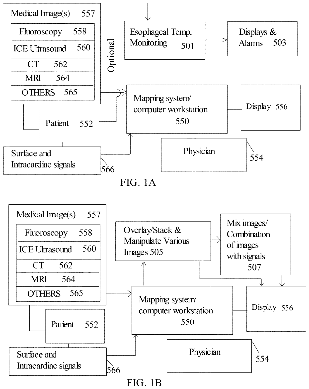
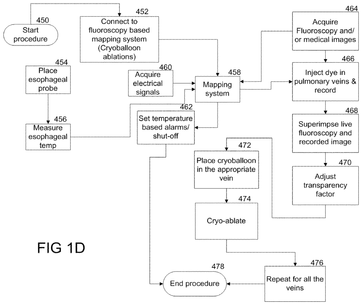
[0191]In the methods and system of this disclosure, medical images based cardiac mapping / electrophysiology tools is disclosed for cardiac ablations for arrhythmias. The methods and system of this disclosure can be employed with a methodology for monitoring esophageal temperature. In another embodiment, the mapping system and mapping methodology can also be used without the use of temperature monitoring.

Definitions

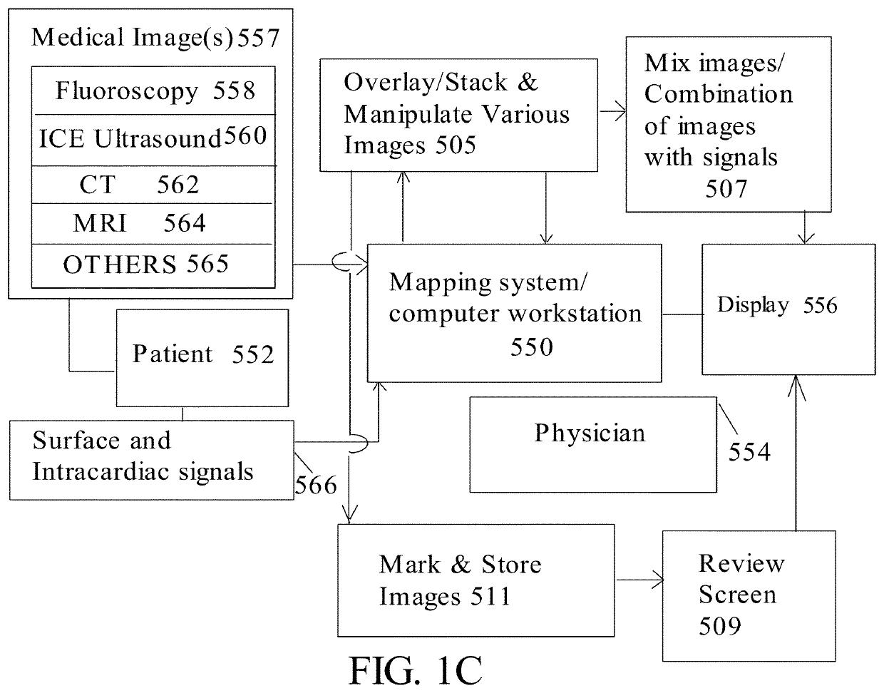
[0192]Cardiac mapping systems are navigation and / or guidance systems used during a cardiac ablation procedure which includes making maps, guiding physicians to the optimal placement of the catheters. Cardiac mapping systems may utilize various types of medical images or a combination of medical images, or overlays of differ...


PUM


Abstract

Methods and system for atrial fibrillation mapping utilizing cardiac mapping based on medical image(s). The methods and system also adapted for any balloon based catheters including cryoballoon catheter, laser balloon catheter, or “hot balloon” catheter, as well as circular catheters. The methods and system includes overlaying of two or more images on top of each other (where the images are of various types or modalities) and aligning the images, where the transparency between the images can be adjusted for navigating and optimal placement of the balloon based catheters or circular catheters. The cardiac system also adopted for guiding optimal placement of the left ventricular (LV) lead placement for cardiac re-synchronization (CRT) therapy, and for mapping and displaying activation times from various branches of the coronary sinus for optimizing CRT therapy.

Description

[0001]This application is a Continuation-In-Part (CIP) of U.S. application Ser. No. 15 / 817,664 filed Nov. 20, 2017, having the projected U.S. Pat. No. 10,413,185 and issue date of Sep. 17, 2019, which is a Continuation-In-Part (CIP) of U.S. application Ser. No. 15 / 209,265 filed Jul. 13, 2016.FIELD OF DISCLOSURE[0002]The present disclosure relates to cardiac mapping for atrial fibrillation ablations, more specifically to methods and system of mapping for atrial fibrillation ablations utilizing a balloon based catheter with optional esophageal temperature monitoring, and for guiding left ventricular (LV) lead placement and mapping.BACKGROUNDGeneral Background of Atrial Fibrillation[0003]Atrial fibrillation (AF) is the most prevalent cardiac arrhythmia. It affects 1% to 2% of the general population with an important increase in incidence with age. In the United States it is estimated that over 5 million people have atrial fibrillation, and because of our aging population the prevalence...

Claims


Application Information

Patent Timeline
Patent Type & Authority Patents(United States)
IPC IPC(8): A61B5/00A61B5/01G06T7/00A61B6/00A61B34/20A61B5/361A61B8/08A61B18/00A61B5/283
CPCA61B5/0044A61B5/0082A61B5/015A61B5/361A61B6/481A61B34/20G06T7/0012A61B5/0035A61B5/283A61B6/485A61B8/5261A61B2018/00577A61B2034/2051A61B6/5247A61B8/0883A61B8/12A61B8/445A61B2018/00351A61B5/367A61B5/6853A61B6/504A61B90/37A61B2090/376A61B2090/378A61B34/25A61B2034/105A61B2018/00839A61B2018/0022A61B2018/0212A61B18/1492A61B18/22A61B2018/00791A61B2018/00023A61F7/123A61F2007/126A61F2007/0056A61F2007/0095A61B2018/00666A61B2018/00714A61B2090/365A61B5/29
Inventor BOVEJA, BIRINDER R.WIDHANY, ANGELY
Owner AMERICAN MEDICAL TECH LLC
Why Eureka
Try Eureka