Optical sampling interface system for in vivo measurement of tissue

a tissue and optical sampling technology, applied in the field of optical sampling interface systems, can solve the problems of death and disability, relatively unskilled operators or users, and the degree of optical sampling reproducibility

Inactive Publication Date: 2003-04-10
GLT ACQUISITION
View PDF34 Cites 36 Cited by
  • Summary
  • Abstract
  • Description
  • Claims
  • Application Information

AI Technical Summary

Problems solved by technology

However, there are many demanding in vivo applications that cannot be performed in a laboratory setting but yet require a high degree of optical sample reproducibility.
Often, a relatively unskilled operator or user must perform the optical measurement.
Diabetes is a leading cause of death and disability worldwide and afflicts an estimated 16 million Americans.
A significant disadvantage of current monitoring techniques is that they discourage regular use due to the inconvenience and pain involved in drawing blood through the skin prior to analysis.
To date, no noninvasive techniques for the self-monitoring of blood glucose have been certified.
In all of these studies, diverse limitations were cited that would affect the acceptance of such a method as a commercial product.
Fundamental to all the studies is the problem of the small signal attributable to glucose, particularly in view of the difficulty in obtaining a reproducible sample of a given tissue volume, as a result of the complex and dynamic nature of the tissue.
The sampling problem is further accentuated by noting that the reported studies were performed under highly controlled conditions using skilled techniques rather than in a home environment by the consumer.
The measurement is further complicated by the heterogeneity of the sample, the multi-layered structure of the skin and the rapid variation related to hydration levels, changes in the volume fraction of blood in the tissue, hormonal stimulation, temperature fluctuations and blood analyte levels.
Therefore, these differences in refractive index between the different layers of the skin are too small to give a noticeable reflection.
However, variations in the physiological state and fluid distribution of tissue profoundly affect the optical properties of tissue layers and compartments over a relatively short period of time.
As a result, relatively small changes in concentration of impermeable solutes in the extracellular fluid, such as glucose, can cause tremendous changes in cell volume.
As herein described, the heterogeneous and dynamic nature of living human skin leads to sampling uncertainty in the in vivo measurement.
Variation in the repeated placement of the optical probe used for sampling at the measuring surface site can lead to sampling errors in two separate ways: first, variations in the location of the probe can cause a different tissue volume to be sampled, and second, varying the amount of pressure applied by the probe on the tissue can alter the optical scattering by the tissue, thereby changing the sampled tissue volume.
Furthermore, air gaps between the surface of the optical probe and the surface of the tissue being sampled give rise to variable surface reflection.
Variable surface reflection leads to a variable light launch into the tissue that in turn gives rise to an increase in nonlinear nature of the spectral measurements.
Certainly, a variable nonlinear measurement would be very difficult to calibrate.
Neither device provides a means for repeatably coupling a fiber optic probe to a tissue measurement site.
Furthermore, the apparatus of Kordis, et al. would not be suitable for repeatably coupling an optical probe to a tissue measurement site.
The Kanne device does not provide any means for repeatably placing a probe guide at a tissue measurement site.
It also has no way of monitoring the surface temperature at a tissue measurement site, or of minimizing surface temperature fluctuations and accumulation of moisture at a tissue measurement site.
Furthermore, the disclosed device allows an operator to immobilize an optical fiber so that it is maintained in a fixed position, but it does not offer a means of repeatably coupling a fiber optic probe to a tissue measurement site.
It also has no way of monitoring the surface temperature at a tissue measurement site, or of minimizing accumulated moisture and temperature fluctuations at the site.
Since they are known carcinogens, chlorofluorocarbons (CFC's) are unsuitable for use in preparations to be used on living tissue.
Furthermore, use of CFC's poses a well-known environmental risk.
Additionally, Messerschmidt's interface medium is formulated with substances that would be likely to leave artifacts in spectroscopic measurements.
Controlling variations in tissue volume sampled due to imprecise placement; and variable stretching of dermal collagen, leading to sampling volume uncertainty;
Sampling errors can be introduced into these measurements because of the difficulty of repeatedly placing the optical probe at the precise location used in preceding measurements, and repeatably producing the same nominal degree of tissue distortion and displacement.
With each small variation in the location of the probe, or variations in the amount of pressure resulting from the repeated probe contact events, a slightly different tissue volume is sampled, thereby introducing sampling errors into the measurements.
The interface between the optical probe and the skin surface at the tissue measurement site can also be a significant source of sampling error.
Since the underlying tissue is not homogenous, the surface skin at the tissue measurement site may be uneven, with frequent irregularities.
Coupling the relatively smooth surface of the optical probe with the irregular skin surface leads to air gaps between the two surfaces.
The air gaps create an interface between the two surfaces that adversely affects the measurement during optical sampling of tissue.
and has poor solvent properties with respect to leaching fatty acids and oils from the skin upon repeated application.
Furthermore, perfluoro compounds of this type are hydrophobic and are poor solvents; therefore they are unlikely to absorb water or other contaminants that will adversely affect the result during optical sampling.
While these compositions have the desired optical characteristics, their toxicity profiles and their solvent characteristics render them less desirable than the previously described perfluoro compounds.

Method used


Image

  • Optical sampling interface system for in vivo measurement of tissue
  • Optical sampling interface system for in vivo measurement of tissue
  • Optical sampling interface system for in vivo measurement of tissue

Examples


example 2

[0113] As a further illustration of the benefit of the guide placement system, sixty measurements were performed on a single subject with and without the guide positioning system. All spectra were collected using a custom built scanning near-infrared spectrometer. The instrument collected intensity spectra in diffuse reflectance from the forearm in the wavelength range 1050-2450 nm. The spectral sampling interval was 1 nm and the signal-to-noise ratio at the peak intensity was approximately 90 dB. The detectors used in the study were a combination of Indium-Gallium-Arsenide (InGaAs) and extended InGaAs detectors. The optical configuration consisted of a simple fiber-optic interface to the skin with a small (<2 mm) distance between the illumination and detection fibers. Reference spectra were recorded prior to each sample measurement by scanning a 99% SPECTRALON reflectance material and absorbance was calculated according to Equation (2). A cradle was developed to position the arm ov...

example 3

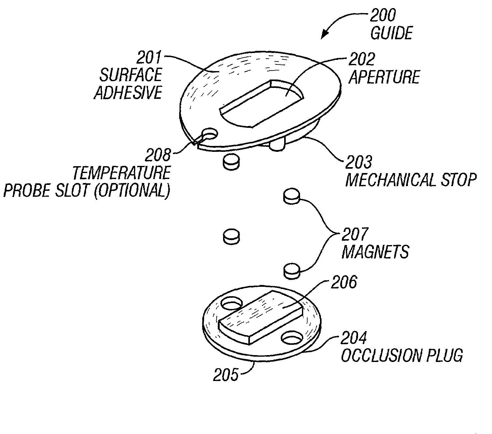
[0114] As a test of the benefit of the method of occlusion, 60 measurements were performed on a single subject using the apparatus described in Example 2. In the first set of measurements, 60 samples were collected using the guide positioning system without occlusion and absorbance was calculated as previously described. In the second set of measurements, 60 samples were collected with the use of both the guide positioning system and the preferred method of occlusion (a plug in the guide aperture). FIG. 6 shows the absorbance spectra collected without occlusion (FIG. 6A) and the absorbance spectra collected after occlusion (FIG. 6B). The decrease in surface variation associated with the water bands demonstrated the improved optical sampling realized as a result of the method of occlusion.

[0115] While the invented optical probe placement guide allows highly repeatable probe placement at a targeted tissue measurement site, the invention may also be used to produce small sampling varia...


PUM

PropertyMeasurementUnit
wavelengthaaaaaaaaaa
thickaaaaaaaaaa
thickaaaaaaaaaa

Abstract

An optical sampling interface system minimizes and compensates error resulting from sampling variations and measurement site state fluctuations. Components include: An optical probe placement guide having an aperture wherein the optical probe is received, facilitates repeatable placement accuracy on surface of a tissue measurement site with minimal, repeatable disturbance to surface tissue. The aperture creates a tissue meniscus that minimizes interference due to surface irregularities and controls variation in tissue volume sampled; an occlusive element placed over the tissue meniscus isolates the meniscus from environmental fluctuations, stabilizing hydration at the site and thus, surface tension; an optical coupling medium eliminates air gaps between skin surface and optical probe; a bias correction element applies a bias correction to spectral measurements, and associated analyte measurements. When the guide is replaced, a new bias correction is determined for measurements done with the new placement. Separate components of system can be individually deployed.

Description

[0001] This application is a Continuation-in-part of U.S. patent application Ser. No. 09 / 563,782, filed on May 2, 2000, hereby incorporated in its entirety by reference.[0002] 1. Field of the Invention[0003] The invention relates to optical sampling of tissue in vivo. More particularly, the invention relates to an optical sampling interface system that includes an optical probe placement guide, a means for stabilizing the sampled tissue, an optical coupler for repeatably sampling a tissue measurement site in vivo, and a means for compensating measurement bias.[0004] 2. Technical Background[0005] In vivo measurement of tissue properties and analytes using optically based analyzers requires that tissue measurement region be positioned and coupled with respect to an optical interface or probe. The requirements of an optical sampling interface system for such placement and coupling would depend upon the nature of the tissue properties and analytes under consideration, the optical techno...

Claims


Application Information

Patent Timeline
Patent Type & Authority Applications(United States)
IPC IPC(8): A61BA61B5/01A61B1/00A61B5/00A61B5/0205A61B5/145A61B5/1455A61K9/00G01N21/01G01N21/35
CPCA61B5/0059A61B5/0071A61B5/0075A61B5/01A61B5/061A61B5/14532A61B2562/146A61B5/1495A61B5/6824A61B5/6833A61B5/6843A61B2560/0233A61B2562/12A61B5/1455
Inventor BLANK, THOMAS B.ACOSTA, GEORGEMATTU, MUTUAMAKAREWICZ, MARCYMONFRE, STEPHEN L.LORENZ, ALEXANDER D.RUCHTI, TIMOTHY L.
Owner GLT ACQUISITION
Why Eureka
Try Eureka