Methods and devices for treating fractured and/or diseased bone

a fracture and diseased bone technology, applied in the field of fractured and/or diseased bone devices, can solve the problems of affecting and significantly weakening healthy bone, degrading bone strength, and bone is especially prone to fracture, collapse and/or is unable to withstand even normal daily stresses

Inactive Publication Date: 2003-11-27
ORTHOPHOENIX
View PDF26 Cites 179 Cited by
  • Summary
  • Abstract
  • Description
  • Claims
  • Application Information

AI Technical Summary

Benefits of technology

[0105] By creating one or more cavities within the cancellous bone 115, the cavity-forming devices of the present invention desirably create preferred flowpaths for the bone filler 180. In addition, the cavity-forming devices can also desirably close and / or block other natural flowpaths out of the cavity, such as veins and / or cracks in the cancellous bone. Moreover, methods and devices disclosed herein can be used to manipulate bone filler already introduced into the bone. Thus, the present invention reduces opportunities for cement leakage outside of the vertebral body and / or improves the distribution of bone filler throughout significant portions of the vertebral body. In addition, the creation of cavities and desired flowpaths described in the present invention permits the placement of biomaterial more safely, under greater control and under lower pressures.
[0106] In addition to the specific uses described above, the cavity-forming devices and methods described herein would also be well-suited for use in treating and / or reinforcing weakened, diseased and / or fractured bones and other organs in various locations throughout the body. For example, the disclosed devices and methods could be used to deliver reinforcing materials and / or medications, such as cancer drugs, replacement bone cells, collagen, bone matrix, demineralized calcium, and other materials / medications, directly to a fractured, weakened and / or diseased bone, thereby increasing the efficacy of the materials, reinforcing the weakened bone and / or speed healing. Moreover, injection of such materials into one bone within a body could permit the medication / material to migrate and / or be transported to other bones and / or organs in the body, thereby improving the quality of bones and / or other organs not directly injected with the materials and / or medications.
[0107] Other embodiments and uses of the invention will be apparent to those skilled in the art from consideration of the specification and practice of the invention disclosed herein. All documents referenced herein are specifically and entirely incorporated by reference. The specification and examples should be considered exemplary only with the true scope and spirit of the invention indicated by the following claims. As will be easily understood by those of ordinary skill in the art, variations and modifications of each of the disclosed embodiments can be easily made within the scope of this invention as defined by the following claims.

Problems solved by technology

However, osteoporosis or a host of other diseases, including such diseases as breast cancer, hemangiomas, osteolytic metastases or spinal myeloma lesions, as well as the long term excessive use of alcohol, tobacco and / or various drugs, can affect and significantly weaken healthy bone over time.
If unchecked, such factors can degrade bone strength to a point where the bone is especially prone to fracture, collapse and / or is unable to withstand even normal daily stresses.
Unfortunately, losses in bone strength are often difficult to discover until bone integrity has already been seriously compromised.
For instance, the effects of osteoporosis are often not discovered until after a bone fracture has already occurred, at which time much of the patient's overall bone strength has typically weakened to dangerous levels.
Moreover, as most bone development occurs primarily during childhood and early adulthood, long-term losses in bone strength are typically irreversible.
In addition, many bone diseases, including osteoporosis, cancer, and other bone-related disorders, are not routinely curable at our current stage of medical development.
For many individuals in our aging world population, undiagnosed and / or untreatable bone strength losses have already weakened these individuals' bones to a point that even normal daily activities pose a significant threat of fracture.
For example, when the bones of the spine are sufficiently weakened, the compressive forces in the spine can often cause fracture and / or deformation of the vertebral bodies.
For sufficiently weakened bone, even normal daily activities like walking down steps or carrying groceries can cause a collapse of one or more spinal bones, much like a piece of chalk collapses under the compressive weight of a human foot.
Fractures such as vertebral compression fractures often result in episodes of pain that are chronic and intense.
Aside from the pain caused by the fracture itself, the involvement of the spinal column can result in pinched and / or damaged nerves, causing paralysis, loss of function, and intense pain which radiates throughout the patient's body.
Even where nerves are not affected, however, the intense pain associated with all types of fractures is debilitating, resulting in a great deal of stress, impaired mobility and other long-term consequences.
For example, progressive spinal fractures can, over time, cause serious deformation of the spine ("kyphosis"), giving an individual a hunched-back appearance, and can also result in significantly reduced lung capacity and increased mortality.
Until recently, treatment options for vertebral compression fractures, as well as other serious fractures and / or losses in bone strength, were extremely limited--mainly pain management with strong oral or intravenous medications, reduced activity, bracing and / or radiation therapy, all with mediocre results.
Because patients with these problems are typically older, and often suffer from various other significant health complications, many of these individuals are unable to tolerate invasive surgery.
In addition, to curb further loss of bone strength, many patients are given hormones and / or vitamin / mineral supplements--again with mediocre results and often with significant side effects.
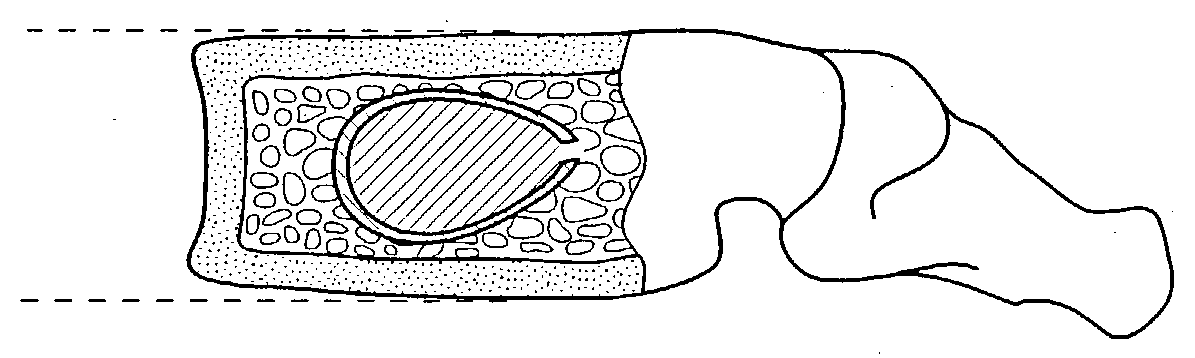
The most significant danger associated with vertebroplasty is the inability of the practitioner to control the flow of liquid bone cement during injection into a vertebral body.
Moreover, because the cancellous bone resists the injection of the bone cement and small diameter needles are typically used in vertebroplasty procedures, extremely high pressures are required to force the bone cement through the needle and into the vertebral body.
Bone cement, which is viscous, is difficult to inject through small diameter needles, and thus many practitioners choose to "thin out" the cement mixture to improve cement injection, which ultimately exacerbates the leakage problems.
In a recent study where 37 patients with bone metastases or multiple myeloma were treated with vertebroplasty, 72.5% of the procedures resulted in leakage of the cement outside the vertebral body.
Moreover, where the practitioner attempts to "thin out" the cement by adding additional liquid monomer to the cement mix, the amount of unpolymerized or "free" monomer increases, which can ultimately be toxic to the patient.
Another drawback of vertebroplasty is due to the inability to visualize (using CT scanning or x-ray fluoroscopy) the various venous and other soft tissue structures existent within the vertebra.
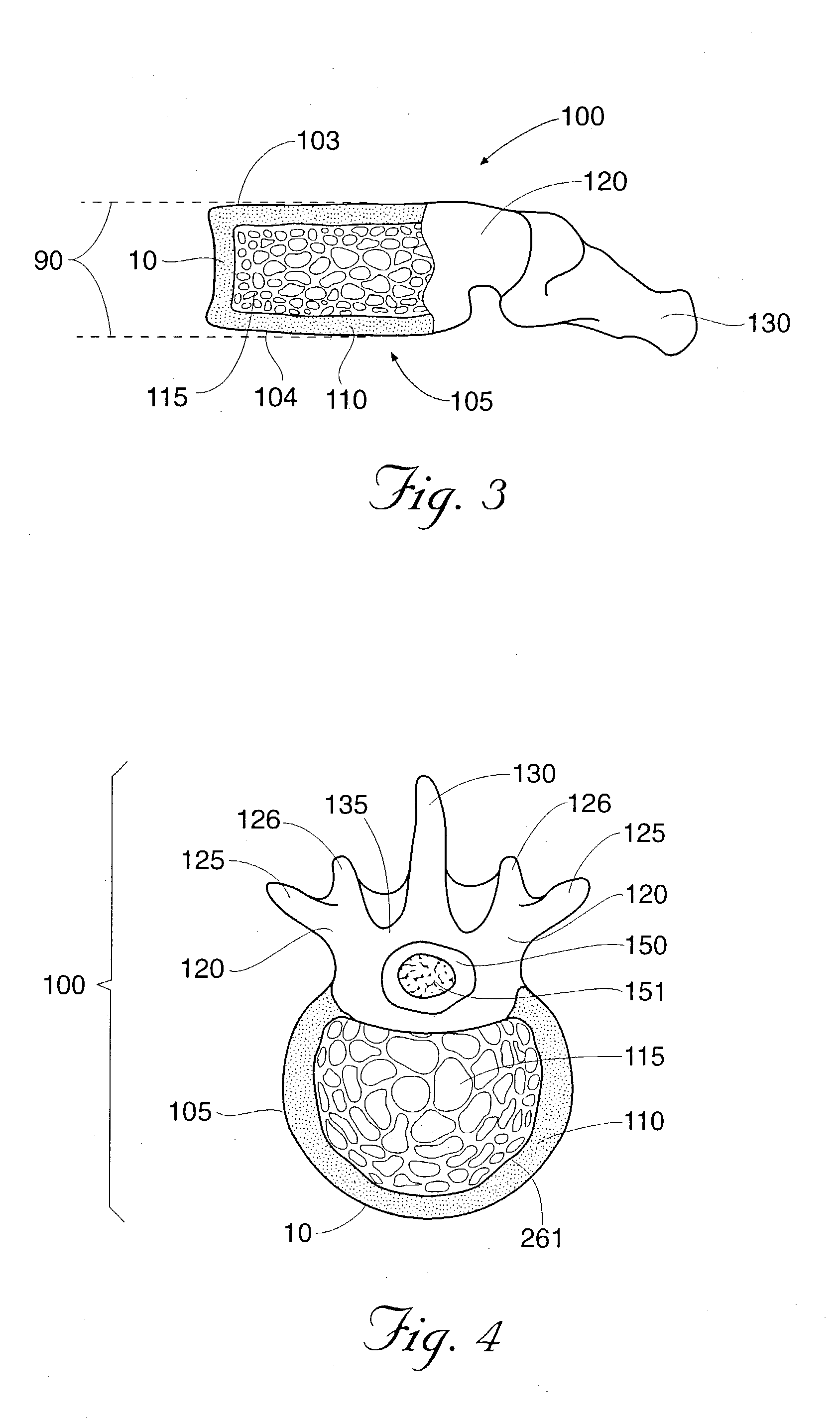
Another significant drawback inherent in vertebroplasty is the inability of this procedure to restore the vertebral body to a pre-fractured condition prior to the injection of the reinforcing material.
Moreover, it is highly unlikely that a traditional vertebroplasty procedure could be capable of restoring significant pre-fracture anatomy--because bone cement flows towards the path of least resistance, any en-masse movement of the cortical bone would likely create gaps in the interior and / or walls of the vertebral body through which the bone cement would then immediately flow.
While Kyphoplasty can restore bones to a pre-fractured condition, and injected bone filler is less likely to leak out of the vertebral body during a Kyphoplasty procedure, Kyphoplasty requires a greater number of surgical tools than a vertebroplasty procedure, at an increased cost.
Moreover, Kyphoplasty tools are typically larger in diameter than vertebroplasty tools, and thus require larger incisions and are generally more invasive.

Method used


Image

  • Methods and devices for treating fractured and/or diseased bone
  • Methods and devices for treating fractured and/or diseased bone
  • Methods and devices for treating fractured and/or diseased bone

Examples


Embodiment Construction

[0063] As embodied and broadly described herein, the present invention is directed to surgical methods for repairing, reinforcing and / or treating weakened, diseased and / or fractured bone. The present invention is further directed to various devices for facilitating such surgical methods.

[0064] FIG. 1 depicts a typical human spine 1, in which a compression fracture 10 has occurred in a lumbar vertebra 100. As best shown in FIG. 3, vertebra 100 has fractured, with the top and bottom plates 103 and 104 depressing generally towards the anterior wall 10 of the vertebra 100 and away from their pre-fracture, normally parallel orientation (indicated generally as parallel lines 90).

[0065] FIG. 4 depicts a coronal (top) view of the vertebra of FIG. 3. Vertebra 100 includes a vertebral body 105, which extends on the anterior (i.e. front or chest) side of the vertebra 100. Vertebral body 105 is approximately the shape of an oval disk, with an anterior wall 10 and a posterior wall 261. The geome...


PUM

PropertyMeasurementUnit
volumeaaaaaaaaaa
volumeaaaaaaaaaa
volumeaaaaaaaaaa

Abstract

The present invention relates to devices and methods for treating fractured and / or diseased bone. More specifically, the present invention relates to devices and methods for repairing, reinforcing and / or treating fractured and / or diseased bone using various devices, including cavity-forming devices.

Description

[0001] This application is a divisional of copending U.S. application Ser. No. 09 / 827,260, filed Apr. 5, 2001, entitled "Methods and Devices for Treating Fractured and / or Diseased Bone," which claims the benefit of provisional application Serial No. 60 / 194,685, filed Apr. 5, 2000.[0002] 1. Field of the Invention[0003] The present invention relates to devices and methods for treating fractured and / or diseased bone. More specifically, the present invention relates to devices and methods for repairing, reinforcing and / or treating fractured and / or diseased bone using various devices, including cavity-forming devices.[0004] 2. Description of the Background[0005] Normal healthy bone is composed of a framework made of proteins, collagen and calcium salts. Healthy bone is typically strong enough to withstand the various stresses experienced by an individual during his or her normal daily activities, and can normally withstand much greater stresses for varying lengths of time before failing....

Claims


Application Information

Patent Timeline
Patent Type & Authority Applications(United States)
IPC IPC(8): A61B17/00A61B17/56A61B17/16A61B17/88A61B19/00A61B19/02A61F2/00A61F2/28A61F2/30A61F2/44A61F2/46A61L27/00A61M25/00
CPCA61B17/1604A61F2310/00353A61B17/3472A61B17/8805A61B17/8855A61B19/0271A61B19/54A61B2017/00261A61B2017/00464A61B2017/00539A61B2017/00557A61B2017/00867A61B2019/0219A61B2019/462A61F2/44A61F2/4601A61F2002/2835A61F2002/3008A61F2002/30581A61F2002/4602A61F2002/4624A61F2002/4635A61F2002/4662A61F2250/0098A61B17/1671A61B2050/0065A61B50/33A61B2090/062A61B90/39A61F2/4603
Inventor OSORIO, REYNALDO A.FOLLMER, MARIALULULAYNE, RICHARD W.BOUCHER, RYAN P.TALMADGE, KAREN D.BASISTA, JOSEPH J.
Owner ORTHOPHOENIX
Why Eureka
Try Eureka