Method and apparatus for the detection of noncovalent interactions by mass spectrometry-based diffusion measurements

Inactive Publication Date: 2003-12-25
UNIV OF WESTERN ONTARIO
View PDF9 Cites 29 Cited by
  • Summary
  • Abstract
  • Description
  • Claims
  • Application Information

AI Technical Summary

Benefits of technology

0133] The present invention addresses the need to accurately assay a large number of potential ligands and targets within a relatively short time frame for their efficacy in forming noncovalent interactions. The present invention is therefore highly advantageous for use in the screening of entire compound libraries, e.g. in the context of HTS. It will be obvious to those skilled in the art that a minia

Problems solved by technology

However, these advances pose challenges of scale in terms of identifying fruitful combinations of molecules.
All of these methods suffer from certain limitations.
Also, it is often difficult to identify unique resonances for compounds that have similar chemical structures.
Affinity chromatography and SPR are relatively time-consuming because they require the chemical immobilization of compounds.
Unfortunately, numerous noncovalent complexes do not remain intact during the ESI process.
However, even in the case of ionic int

Method used


Image

  • Method and apparatus for the detection of noncovalent interactions by mass spectrometry-based diffusion measurements
  • Method and apparatus for the detection of noncovalent interactions by mass spectrometry-based diffusion measurements
  • Method and apparatus for the detection of noncovalent interactions by mass spectrometry-based diffusion measurements

Examples


example 2

[0114] Myoglobin in the laminar flow tube is exposed to denaturing conditions (50% acetonitirile, pH 10). Under these conditions the heme group is not expected to bind to the protein. Referring to FIG. 5, dispersion profiles of the protein in myoglobin (A), and of the heme in myoglobin (B) were recorded. Panel (C) shows the dispersion profile of heme recorded under the same solvent conditions but in the absence of protein. The fitted diffusion coefficients D are indicated in each panel. Solid lines are fits to the experimental data based on equation 17. These dispersion profiles reveal a small diffusion coefficient for the protein, and a much larger diffusion coefficient for the heme, as expected. The diffusion coefficient D of heme in the protein solution is almost as large as that of heme in the protein-free solution (considering the experimental uncertainty in the measured value of D), thus confirming that noncovalent interactions between heme and the protein are absent or extrem...

example 3

[0115] Myoglobin in the laminar flow tube is exposed to "semi-denaturing" conditions (30% acetonitrile, pH 10). FIG. 6 shows the dispersion profiles of the protein in myoglobin (A), and of the heme in myoglobin (B) recorded under these solvent conditions. Panel (C) shows the dispersion profile of heme recorded under the same solvent conditions but in the absence of protein. The fitted diffusion coefficients D are indicated in each panel. Solid lines are fits to the experimental data based on equation 17. The diffusion coefficients of heme and protein in the myoglobin solution are almost identical. A much larger diffusion coefficient is measured for heme in the absence of protein. These results show that under these semi-denaturing conditions, heme and protein are still bound to each other.

[0116] The findings presented in Examples 1, 2, and 3 are in agreement with the results of optical control experiments. It is pointed out that standard ESI-MS fails to detect the different noncoval...

example 4

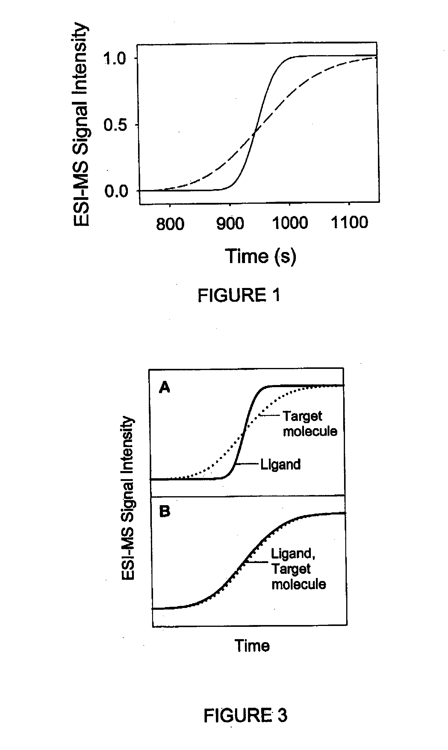
[0117] It will now be described how the present invention can be generalized to screen a number of potential ligands for binding to a particular target. The principle of this approach is schematically depicted in FIG. 7. An ESI mass spectrometer is used to monitor the dispersion profiles of a number of potential ligands simultaneously. All of these potential ligands are mixed in the same solution, initially in the absence of the target, resulting in the dispersion profiles shown in FIG. 7A. Note that all of the profiles are steep, due to the relatively small molecular size of the potential ligands. FIG. 7B shows a scenario where the experiment is repeated in the presence of the target. It is assumed that one of the ligands binds noncovalently to the target. The dispersion profile of this ligand (solid line in FIG. 7B) is much more extended than that of the other potential ligands, and it is also much more extended than the profile of this ligand recorded in the absence of the protei...


PUM


Abstract

The present invention provides a method and apparatus for detecting the noncovalent binding of a potential ligand (such as a drug candidate) to a target, e.g. a biochemical macromolecule such as a protein. The method is based on the Taylor dispersion of an initially sharp boundary between a carrier solution, and an analyte solution that contains the potential ligand(s) and the target. Dispersion profiles of one or more potential ligands are monitored by mass spectrometry at the exit of the laminar flow tube. Potential ligands will usually be relatively small molecules that have large diffusion coefficients. In the absence of any noncovalent interactions in solution, very steep dispersion profiles are expected for these potential ligands. However, a ligand that binds to a large target in solution, will show an apparent diffusion coefficient that is significantly reduced, thus resulting in a more extended dispersion profile. Noncovalent binding can therefore be detected by monitoring dispersion profiles of potential ligands in the presence and in the absence of the target. In contrast to other mass spectrometry-based methods for detecting noncovalent interactions, this method does not rely on the preservation of specific noncovalent interactions in the gas phase. This method has an excellent sensitivity and selectivity, therefore it can be used for testing multiple potential ligands simultaneously. The method is therefore useful for the high throughput screening of compound libraries.

Description

CROSS REFERENCE TO RELATED FOREIGN PATENT APPLICATION[0001] This application claims the benefit of priority from Canadian patent application Serial No. 2,387,316 filed on May 31, 2002, entitled METHOD AND APPARATUS FOR THE DETECTION OF NONCOVALENT INTERACTIONS BY MASS SPECTROMETRY-BASED DIFFUSION MEASUREMENTS, which was filed in English.[0002] The present invention provides a method and apparatus for the detection of noncovalent interactions between analyte species in the liquid phase by mass spectrometry-based diffusion measurements, and more particularly the present invention relates to a method and apparatus using electrospray ionization (ESI) or atmospheric pressure chemical ionization (APCI) mass spectrometry (MS) for the detection of noncovalent interactions.[0003] Noncovalent interactions play a central role for numerous physiological processes. Of particular importance is the noncovalent binding of small molecules to biological macromolecules such as proteins or nucleic acid...

Claims


Application Information

Patent Timeline
IPC IPC(8): G01N13/00H01J49/04
CPCG01N13/00G01N33/487H01J49/165H01J49/0431H01J49/0031
Inventor KONERMANN, LARSCLARK, SONYA M.
Owner UNIV OF WESTERN ONTARIO
Why Eureka
Try Eureka