Neo-cartilage constructs and a method for preparation thereof

Inactive Publication Date: 2004-08-05
TAKAGI IND CO LTD +1
View PDF20 Cites 42 Cited by
  • Summary
  • Abstract
  • Description
  • Claims
  • Application Information

AI Technical Summary

Problems solved by technology

Damage to the articular cartilage which occurs in active individuals and older generation adults as a result of either acute or repetitive traumatic injury or aging is quite common.
Such damaged cartilage leads to pain, affects mo

Method used


Image

  • Neo-cartilage constructs and a method for preparation thereof
  • Neo-cartilage constructs and a method for preparation thereof
  • Neo-cartilage constructs and a method for preparation thereof

Examples


example 2

The Production of Human Neo-Cartilage Construct

[0358] This example describes conditions for production of neo-cartilage for human use.

[0359] The patient undergoes arthroscopic biopsy of a small (200-500 mg) piece of healthy cartilage from the ipsilateral knee. The biopsy is taken from the non-weight bearing portion of the femoral condyle or from the femoral notch as deemed most appropriate for the patient. The biopsy sample is placed into a sterile, non-cytotoxic, non-pyrogenic specimen container which is packaged and shipped to the laboratory.

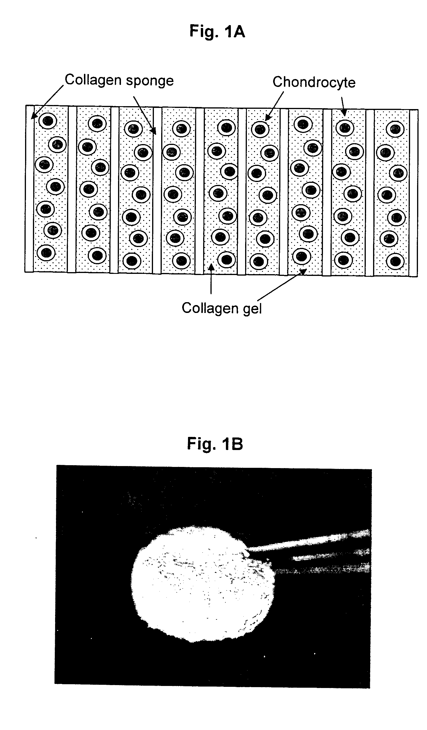
[0360] At the laboratory the biopsy sample is examined against acceptance criteria and then transferred to the chondrocyte isolation and expansion area. Samples from the biopsy specimen transport buffer are tested for sterility and for mycoplasma. The expanded chondrocytes are suspended in VITROGEN.RTM. gellable collagen solution, commercially available from Cohesion Corp., Palo Alto, Calif. A pre-formed collagen sponge (22.times.22 mm square ...

example 3

Preparation of Support Matrices

[0363] This example illustrates preparation of the cellular support matrix, also called the TESS matrix.

[0364] 300 grams of a 1% aqueous atelocollagen solution (VITROGEN.RTM.), maintained at pH 3.0, is poured into a 10.times.20 cm tray. This tray is then placed in a 5 liter container. A 50 ml open container containing 30 ml of a 3% aqueous ammonia solution is then placed next to the tray, in the 5 liter chamber, containing 300 grams of said 1% aqueous solution of atelocollagen. The 5 liter container containing the open trays of atelocollagen and ammonia is then sealed and left to stand at room temperature for 12 hours. During this period the ammonia gas, released from the open container of aqueous ammonia and confined within the sealed 5 liter container, is reacted with the aqueous atelocollagen resulting in gelling said aqueous solution of atelocollagen.

[0365] The collagenous gel is then washed with water overnight and, subsequently, freeze-dried to y...

example 4

Seeding Cells in the TESS Matrix

[0369] This example describes procedures used for seeding cells in the TESS matrix.

[0370] Isolated chondrocytes were incubated for a period of five days at 37.degree. C. in a standard incubator. Cells were then collected by trypsinization.

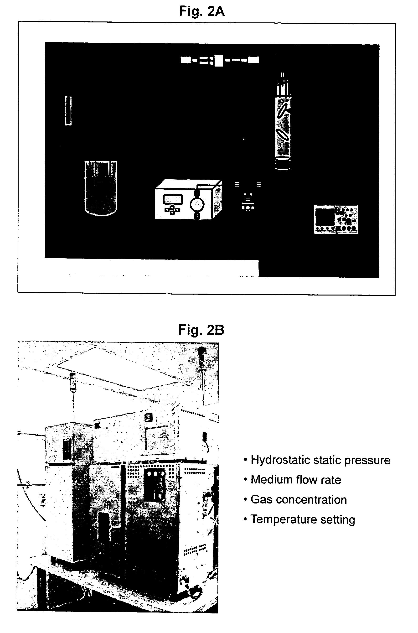
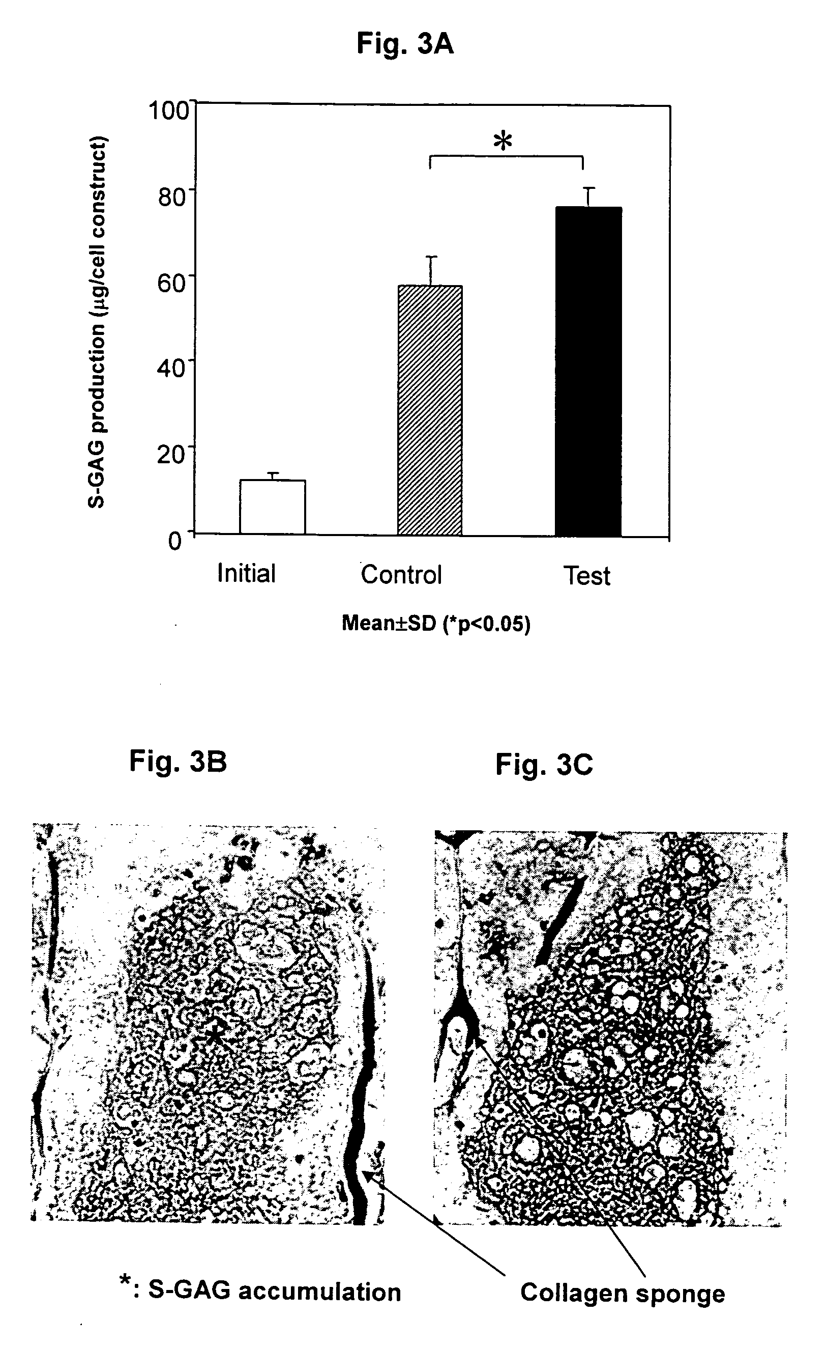
[0371] A cell suspension of 150,000 cells in 18 .mu.l of VITROGEN solution was seeded per matrix having an approximate volume of 19 .mu.l, with nine matrices per group. The seeded matrix (collagen sponge 4 mm in diameter and 1.5 mm in thickness) may be scaled-up to an increased volume, where approximately 1 .mu.l of the above described cell suspension is seeded in 1 .mu.l of matrix. The control group matrices were incubated in a 37.degree. C. incubator and the test group was incubated in the TESS.

[0372] In alternative set-up, isolated chondrocytes were incubated for a period of five days at 37.degree. C. in a standard incubator. Cells were then collected by trypsinization. A cell suspension of 300,000 cells in 18 .mu...


PUM

PropertyMeasurementUnit
Fractionaaaaaaaaaa
Fractionaaaaaaaaaa
Fractionaaaaaaaaaa

Abstract

Neo-cartilage constructs suitable for implantation into a joint cartilage lesion in situ and a method for repair and restoration of function of injured, traumatized, aged or diseased cartilage. The construct comprises at least chondrocytes incorporated into a support matrix processed according to the algorithm comprising variable hydrostatic or atmospheric pressure or non-pressure conditions, variable rate of perfusion, variable medium composition, variable temperature, variable cell density and variable time to which the chondrocytes are subjected.

Description

[0001] This application is a continuation-in-part application of Ser. No. 10 / 104,677 filed on Mar. 22, 2002 and is based on and claims priority of the Provisional application Ser. No. 60 / 425,696 filed on Nov. 12, 2002 and Provisional application Ser. No. 60 / 427,627 filed on Nov. 18, 2002.[0002] 1. Field of Invention[0003] The current invention concerns neo-cartilage constructs suitable for implantation into a joint cartilage lesion in situ and a method for repair and restoration of function of injured, traumatized, aged or diseased cartilage. The construct of the invention comprises at least chondrocytes incorporated into a support matrix processed according to the algorithm of the invention. In particular, the invention concerns a neo-cartilage construct for in situ implanting into a cartilage lesion wherein said construct comprises a cultured differentiated autologous or heterologous chondrocytes or cells which could be differentiated into chondrocytes incorporated into a support ...

Claims


Application Information

Patent Timeline
IPC IPC(8): A61F2/00A61K35/12A61L27/24A61L27/38A61L27/56C12N5/077
CPCA61F2310/00365A61L2430/06A61L27/24A61L27/3633A61L27/3817A61L27/3843A61L27/3852A61L27/3895A61L27/56C12N5/0655C12N2533/54A61K35/12A61K35/32A61K9/0024A61L27/38A61F2/28A61L27/54A61L2300/64C12N2521/00
Inventor MIZUNO, SHUICHIKUSANAGI, AKIHIKOTARRANT, LAURENCE J. B.TOKUNO, TOSHIMASASMITH, ROBERT LANE
Owner TAKAGI IND CO LTD
Why Eureka
Try Eureka