
Diagnosis of mould infection
a mould infection and mould technology, applied in the field of microbiology and pathology, can solve the problems of difficult diagnosis, difficult diagnosis, and inability to mount an effective immune response in immunocompromised patients, and achieve the effects of aggressive diagnosis and treatment, high mortality rate, and increased prevalen
- Summary
- Abstract
- Description
- Claims
- Application Information
AI Technical Summary
Benefits of technology
Problems solved by technology
Method used
Image

Examples
example 1
Materials and Methods
[0186] Case Definition and Serum Samples.
[0187] As described earlier, cases of IMI were defined according to the criteria established by EORTC and Mycoses Study Group (Ascioglu et al., 2002). Depending on the degree of diagnostic certainty, the cases are defined as “definitive”, “probable”, “possible”, and “unlikely” IMI. “Proven” IMI represented a tissue diagnosis where branched septate hyphae, inflammation, and necrosis were seen microscopically and / or the fungus was successfully cultured from the tissue. Most of the patients in this group had pulmonary IMI and were typically neutropenic and / or immunosuppressed for an extended period of time. They exhibited prolonged pneumonia unresponsive to anti-bacterial therapy with nodular and / or cavitory lesions in the lung radiologically. Of the 13 patients with documented IMI in this study, tissue diagnoses were rendered in 12 patients by surgery or biopsy and one by autopsy. Patients with “probable IMI” typically we...
example 2
Results
[0207] Specificity and Detection Range.
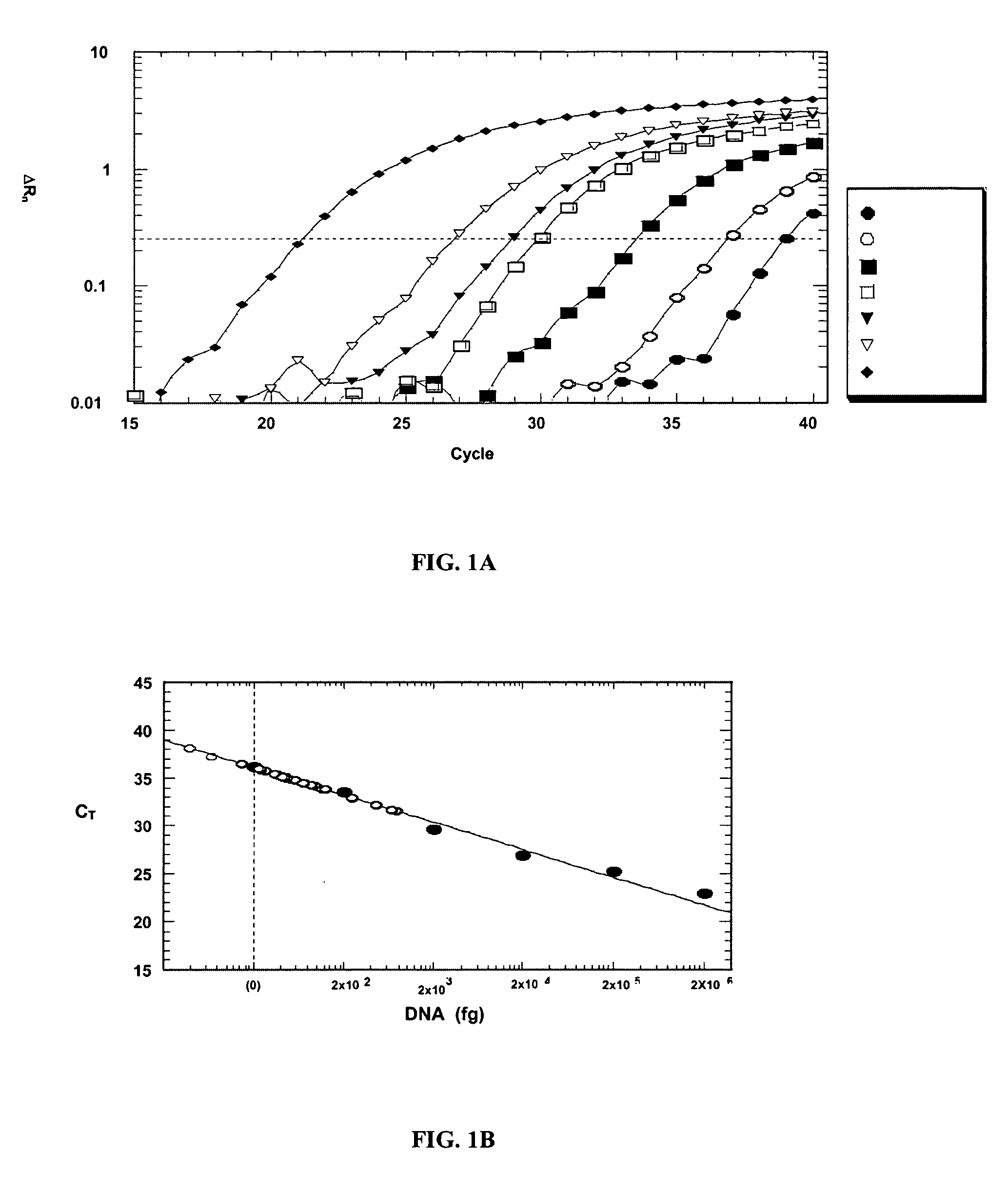
[0208] The specificity and detection range of the real-time PCR were assessed with purified Aspergillus, human, and candidal DNA. Neither human nor candidal DNA was amplified (data not shown). With normal human DNA as a background, purified Aspergillus DNA from 20 ng to 200 fg (5-log range) was detected at various amplification cycles (FIG. 1A). A logarithmic plot of the DNA quantity correlated linearly with the number of cycles (FIG. 1B), thus providing a basis for quantitative analysis of patient specimens.
[0209] Test of Sera.
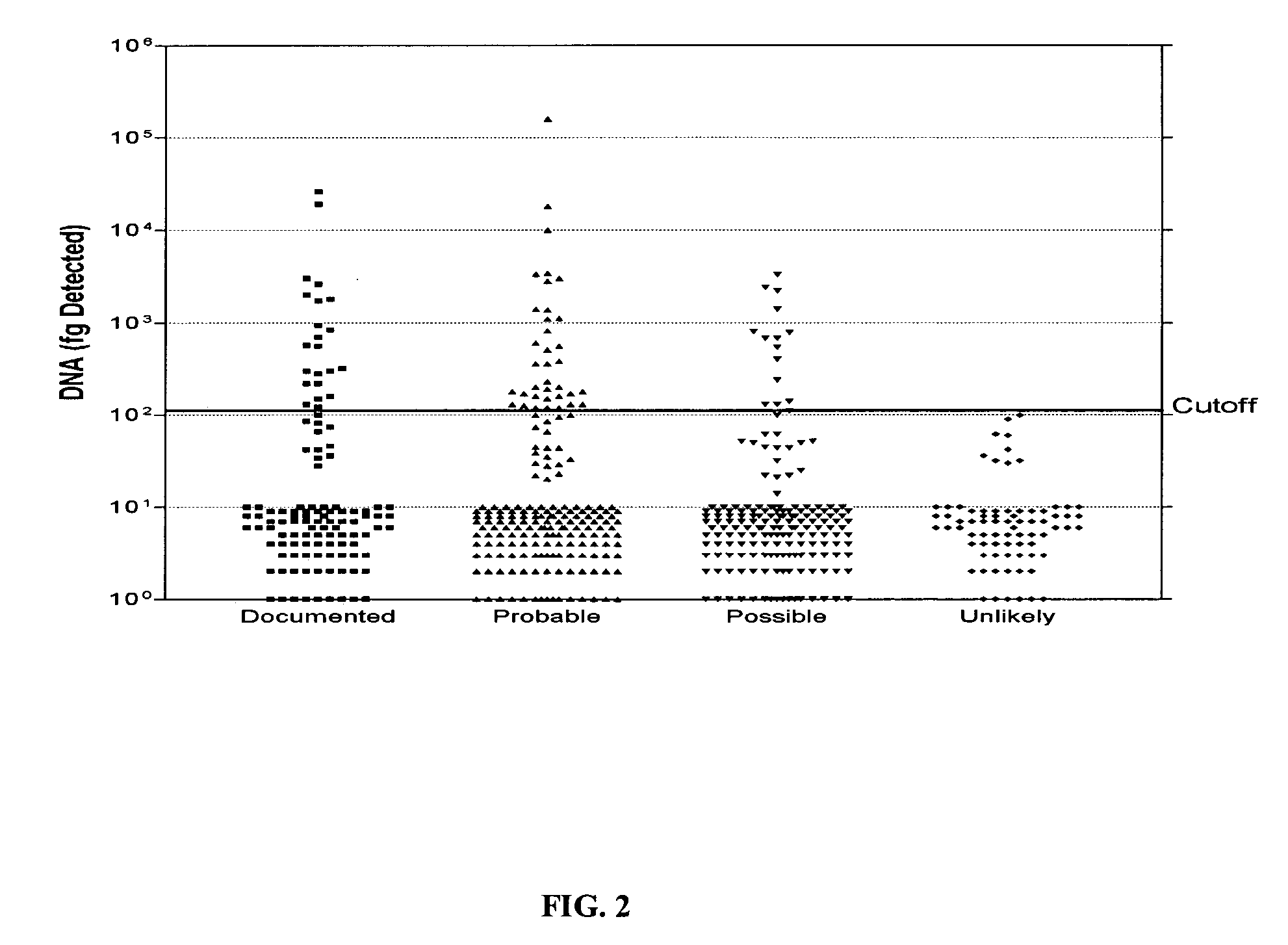
[0210] A total of 559 serum samples from 106 patients were tested with this real-time PCR assay and the results are shown in FIG. 2. All 76 sera from 35 patients with no evidence of IMI showed undetectable (less than negative control, 10,000 fg) positivity (FIG. 2). At this cutoff, sera from patients with documented and suspected (probable and possible) IMI showed varying percentages of positivity that correla...
PUM
| Property | Measurement | Unit |
|---|---|---|
| size | aaaaa | aaaaa |
| particle sizes | aaaaa | aaaaa |
| melting temperatures | aaaaa | aaaaa |
Abstract
Description
Claims
Application Information