Noninvasive tissue assessment

a tissue assessment and non-invasive technology, applied in the field of tissue assessment, can solve the problems of loss of mechanical strength, increased risk of fracture, degradation of the mechanical strength of the bones, etc., and achieve the effect of reducing measurement errors

Inactive Publication Date: 2005-05-26
RICE UNIV
View PDF32 Cites 103 Cited by
  • Summary
  • Abstract
  • Description
  • Claims
  • Application Information

AI Technical Summary

Benefits of technology

[0013] In another embodiment of the invention, a device (as described herein) is pressed against the skin in an anatomic region with a relatively thin flesh layer. Choosing such a region will reduce measurement errors due to the interaction between soft and hard tissue. Although, measurement methodologies presented herein are intended to circumvent this problem. Constant contact pressure is either monitored through a mechanical constant spring system or via a software and load-cell system. A mechanical or software trigger mechanism starts the testing procedure when adequate pressure is applied. The test will run for a duration of about 1 to about 30 seconds, preferably about 5 seconds. Preliminary data analysis calculating signal to noise ratio and signal strength will indicate if the test will need to be repeated. The time signals from the electromagnetic shaker (driver), the accelerometer (output) and the load cell (input), will be recorded and at a give time transferred to a computer or microprocessor for analysis.

Problems solved by technology

Osteoporosis may be defined as a metabolic disease causing an unbalance in the natural process of bone resorption and bone formation with the result of a loss of mechanical strength and increased risk of fractures.
The degradation of the mechanical strength of the bones may proceed to a stage where even minimal trauma result in bone fractures.
An accurate assessment of osteoporosis is difficult.
The risk of long-term effects from X-ray radiation is compounded by multiple exposures whenever the patient is to be reevaluated.
Typical X-ray scanners are very expensive and require extensively trained technicians to operate.
In addition, the expense and / or inconvenience of existing technology that can only be accessed with an office visit is not conducive to early detection because it limits the number of times a patient will actually be checked.
Being tested only every other year or less for the early stages of osteoporosis may not be enough.
Further, the existing technology may report only bone areal density, and do not directly indicate bone strength or tendency for bone loss, nor do they take into account differences in body build, body weight, patient height, or loading history.
Unfortunately, these tests are limited to bones, such as the calcaneus and patella, which suffer from osteoporosis to a negligible extend and are only weakly indicative of risk of fracture.
Traditional bone mass measurements, by their very nature, are unable to predict bone loss prior to its occurrence and can only chart the course of bone loss over an extended period of time.
Further, these diagnostics only consider bone mass, and fail to consider other factors such as tendency to fall, or ability to protect oneself during falling.
In addition, the capital expense associated with this type of technology can be greater than $7,500.

Method used


Image

  • Noninvasive tissue assessment
  • Noninvasive tissue assessment
  • Noninvasive tissue assessment

Examples


Embodiment Construction

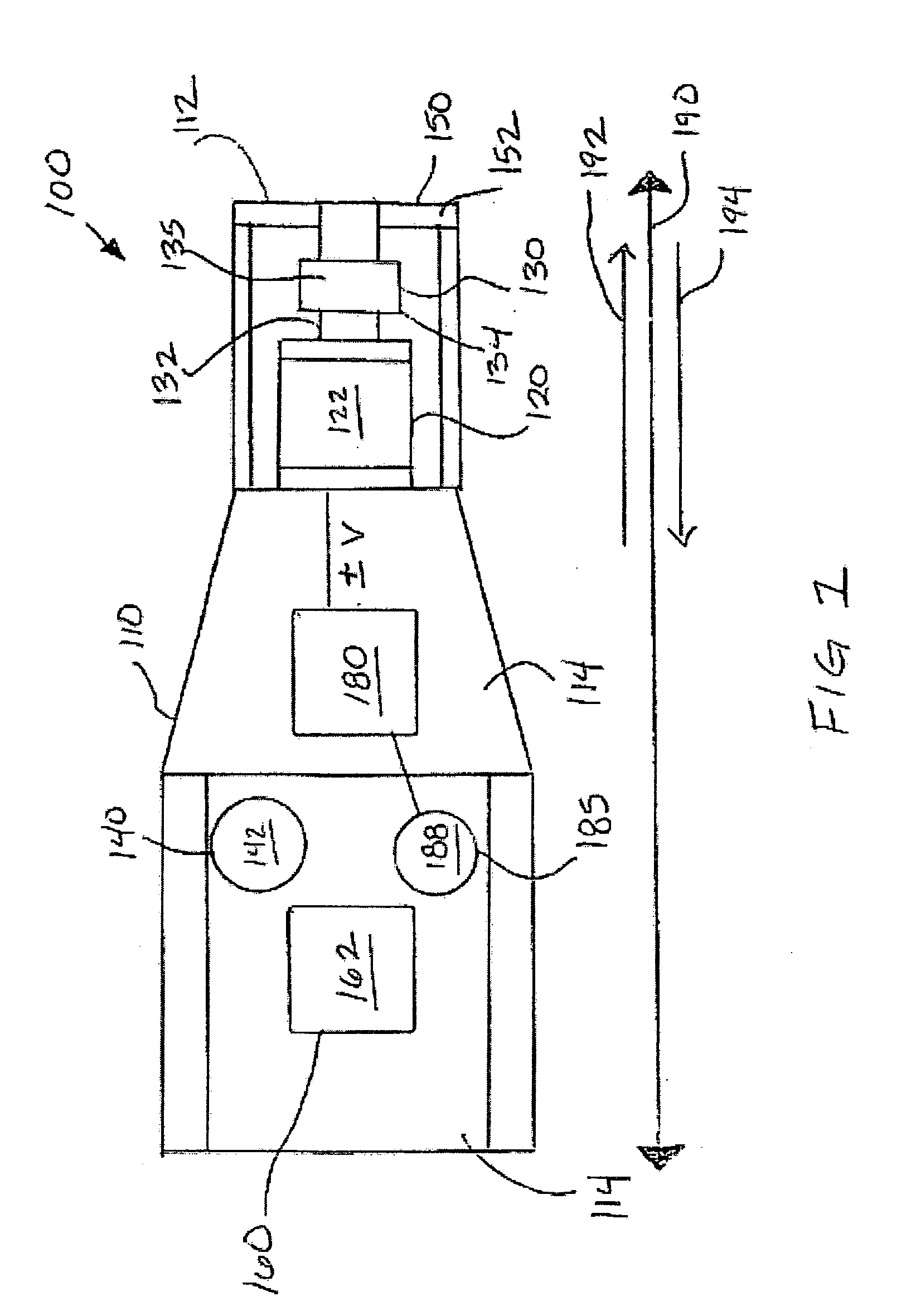
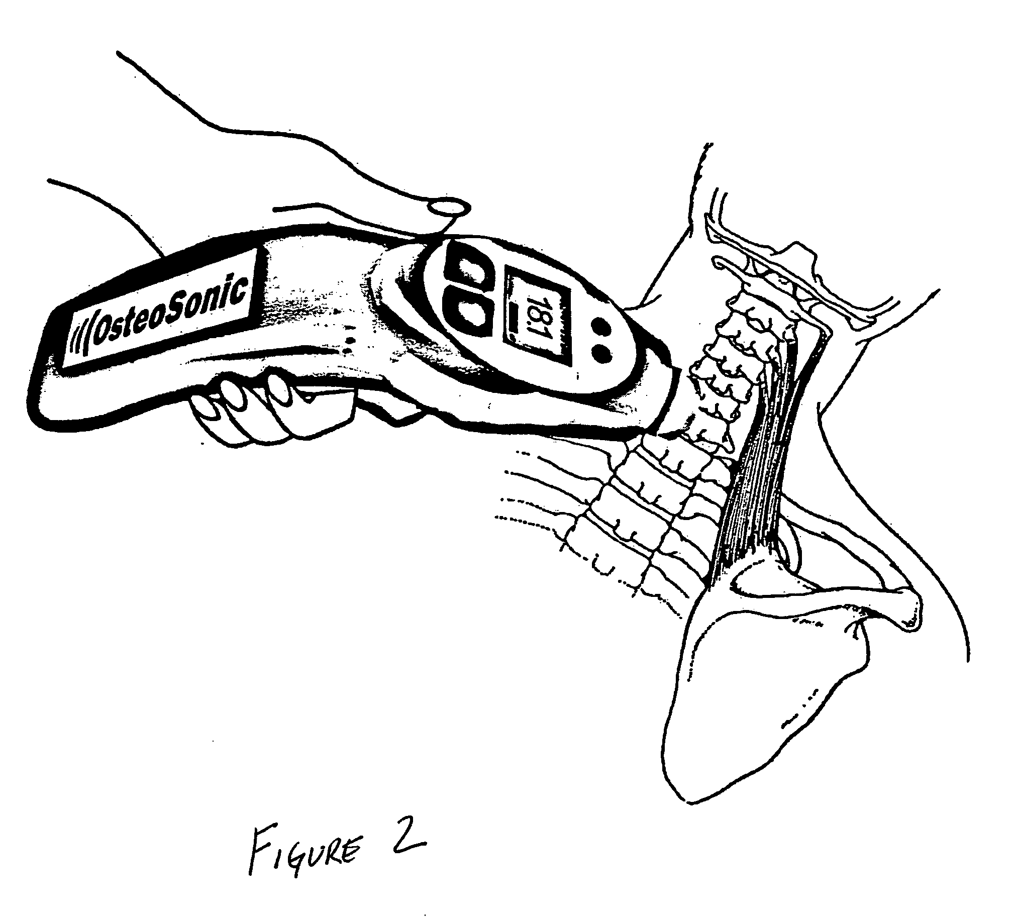
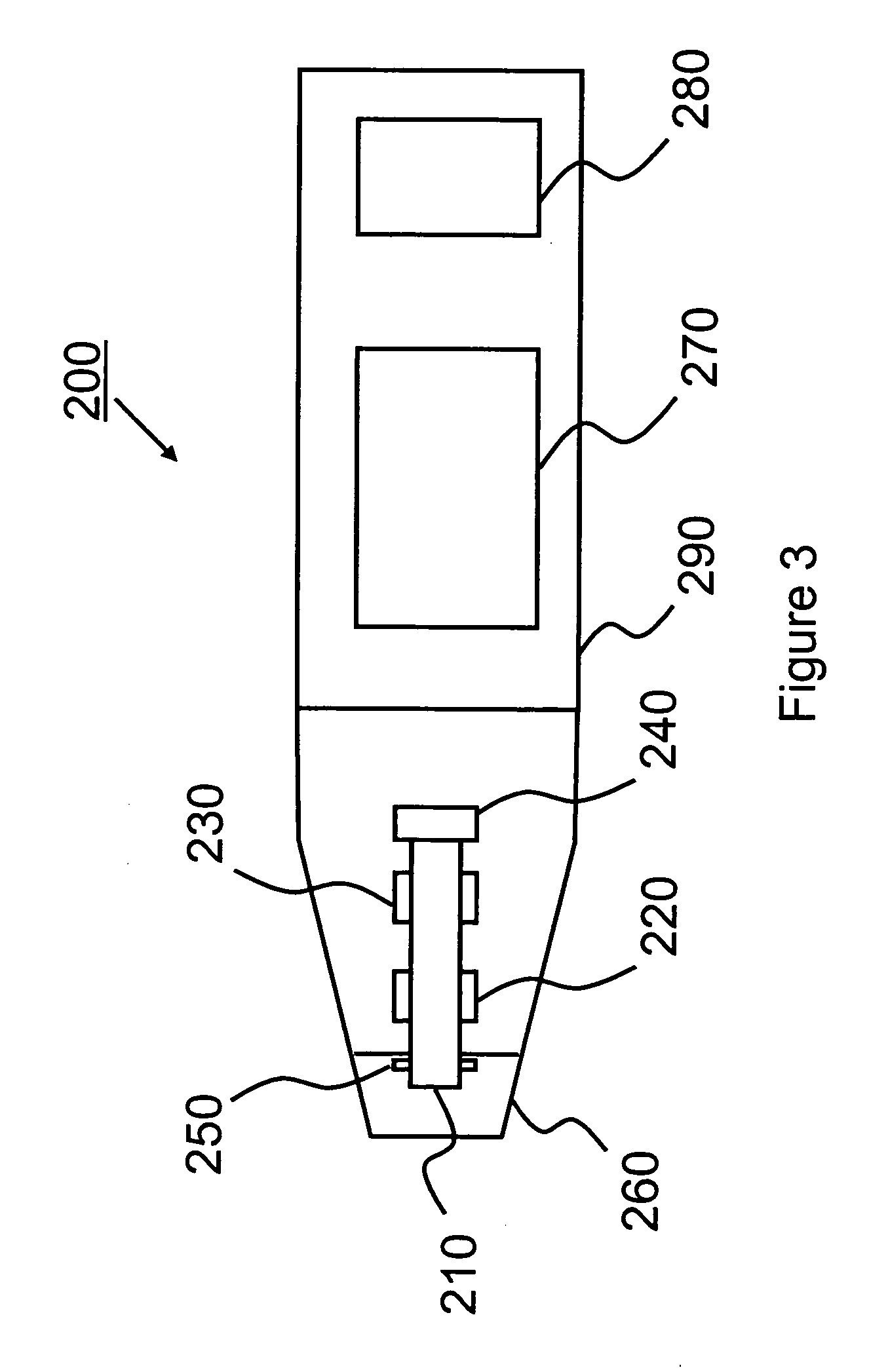
[0027] As can be appreciated, osteopenia and osteoporosis, i.e., the loss of bone mass, arises to a large degree from the natural aging process, but also to a lesser degree from a decrease in muscle activity, such as due to bed rest. This loss can be detected using vibrational stimulation arising from a source external to the body, which creates mechanical, frequency specific, low level oscillations in the subjacent bones. Compared to imaging techniques such as DXA that neglect the volume of the measured bones, or ultrasonometer that measure localized speed of sound, acoustic vibrations around the natural frequency of an object excites the whole object, independent of size and shape. Changes in shape and bone mass content will alter its responsive frequency, and can thus be detected by the disclosed device and / or using the disclosed methods.

[0028] In more technical terms if one considers trabecular bone as a random network of struts, it is known from percolation theory that when a ...


PUM

PropertyMeasurementUnit
frequencyaaaaaaaaaa
frequencyaaaaaaaaaa
frequencyaaaaaaaaaa

Abstract

Methods and apparatus for non-invasively assessing physiological hard of soft tissue of human and other species are described. In a preferred embodiment, tissue is vibrationally stimulated in vivo through a frequency spectrum. The tissue reacts against the stimulus and the reaction is preferably measured and recorded. Based on analytical algorithms or comparisons with previously taken measurements, changes within the tissue can be detected and used for diagnostic purposes. Further embodiments describe the usage of the device and methods for in vivo intra-operative and post-operative implant evaluations and as a therapeutic tool.

Description

CROSS-REFERENCE TO RELATED APPLICATIONS [0001] The present application claims the benefit of 35 U.S.C. 111(b) US Provisional Application Ser. No. 60 / 500,796 filed Sep. 5, 2003, entitled “Noninvasive Bone and Joint Damage Detection Device” and U.S. Provisional Application Ser. No. 60 / 553,670 filed Mar. 16, 2004, which are hereby incorporated by reference herein for all purposes.STATEMENT REGARDING FEDERALLY SPONSORED RESEARCH OR DEVELOPMENT [0002] None FIELD OF THE INVENTION [0003] The present invention relates to a method and apparatus for in-vivo investigation and characterization of tissue. In particular, the present invention relates to the use of vibration for assessment of tissue. BACKGROUND OF THE INVENTION [0004] The investigation of the mechanical properties of bone in vivo is of great interest in view of the occurrence of fracture risk in osteopenia or osteoporosis. Osteoporosis may be defined as a metabolic disease causing an unbalance in the natural process of bone resorp...

Claims


Application Information

Patent Timeline
Patent Type & Authority Applications(United States)
IPC IPC(8): A61BA61B5/103A61B8/14C12M1/34
CPCA61B5/0048A61B5/4504A61B5/4509A61B5/4514Y10S128/923A61B5/4523A61B5/4533A61B9/00Y10S128/92A61B5/4519A61B5/0051
Inventor LIEBSCHNER, MICHAEL A. K.
Owner RICE UNIV
Why Eureka
Try Eureka