Method of using fibrin-bound angiogenic factors to stimulate vascularization of transplant site of encapsulated cells

a technology of fibrin-bound angiogenic factors and vascularization of the transplant site, which is applied in the field of cell-based therapy, can solve the problems of increased portal venous pressure, portal venous thrombosis or hemorrhage, and the technique still has to overcome problems, so as to enhance the survival and function of implanted cells

Inactive Publication Date: 2005-08-18
NOVOCELL
View PDF93 Cites 55 Cited by
  • Summary
  • Abstract
  • Description
  • Claims
  • Application Information

AI Technical Summary

Benefits of technology

[0017] This invention combines the implantation of cells, tissues or organs [stem cell, autologous, allogeneic, xenogeneic or genetically-modified], either unencapsulated, or encapsulated in macrodevices, microcapsules, or c

Problems solved by technology

This technique still has to overcome problems related to the inconsistencies in the achievement of insulin independence.
The islet supply and side effects related to systemic immunosuppression currently restrict the clinical application to a limited number of Type I diabetic patients.
A major risk of this procedure is increased portal venous pressures, depending on the rate of infusion and the amount infused, due to the injection of the islets into the portal vein.
Also, portal venous thrombosis or hemorrhage is a complication of this procedure.
This reaction can lead to additional long-term problems, such as, bowel obstruction.
Due to its relatively large size, there are few sites in the body able to accommodate a flat sheet macrodevice to treat a disease like diabetes.
It has proven quite difficult to flush and reload cells, while at the same time maintaining the critical cell compartment distance for oxygen diffusion.
For the diabetes product, it has been quite difficult to place this device into the intraperitoneal cavity of large animals, while maintaining its integrity.
This type has shown efficacy in large animal diabetic trials, but has been plagued with problems in va

Method used


Image

  • Method of using fibrin-bound angiogenic factors to stimulate vascularization of transplant site of encapsulated cells
  • Method of using fibrin-bound angiogenic factors to stimulate vascularization of transplant site of encapsulated cells
  • Method of using fibrin-bound angiogenic factors to stimulate vascularization of transplant site of encapsulated cells

Examples


example 1

Subcutaneous Implant of Encapsulated Islets in Baboons

[0154] Surgical Procedures: Baboon pancreata were removed from donors, cannulated, and flushed with pancreas preservation solution and then shipped to Novocell for islet preparation and encapsulation. They were subsequently cultured, shipped to the holding facility for implantation, and then prepared for surgical implantation by suspension in culture medium, using similar protocols as are proposed for human islet preparation. The baboons were anesthetized, and a 16-gauge catheter was placed into the subcutaneous site of the anterior abdomen. A trochar was inserted through the implanted catheter to create a “fan shaped” area of 1-10 subcutaneous tracts (˜1″-5″ each in length) under the skin of the abdomen. The test material (in 1 to 10 ml volume) was gently suspended, pulled into a syringe, and deposited along the subcutaneous tracts (or “pockets”) with an even pattern of deposition throughout the pockets. The needle insertion s...

example 2

Encapsulated Islet Allograft without VEGF in Streptozotocin Diabetic Baboons

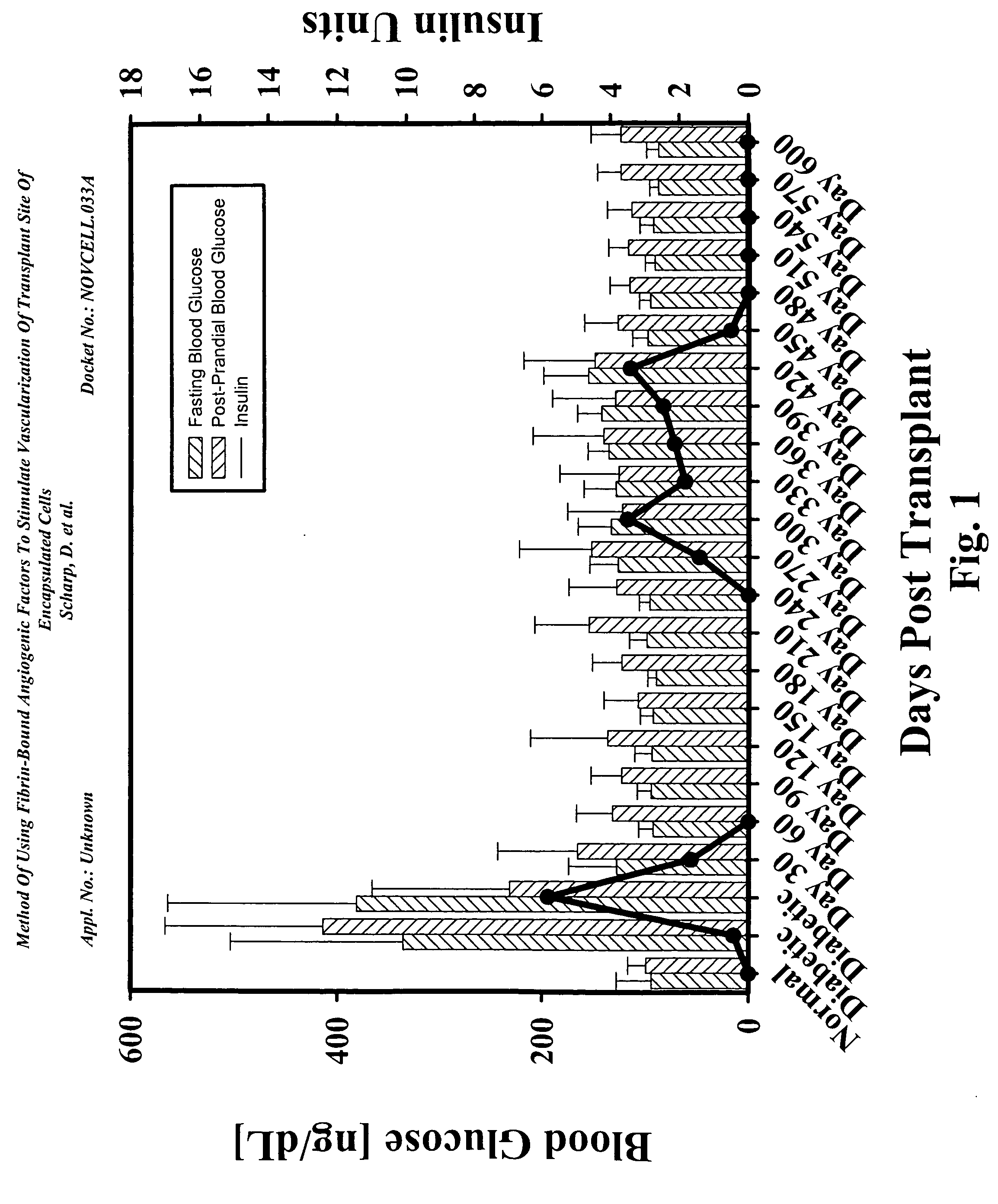
[0158]FIG. 1 shows the results of a diabetic baboon implanted with encapsulated islet allografts. This diabetic baboon recipient showed the ability to achieve insulin independence within 17 days after subcutaneous implantation of encapsulated islet allografts. This was in contrast to Cynomolgus primate diabetics where insulin independence was only achieved 30 days after islet implantation.

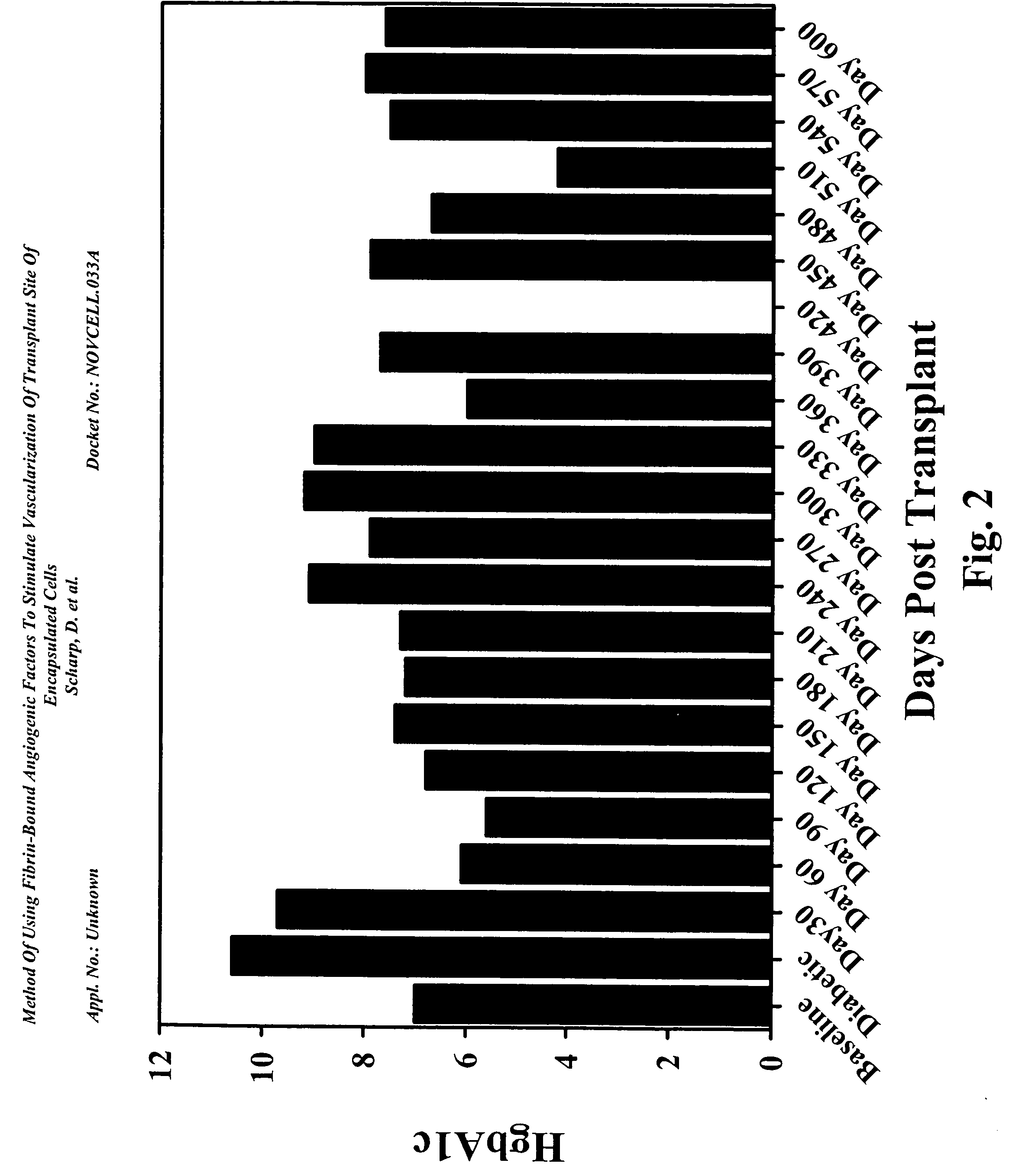
[0159] The baboon diabetic model used IM injection of cyclosporine for administration of the drug to these large animals. This eliminated the variances in the 24-hour trough levels. FIG. 2 shows this recipient achieved normal Hemoglobin A1c levels by 60 days post-implant and remained in the normal level through 210 days without insulin. There were slight increases followed by decrease between day 210 and day 360. The animal returned to partial graft function at day 240 requiring low doses of insulin.

[0160] At day 420, t...

example 3

Subcutaneous Site Enhancement by Implanting Encapsulated Islet Allografts within Fibrin Glue Conjugated with VEGF

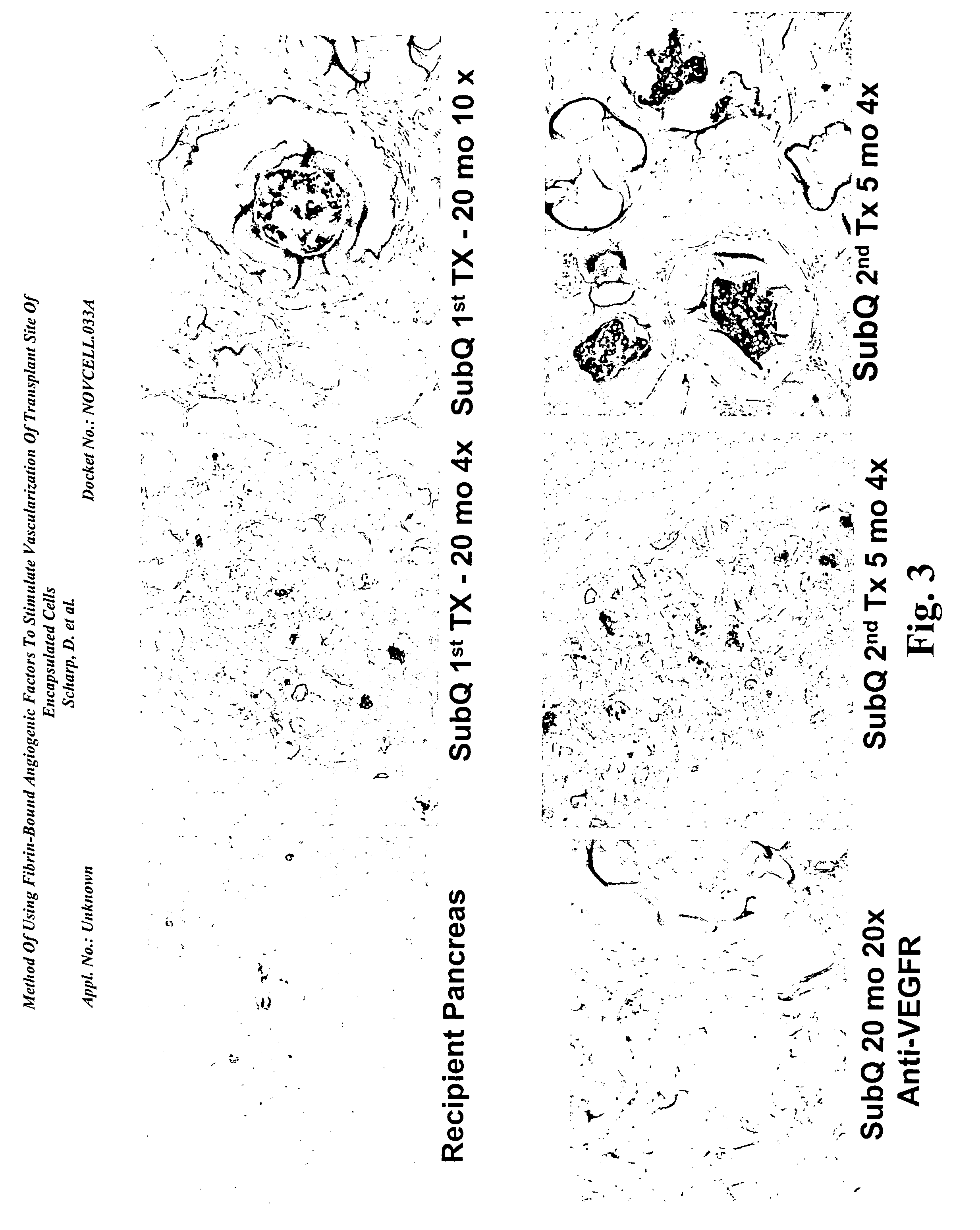
[0170] There was a continual finding of new blood vessel formation surrounding the PEG encapsulated islets in the subcutaneous site several weeks following implant. The subcutaneous site was enhanced by an angiogenic growth factor at the time of implant to increase the survival of the encapsulated islets during the first few weeks post-implant. This approach reduced the number of isolated islets required to eliminate diabetes.

[0171] A new angiogenic reagent has been developed that is used for wound healing. A human fibrin glue product is used as a base for adding VEGF. The conjugated VEGF breaks down at the same time as the fibrin clot breaks down in the body. A slow release of VEGF at the site causes the development of normal capillaries.

[0172] These results were confirmed by transplanting empty PEG beads, as well as, PEG conformally coated islets in diabetic mice an...


PUM

PropertyMeasurementUnit
Timeaaaaaaaaaa
Timeaaaaaaaaaa
Biological propertiesaaaaaaaaaa

Abstract

The present invention relates to compositions and methods of treating a disease, such as diabetes, by implanting encapsulated biological material with a growth factor and conjugate into a patient in need of treatment. Several methods are presented to accomplish transplanting several different types of biological materials. This invention also provides methods of utilizing these encapsulated biological materials to treat different human and animal diseases or disorders by implanting them into several areas in the body including the subcutaneous site.

Description

RELATED APPLICATIONS [0001] This application claims priority to U.S. Application No. 60 / 537,373; filed Jan. 16, 2003, which is incorporated herein by reference. [0002] All publications, patents, patent applications, databases and other references cited in this application, all related applications referenced herein, and all references cited therein, are incorporated by reference in their entirety as if restated here in full and as if each individual publication, patent, patent application, database or other reference were specifically and individually indicated to be incorporated by reference.BACKGROUND OF THE INVENTION [0003] 1. Field of the Invention [0004] The present invention relates generally to cell-based therapy, in particular, methods of implanting cells with sufficient access to oxygen and other nutrients at the implant site by stimulating neovascularization. [0005] 2. Description of the Related Art [0006] Pancreatic islet transplantation is an attractive procedure for the...

Claims


Application Information

Patent Timeline
IPC IPC(8): A61K33/06A61K35/12A61K35/39A61K38/18A61K38/19A61K38/36A61K38/48A61K45/00A61L24/10A61L27/38A61L27/50C12N5/071
CPCA61K35/12A61K38/363A61K35/39A61K38/37A61K38/1866C12N5/0677A61L27/507A61L27/3804A61L24/10A61K38/4833A61K2300/00
Inventor SCHARP, DAVIDLATTA, PAULYU, XIAOJIEHUBBELL, JEFFREY
Owner NOVOCELL
Why Eureka
Try Eureka