Diagnosis of body metabolic emergency state

a metabolic emergency and early diagnosis technology, applied in diagnostics, medical science, catheters, etc., can solve the problems of morbidity and mortality in a large number of patients, decreased atp production, and cellular function affected

Inactive Publication Date: 2005-10-20
CRITISENSE
View PDF22 Cites 31 Cited by
  • Summary
  • Abstract
  • Description
  • Claims
  • Application Information

AI Technical Summary

Problems solved by technology

When O2 supply will be restricted or limited the production of ATP will decrease and as a result, cellular functions will be affected (inhibition of pumping activity for example).
In a large segment of critically ill patients the changes in O2 balance is a slow process developed in one or more areas of the body and may end up in morbidity and mortality in large number of patients.
A typical example of such a pathological state is sepsis, which is a major cause of death in many patients hospitalized in intensive care units (ICUs) or even in regular hospital departments.
They concluded that “Severe sepsis is a common, expensive and frequently fatal condition, with as many deaths annually as those from acute myocardial infarction”.
Sepsis represents a large group of patient illnesses in which the deterioration of O2 balance is very hard to identify and the consequences in many cases are fatal.
The various groups of “slow killing” pathologies (like sepsis) are very hard to diagnose due to lack of specific and sensitive enough techniques or methods.
The same problem of O2 imbalance may develop in preterm or full term infants hospitalized in the neonatal ICU or even in the newborn during delivery.
Under moderate or severe O2 imbalance or the development of emergency pathological state, such as sepsis or severe hypotension for example, the body will not be able to compensate completely the deficit of O2.
During this phase the fetus will present an extreme response to the increasing hypoxemia and the fetal heart function will be impaired.
This stage, known as the decompensatory phase may lead to brain damage including edema.
During the initial phase the Doppler technique is not a sensitive tool for detecting changes in the uteroplacental vascular bed or gas exchange and metabolites.
The disadvantage of this approach is that the information obtained is not in real time mode.
The same pattern of pathophysiological cascade of events may occur in many emergency clinical situations in adult patients and may lead to morbidity and mortality.
Under all those situations the metabolic state of the body will be deteriorated and energy failure will develop.
Fink wrote that the effort to improve outcome in patients with sepsis by monitoring and manipulating cardiac output, systemic DO2, and regional blood flow are doomed to failure; instead, the focus should be on developing pharmacological strategies to restore normal mitochondrial function and cellular energetics.
Nevertheless, Fink does not suggest that any particular parameters should be monitored to achieve such a goal, less so how to monitor such parameters
None of these patents discloses or suggests the parametric monitoring for determining early diagnosis of body metabolic emergency state that may develop in many acute or chronic clinical conditions.
Monitoring of pH has the same problematic disadvantage due to the various processes taking place in the acid-base balance.
Therefore monitoring each one of them alone is not sufficient due to coupling or uncoupling processes between the two parameters.
An increase in the level of NADH with respect to NAD and the resulting increase in fluorescence intensity indicate that insufficient Oxygen is being supplied to the tissue.
A particular drawback encountered in NADH measurements is the Haemodynamic Artifact.
This refers to an artifact in which NADH fluorescence measurements in-vivo are underestimated or overestimated due to the haemoglobin present in blood circulation, which absorbs radiation at the same wavelengths as NADH, and therefore interferes with the ability of the light to reach the NADH molecules.
However, U.S. Pat. No. 4,449,535 has at least two major drawbacks; firstly, and as acknowledged therein, using a single optical fiber to illuminate the organ, as well as to receive emissions therefrom causes interference between the outgoing and incoming signals, and certain solutions with different degrees of effectiveness are proposed.
This results in measurements that are incompatible one with the other, the blood volume measurement relating to a greater depth of tissue than the NADH measurement.
Therefore, the device disclosed by this reference does not enable adequate compensation of NADH to be effected using the simultaneous, though inappropriate, blood volume measurement.
Further, there is no indication of how to measure other parameters such as blood flow rate, Fp level or blood oxygenation level using the claimed apparatus.
(i) The oxidation level of the blood will introduce artifacts, affecting the microcirculatory blood volume (MBV) since these patents do not specify how to compensate for the oxygenation state of the blood in the tissue, i.e., the relative quantities of oxygenated blood to deoxygenated blood in the tissue.
Using a relatively high intensity UV laser illumination source as proposed raises safety issues, especially for long-term monitoring.
An additional problem of NADH photo-bleaching arises since high intensity UV laser is used.
This method of oxygenation measurement may be of limited use while the fluorescence signals are fading.
In any case, apparatuses that incorporate a laser light source are generally required to comply with relevant laser safety standards, since there is some possibility of harm to tissue from exposure to extensive radiation.
This requirement implies a severe limitation on the light intensity emitted by the distal tip of the fiber optic probe, particularly when shorter wavelength, higher intensity radiation is used.
Therefore this has led to the search for a special light source that may be used for as many parameters as possible, and resulted in the evolution of special expensive low noise UV light sources for Doppler measurement.
Measurement of Laser Doppler at UV wavelengths raise additional safety aspects that significantly complicate the device.
Measurement of Laser Doppler at UV wavelengths raises additional safety aspects that significantly complicate the device.

Method used


Image

  • Diagnosis of body metabolic emergency state
  • Diagnosis of body metabolic emergency state
  • Diagnosis of body metabolic emergency state

Examples


first embodiment

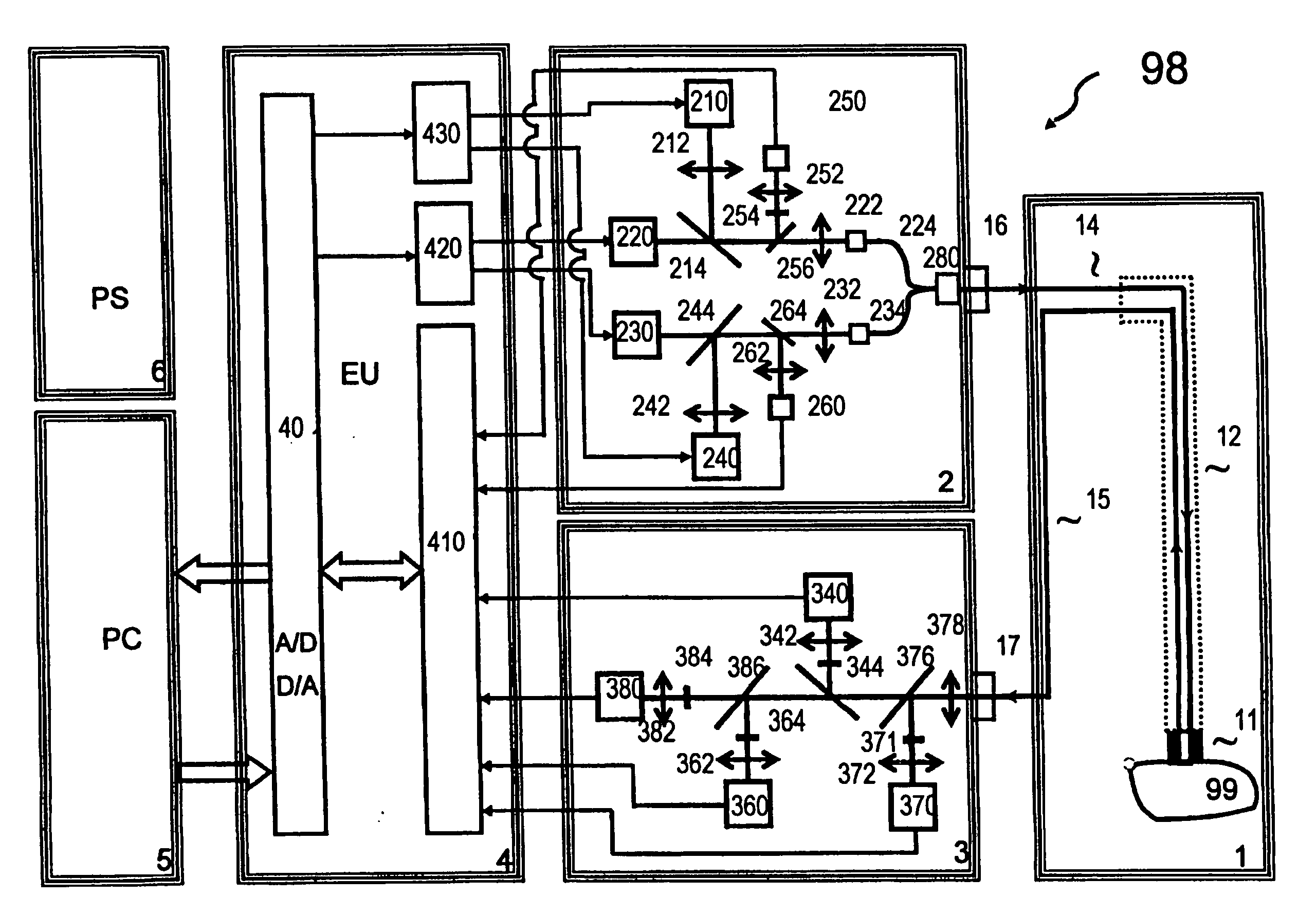
[0220] Referring to FIG. 19, the probe, generally designated (101), comprises all the elements and features of the probe (1) as described herein, mutatis mutandis. In addition, the probe (101) is particularly adapted for optical measurement of low-radiance signals from the inner surface of elastic body vessels, during catheterization. This is achieved by bending the fibers, which are laid along the internal axis of the catheter, at an angle 90 degrees, or close thereto. In this manner, the distal end of the fibers, i.e., the end from which light is emitted or received, comes into direct contact with the tissue, which is typically concentric with the probe (101). This arrangement for the fiber distal ends eliminates the costly and complicated treatment which would otherwise be required to be performed to the fibers ends, to convert these into “side firing” or “side facing” fibers. Moreover, this arrangement is superior to such side facing fibers, in which the coupling efficiency betw...

second embodiment

[0223] Referring to FIG. 20(a) and FIG. 20(b), the probe, generally designated (201), comprises all the elements and features of the probe (1) as described herein, mutatis mutandis. In addition, the probe (201) is particularly adapted for surface attachment to neonates' skin, using adhesive gels. Such gels (such as Ten20, by DO Weaver and Co. CO, USA, for example) are widely used with non-disposable neurodiagnostic electrodes. Thus, the head (21) is in the form of a cylindrical button, typically made from a suitable plastic, and comprises a fiber optic bundle (23). The distal end of the fiber bundle (23) is located at the center of the contact face (30) of the head (21), which also comprises two concentric open channels (25), (27). After slightly overfilling the outermost channel (25) with adhesive gel, the head (21) is pressed towards the skin and adhered thereto. The bundle (23) and plastic guide (28) which projects distally at the distal tip thereof form the contact surface (A2),...

third embodiment

[0225] Referring to FIG. 21(a) and FIG. 21(b), the probe, generally designated (301), comprises all the elements and features of the probe (1) as described herein, mutatis mutandis. In addition, the probe (301) is particularly adapted for surface attachment to neonates' skin, using adhesive tape or the like. Thus, the head (31) is in the form of a mouse, and made of relatively soft (30 to 40 Shore A) RTV Silicone or silicone rubber, such that its contact surface (A3) may easily adapt to the skin contour of the patient. A fiberoptic bundle (34) is embedded into the head (31), forming an internal bend (35) of obtuse angle. Since the silicone is elastic with respect to the fibers, it can not force the required additional sharp angled bend upon the fibers. Thus, the head (31) comprises a pre-molded stiff kernel (32), made from medical grade epoxy for example, wherein to form and maintain the bend (35). The kernel (32) is then embedded into a silicone body, thereby positioning the fibers...


PUM


Abstract

An apparatus, system and method are provided for diagnosing the degree of body metabolic emergency state. A non-vital organ with respect to the metabolic emergency state is first chosen. One or more tissue viability parameters including at least one of NADH and Flavoprotein (Fp) concentration are monitored in the non-vital organ, whereupon the degree of body metabolic emergency state may be determined based on the monitored tissue viability parameters.

Description

FIELD OF THE INVENTION [0001] The present invention relates to the early diagnosis of body metabolic emergency state that may develop in many acute or chronic clinical conditions. [0002] In particular, the invention is directed to a multiparametric method, system and apparatus for the monitoring of tissue blood flow, tissue reflectance, tissue oxy-deoxy hemoglobin saturation and mitochondrial (NADH) Nicotin-amide adenine dinucleotide and Flavoproteins (Fp) redox state. More particularly, the invention is concerned with measuring such parameters in real time for the most sensitive tissue or organ in the body to stress situations and sympathetic stimulation, in order to enable early diagnosis of emergency metabolic state of the body, particularly useful in facilitating formulation and navigation of the treatment given to the patient. BACKGROUND OF THE INVENTION [0003] The human body is totally dependent upon continuous supply of Oxygen (O2) utilized by various tissues and cells in ord...

Claims


Application Information

Patent Timeline
Patent Type & Authority Applications(United States)
IPC IPC(8): A61B5/00
CPCA61B5/0059A61B5/0084A61B5/4519A61B5/412A61B5/14546
Inventor MAYEVSKY, AVRAHAMPEWZNER, ELIAHUDEUTSCH, ASSAFMANOR, TAMAR
Owner CRITISENSE
Why Eureka
Try Eureka