Method and apparatus for viable and nonviable prokaryotic and eukaryotic cell quantitation

a prokaryotic and eukaryotic cell and quantitation method technology, applied in chemical methods analysis, instruments, enzymology, etc., can solve the problems of difficult to interpret colony counts, inaccurate estimations, and more severe problems, so as to speed up the conversion rate and increase the uptake rate of dye into cells

Inactive Publication Date: 2005-12-22
GENPRIME
View PDF10 Cites 3 Cited by
  • Summary
  • Abstract
  • Description
  • Claims
  • Application Information

AI Technical Summary

Benefits of technology

[0026] In other embodiments the kits may further comprise a third solution containing a compound or compounds that increase the rate of uptake of dye into cells or speeds up the rate of conversion the detectable fluorescent form of the dye inside said cells in second solution.

Problems solved by technology

However, colony counts are difficult to interpret since bacteria often clump or form chains that can give rise to significantly inaccurate estimations of the total number of viable organisms in a sample.
This problem is more severe when selective media are used.
Thus, the standard plate count does not provide a definitive count of viable cells in a sample, which may be very important for certain purposes.
Moreover, the technique is not useful when rapid determination of cell counts is required since it often requires over 24 hours to obtain results.
The technique is widely employed but nevertheless suffers from factors that must be held constant during the assay, e.g., medium used, chemical conditions, temperature and the types of cells being examined.
Also, dye reduction tests that incorporate microscopic counting techniques require trained technical personnel and often depend on subjective interpretations.
While the dye may enter the interior of both living and dead cells, dead cells are not capable of actively pumping the dye out under the conditions normally used.
It is not applicable to certain microbes and it yields incorrect viable counts with stressed cells.
Unfortunately, the long incubation time makes it unacceptable as a routine method.
Unfortunately, the technique requires the use of an expensive microscope and a trained individual or an expensive automated system (Pettipher et al., 1989).
The technique requires, and is limited by, expensive equipment.
Some flow cytometric devices have been used by the food and dairy industry, but their application has been limited by the high cost of instrumentation.
ATP levels vary in a single cell over more than two orders of magnitude, making this method a relatively inaccurate technique for the enumeration of viable organisms in a sample.
The method is rapid and simple but is highly inaccurate since all cells, particles and substances, including non-living particulate matter, interfere with the interpretation of the results.
The methylene blue test is rapid; however, the accuracy is unacceptable for cultures that are less than 90% viable.
The slide viability test is accurate for large viability ranges but the time required for results is not suitable for routine use.

Method used


Image

  • Method and apparatus for viable and nonviable prokaryotic and eukaryotic cell quantitation
  • Method and apparatus for viable and nonviable prokaryotic and eukaryotic cell quantitation
  • Method and apparatus for viable and nonviable prokaryotic and eukaryotic cell quantitation

Examples


example i

Materials and Calibration

[0058] The following shows examples of solutions, volumes and concentrations that can be used to carry out the invention. Other concentrations and volumes and different buffers with different pH values may be used. The samples and reaction solutions may be provided in any volume necessary for detection, however, in the present embodiment, small volumes (less than 1 ml) are utilized such that reagents and sample amounts are kept to a minimum. The volumes and concentrations to be utilized are any that are convenient to the practitioner of the methodology. In certain embodiments, the sample and reagent amounts range from 1 to 10,000 micro liters.

[0059] The reader used in the present embodiment is a “picofluor” hand-held fluorometer from Turner Designs, Sunnyvale, Calif. 94086. Any fluorometer that can measure fluorescence at specific wavelengths for detection of the dyes or cells can be employed in the invention. Ideally, the fluorometer can switch back and f...

example ii

Total Cell Counts

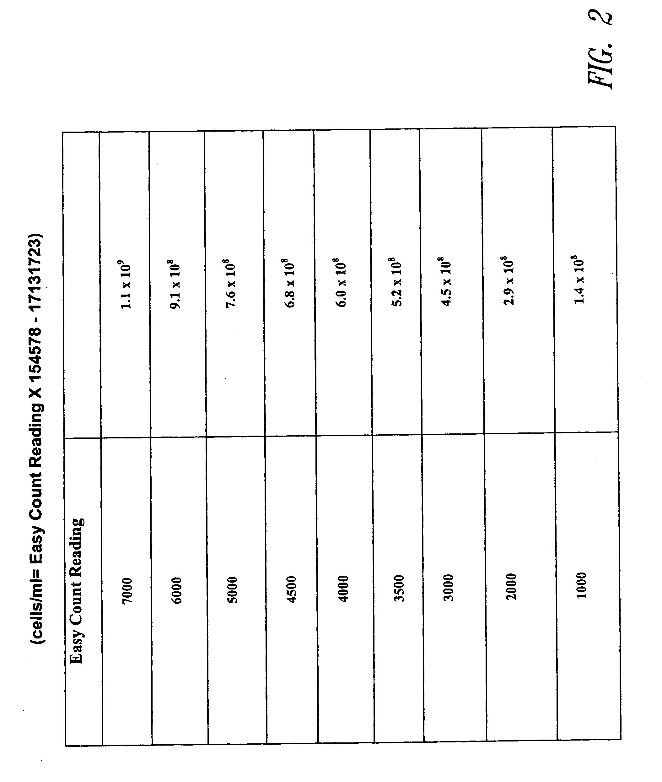
[0085] Total cell counts can be determined by a variety of methodologies, including using the fluorometer noted above. UV operation mode is selected. A fixed volume (200 microliters) of Cell preparation solution is added to the glass sample vial. 5 microliters of sample (yeast cells) is then added to the Cell preparation solution in the vial and centrifuged for about 30 seconds to sediment the a cell pellet. The cell pellet is resuspended in about 100 microliters of Cell preparation solution to the sample vial. The sample vial is then read in fluorometer. See FIG. 2.

example iii

Viable Cell Counts

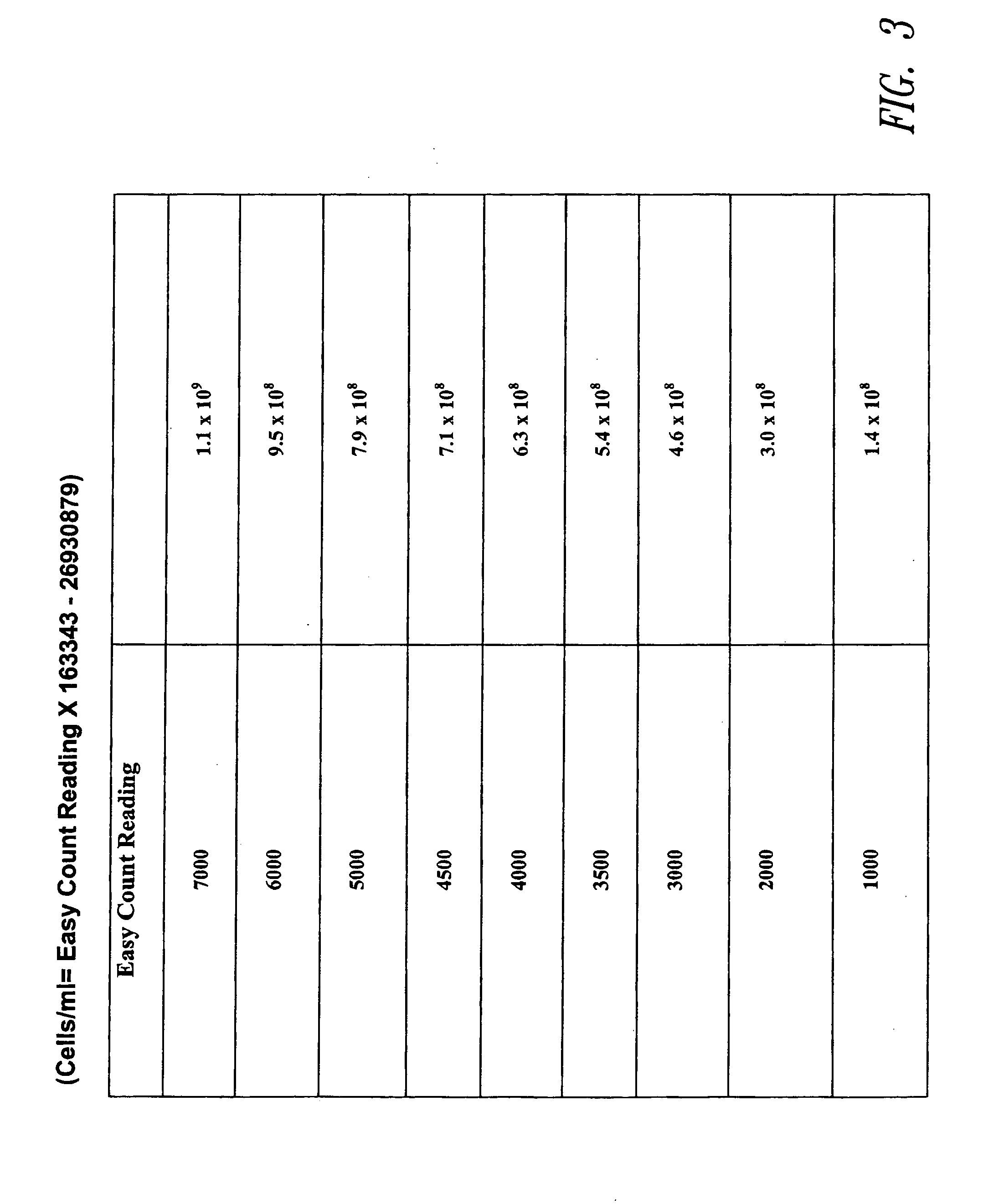
[0086] Live cells are quantitated utilizing a dye that is detectably altered by an intracellular enzyme. For example, the fluorometer noted above is set in visible mode and a fixed volume (200 microliters) of Cell preparation solution is added to the sample vial. 5 microliters of sample is added to the solution A in the sample vial and centrifuged for 30 seconds. 100 microliters of Suspension solution is added to the sample vial and 5 microliters of Reaction solution is added to the sample vial. The sample is mixed and placed in the reader and fluorescence determined at time zero and again at 15 minutes. This is the value that will be compared to the Easy Count correlation chart for conversion to cells / ml. See FIG. 3.


PUM

PropertyMeasurementUnit
volumesaaaaaaaaaa
temperatureaaaaaaaaaa
fluorescenceaaaaaaaaaa

Abstract

A rapid method for the quantitation of various live cell types is described. The method may include a variety of steps including: 1) suspending the cells in a detergent-like compound, 2) isolating the washed cells by centrifugation or filtration, 3) resuspending the cells in a solution that contains a preservative, a fluorescent dye and a compound such as dequalinium which can be taken up by the cells, 4) measuring the fluorescence increase over time of the cell-dye mixture with a simple fluorometer, and 5) measuring the native fluorescence of the cells. This new cell fluorescence method correlates with other methods of enumerating cells such as the standard plate count, the methylene blue method and the slide viability technique. The method is particularly useful in several applications such as: a) quantitating bacteria in milk, yogurt, cheese, meat and other foods, b) quantitating yeast cells in brewing, fermentation and bread making, c) quantitating mammalian cells in research, food and clinical settings. The method is especially useful when both total and viable cell counts are required such as in the brewing industry. The method can also be employed to determine the metabolic activity of cells in a sample. The apparatus, device, and/or system used for cell quantitation is also disclosed.

Description

CROSS REFERENCE TO RELATED APPLICATION [0001] The present application claims priority to U.S. Provisional Application No. 60 / 220,298, filed Jul. 24, 2000.TECHNICAL FIELD [0002] The present invention relates to methods for the rapid quantitation of both viable and nonviable cells. More specifically, the invention involves incubating cells with a metabolically activated visible fluorescent dye and measuring the fluorescence generated by viable cells. Total cell populations (viable+nonviable) are separately determined by measuring the native UV fluorescence of the cells. The two fluorescence readings are directly related to the number of viable and nonviable cells. This permits the user to determine the percent viability of a mixed population of live and dead cells. BACKGROUND OF THE INVENTION [0003] The ability to quantify living cells is vitally important to the food, beverage, pharmaceutical, environmental, manufacturing and clinical industries. Several methods are currently employe...

Claims


Application Information

Patent Timeline
Patent Type & Authority Applications(United States)
IPC IPC(8): C12Q1/06
CPCY10T436/101666C12Q1/06
Inventor FLEMING, JAMES E.SOMES, JASON BUCKMCLEAN, DARBYHOLCOMB, JERAD R.
Owner GENPRIME
Why Eureka
Try Eureka