Vacsularized tissue for transplantation

Inactive Publication Date: 2006-01-26
RGT UNIV OF CALIFORNIA
View PDF0 Cites 40 Cited by
  • Summary
  • Abstract
  • Description
  • Claims
  • Application Information

AI Technical Summary

Benefits of technology

[0021] Also, rather than dictating the vascular branching pattern and geometry, the vascular network in the tissue constructs of the invention develops naturally based on intrinsic biological signals, and the microfabrication technology described herein carefully controls the delivery of essential nutrients and oxygen in a temporary biodegradable microfluidic network.

Problems solved by technology

Second, the tissue construct is transplanted to the endogenous wound bed where vessels of the tissue construct and the host rapidly anastomose.

Method used


Image

  • Vacsularized tissue for transplantation
  • Vacsularized tissue for transplantation
  • Vacsularized tissue for transplantation

Examples


example 1

In Vitro Formation of Vascular Networks

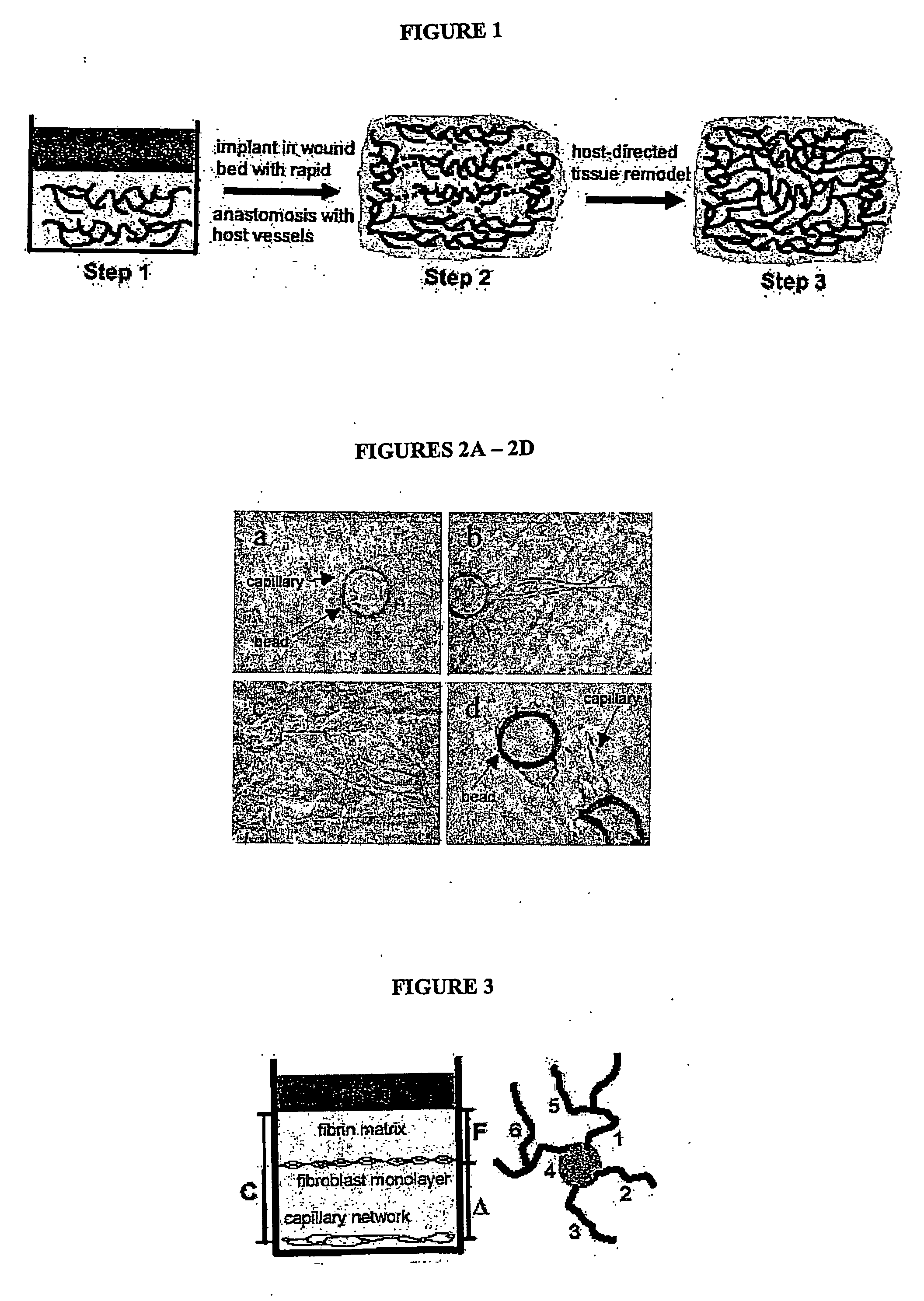
[0036] To provide the tissue construct of the invention with sufficient oxygenation, nutrient delivery and overall viability, endothelial cells [EC] can be induced to form complex, anastomosing capillary-like networks in vitro. The networks generated are stable (no apopotosis), exhibit long-term survival (several weeks), and readily anastomose to networks of capillaries with patent lumens. By controlling gel pH at approximately 7.4 (which affects rigidity), growth factor (VEGF, bFGF) concentrations (which affects vessel length and diameter), bead concentration at approximately 200 beads per ml of tissue (which affects degree of anastomosis and complexity of the network) and gel composition at approximately 2.5 mg / ml of Fibrin (the presence of fibronectin stimulates sprouting) capillary network formation is optimized.

[0037] Briefly, human umbilical vein endothelial cells are harvested and passaged twice, then seeded onto 150 micron diameter Cy...

example 2

Transplanted, In Vitro-Derived Capillary Beds Anastomose with Host Vasculature

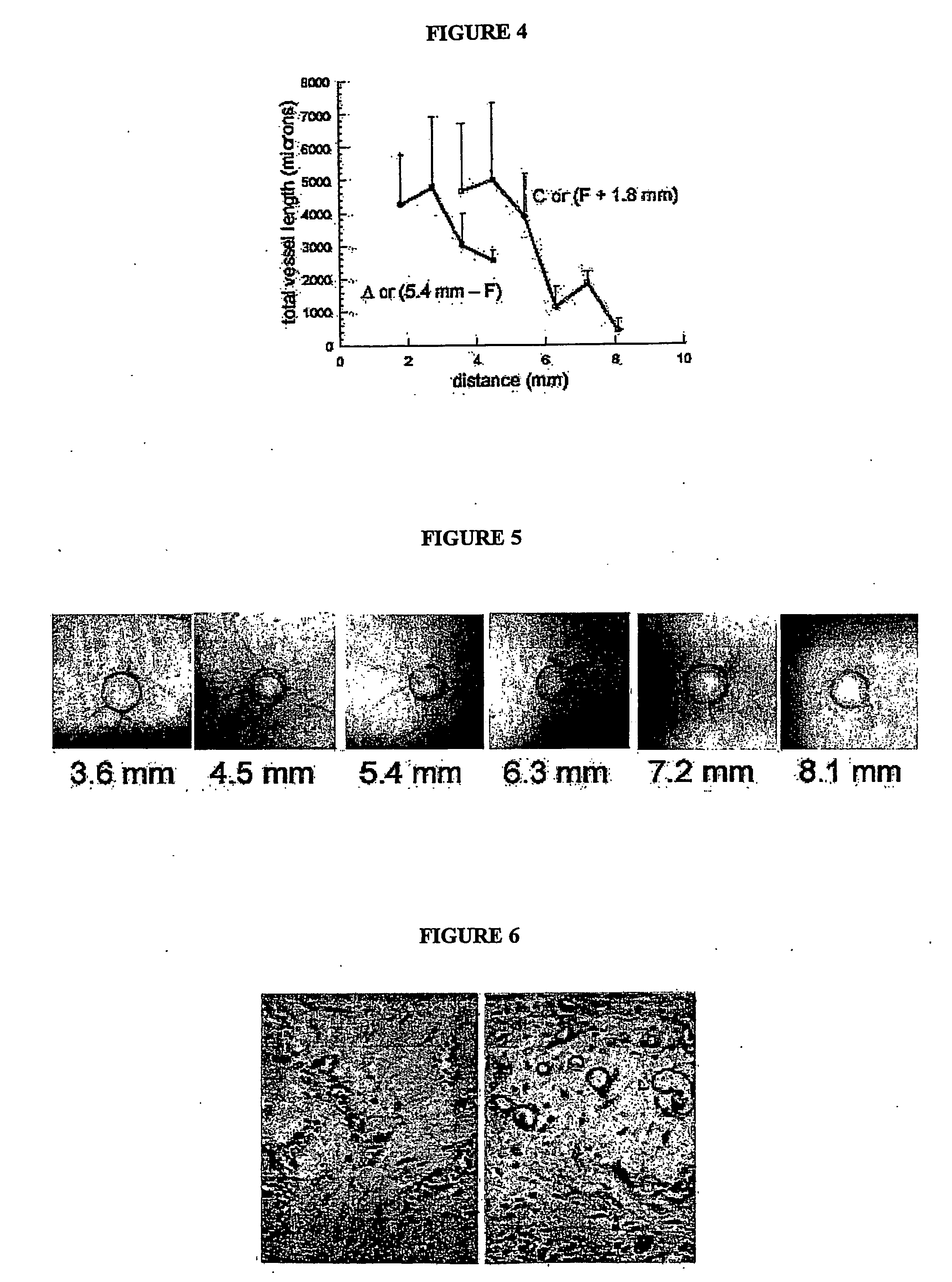
[0042] A crucial concern in transplanting a vascularized tissue into a host is whether the two vascular networks will “hook-up” correctly, allowing perfusion of the transplanted vessels. In two different systems that transplanted vascular beds in tissue constructs spontaneously anastomose with host vasculature. In the first, human epidermis, including the superficial vascular plexus, was transplanted onto the back of an immunocompromised (SCID) mouse. After only three days, anastomoses between human and mouse vasculature could be identified using species-specific antibodies, and moreover, the skin survived through perfusion of human vessels, not through ingrowth of host vessels. Referring to FIG. 6 in the left panel, an H&E-stained section showing human in vitro-derived capillaries within a collagen gel, inserted under a skin flap on the back of a SCID mouse. Note that the capillaries are filled with bloo...

example 3

Manipulation of Gelatin at Micron Dimensions

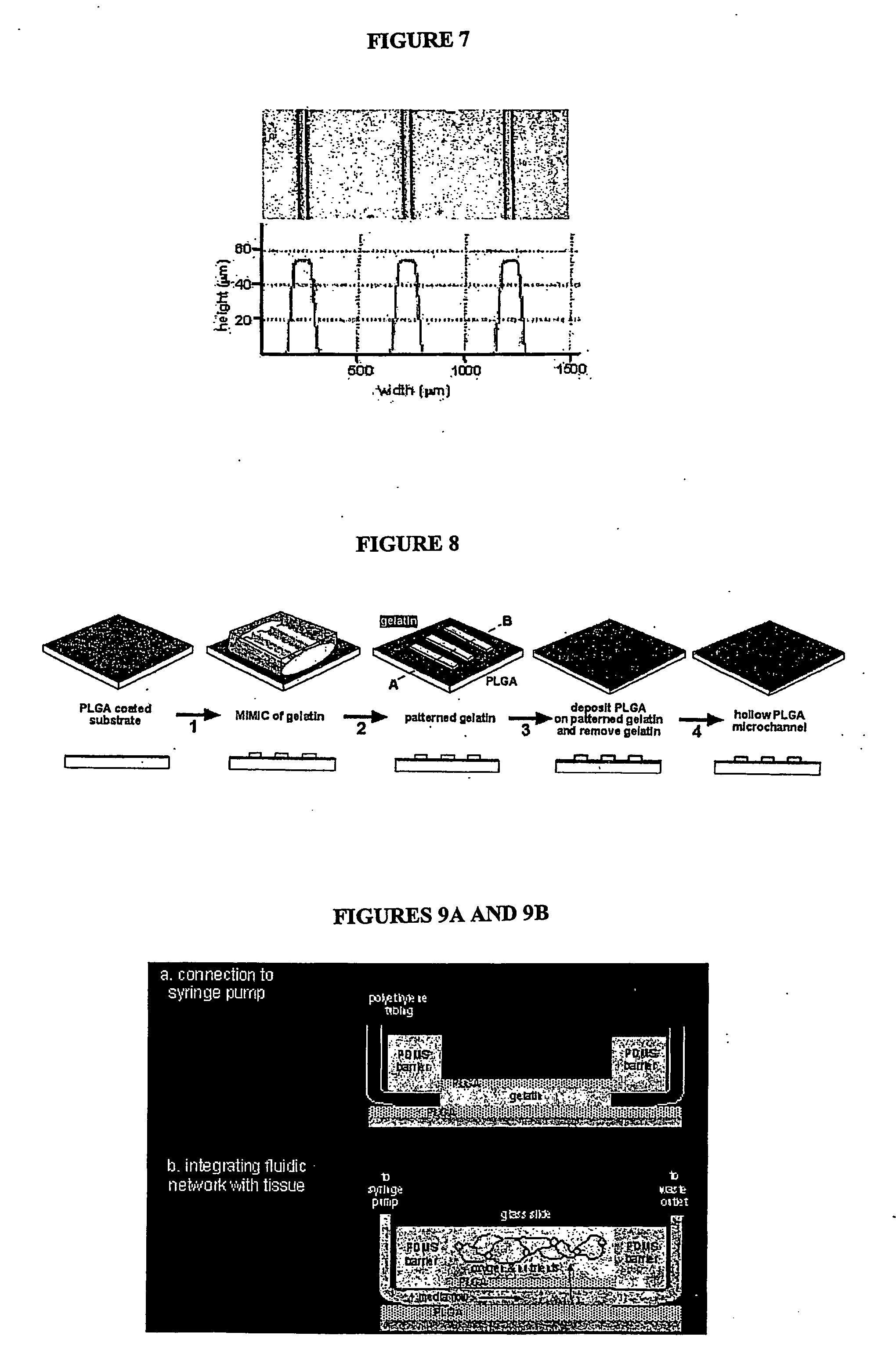
[0044] A combination of soft lithography and micromolding in capillaries (MMIC) generates rows of gelatin with dimensions on the micron scale. A high-resolution photomask is generated and used to selectively expose photoresist by contact photolithography. The unexposed photoresist is removed leaving a positive relief that serves as the master mold. Prepolymer of poly(dimethlysiloxane) (PDMS) is cast on the master and cured to obtain a PDMS replica with embedded channels. A solution of gelatin (appropriate viscosity) is then wicked into the PDMS mold by capillary action MIMIC) and allowed to gel. The PDMS mold is removed leaving the micropatterned gelatin rows (FIG. 4). Referring to FIG. 7, the rows are approximately 100 microns in diameter, 55 microns in height, and 455 microns apart (bottom panel) as determined by a surface profilometer. The gelatin rows serve as the mold for the lumen of the microfluidic channels. Following coding with ...


PUM

PropertyMeasurementUnit
thicknessaaaaaaaaaa
thicknessaaaaaaaaaa
total volumeaaaaaaaaaa

Abstract

Tissue engineering holds enormous potential to replace or restore function to a wide range of tissues. However, the most successful applications have been limited to thin avascular tissues in which delivery of essential nutrients occurs primarily by diffusion. Pursuant to the present invention, a prevascularized, thick tissue construct is created having a network of capillaries with lumens capable of nutrient and origin delivery and forming anastamoses to host vasculature. A tissue transplantation strategy is comprised of (1) in vitro vascularization of a tisue construct, (2) transplantation of prevascularized tissue to wound bed of host where vessels of implantable tissue and host rapidly anastomose, and (3) host-directed remodeling and reorganization of the tissue and vascular network.

Description

FIELD OF THE INVENTION [0001] This application relates to engineered tissue having an engineered vascular network for forming anastamoses to endogenous vasculature after transplantation and methods to produce the tissue in vitro. BACKGROUND [0002] Tissue transplantation is critically necessary in many clinical situations, including reconstructive surgery, wound healing, cardiovascular treatment and many others. The first examples of tissue trasplantation were “autologous,” meaning that the tissue was simply removed from a donor site in the patient and then re-inserted at another target site. Although autologous tissue transfers are well known, autologous transfers have certain drawbacks that cannot be overcome. Significantly, autologous transfers compromise the donor site and carry the risk of infection and loss of function. In a surgical setting, a second procedure to remove an autologous tissue graft always carries a finite risk and unavoidably adds to patient discomfort and expen...

Claims


Application Information

Patent Timeline
Patent Type & Authority Applications(United States)
IPC IPC(8): A61K48/00A61L9/04A61K35/12A61L27/38C12N5/00C12N5/071
CPCA61K35/12C12N2535/10A61L27/3808A61L27/3895C12N5/0068C12N5/0691C12N2500/60C12N2501/115C12N2501/165C12N2531/00C12N2533/40C12N2533/52C12N2533/54C12N2533/56A61L27/3804C12M25/16
Inventor GEORGE, STEVENCJEON, NOO LIHUGHES, CHRIS C.W.
Owner RGT UNIV OF CALIFORNIA
Why Eureka
Try Eureka