
Unique recognition sequences and methods of use thereof in protein analysis

a recognition sequence and protein technology, applied in the field of unique recognition sequences and methods of use in protein analysis, can solve the problems of difficult reagent generation, less specificity of detection agents against native proteins, and a lot of time (years) and resources

Inactive Publication Date: 2006-02-16
MILLIPORE CORP
View PDF22 Cites 38 Cited by
  • Summary
  • Abstract
  • Description
  • Claims
  • Application Information

AI Technical Summary

Benefits of technology

[0014] Embodiments of the present invention also overcome the imprecisions in detection methods caused by: the existence of proteins in multiple forms in a sample (e.g., various post-translationally modified forms or various complexed or aggregated forms); the variability in sample handling and protein stability in a sample, such as plasma or serum; and the presence of autoantibodies in samples. In certain embodiments, using a targeted fragmentation protocol, the methods of the present invention assure that a binding site on a protein of interest, which may have been masked due to one of the foregoing reasons, is made available to interact with a capture agent. In other embodiments, the sample proteins are subjected to conditions in which they are denatured, and optionally are alkylated, so as to render buried (or otherwise cryptic) URS moieties accessible to solvent and interaction with capture agents. As a result, the present invention allows for detection methods having increased sensitivity and more accurate protein quantitation capabilities. This advantage of the present invention will be particularly useful in, for example, protein marker-type disease detection assays (e.g., PSA or Cyclin E based assays) as it will allow for an improvement in the predictive value, sensitivity, and reproducibility of these assays. The present invention can standardize detection and measurement assays for all proteins from all samples.
[0015] The present invention is based, at least in part, on the realization that exploitation of unique recognition sequences (URSs) present within individual proteins can enable reproducible detection and quantitation of individual proteins in parallel in a milieu of proteins in a biological sample. As a result of this unique recognition sequence-based approach, the methods of the invention detect specific proteins in a manner that does not require preservation of the whole protein, nor even its native tertiary structure, for analysis. Moreover, the methods of the invention are suitable for the detection of most or all proteins in a sample, including insoluble proteins such as cell membrane bound and organelle membrane bound proteins.
[0016] The present invention is also based, at least in part, on the realization that unique recognition sequences can serve as Proteome Epitope Tags characteristic of a specific organism's proteome and can enable the recognition and detection of a specific organism.
[0017] The present invention is also based, at least in part, on the realization that high-affinity agents (such as antibodies) with predefined specificity can be generated for defined, short length peptides and when antibodies recognize protein or peptide epitopes, only 4-6 (on average) amino acids are critical. See, for example, Lerner R A (1984) Advances In Immunology. 36:1-45.
[0018] The present invention is also based, at least in part, on the realization that by denaturing and / or fragmenting all proteins in a sample to produce a soluble set of protein analytes, e.g., in which even otherwise buried URS's are solvent accessible, the subject method provides a reproducible and accurate (intra-assay and inter-assay) measurement of proteins.
[0019] Accordingly, in one aspect, the present invention provides a method for globally detecting the presence of a protein(s) (e.g., membrane bound protein(s)) in an organism's proteome. The method includes providing a sample which has been denatured and / or fragmented to generate a collection of soluble polypeptide analytes; contacting the polypeptide analytes with a plurality of capture agents (e.g., capture agents immobilized on a solid support such as an array) under conditions such that interaction of the capture agents with corresponding unique recognition sequences occurs, thereby globally detecting the presence of protein(s) in an organism's proteome.

Problems solved by technology

There are several problems with the current approaches to massively parallel, e.g., cell-wide or proteome wide, protein detection.
First, reagent generation is difficult: One needs to first isolate every individual target protein in order to isolate a detection agent against every protein in an organism and then develop detection agents against the purified protein.
Since the number of proteins in the human organism is currently estimated to be about 30,000 this requires a lot of time (years) and resources.
Furthermore, detection agents against native proteins have less defined specificity since it is a difficult task to know which part of the proteins the detection agents recognize.
This prolem causes considerable cross-reactivity of when multiple detection agents are arrayed together, making large-scale protein detection array difficult to construct.
Second, current methods achieve poor coverage of all possible proteins in an organism.
Third, current methods are not general to all proteins or to all types of biological samples.
Physiological fluids like urine and blood serum are relatively simple, but biopsy tissue samples are very complex.
Current detection methods are either not effective over all proteins uniformly or cannot be highly multiplexed to enable simultaneous detection of a large number of proteins (e.g., >5,000).
Optical detection methods would be most cost effective but suffer from lack of uniformity over different proteins.
Proteins in a sample have to be labeled with dye molecules and the different chemical character of proteins leads to inconsistency in efficiency of labeling.
Labels may also interfere with the interactions between the detection agents and the analyte protein leading to further errors in quantitation.
Non-optical detection methods have been developed but are quite expensive in instrumentation and are very difficult to multiplex for parallel detection of even moderately large samples (e.g., >100 samples).
Another problem with current technologies is that they are burdened by intracellular life processes involving a complex web of protein complex formation, multiple enzymatic reactions altering protein structure, and protein conformational changes.
(1998) Clinical Chemistry 44(6):1325-1333, standarizing immunoassays is difficult due to the variability in sample handling and protein stability in plasma or serum.
Finally, current technologies are burdened by the presence of autoantibodies which affect the outcome of immunoassays in unpredictable ways, e.g., by leading to analytical errors (Fitzmaurice T. F. et al.
These problems prompted the question whether it is even possible to standardize immunoassays for hetergenous protein antigens.

Method used


Image

  • Unique recognition sequences and methods of use thereof in protein analysis
  • Unique recognition sequences and methods of use thereof in protein analysis
  • Unique recognition sequences and methods of use thereof in protein analysis

Examples


example 1

Identification of Unique Recognition Equences within the Human Proteome

[0264] As any one of the total 20 amino acids could be at one specific position of a peptide, the total possible combination for a tetramer (a peptide containing 4 amino acid residues) is 204; the total possible combination for a pentamer (a peptide containing 5 amino acid residues) is 205 and the total possible combination for a hexamer (a peptide containing 6 amino acid residues) is 206. In order to identify unique recognition sequences within the human proteome, each possible tetramer, pentamer or hexamer was searched against the human proteome (total number: 29,076; Source of human proteome: EBI Ensembl project release v 4.28.1 on Mar. 12, 2002, http: / / www.ensembl.org / Homo—sapiens / ).

[0265] The results of this analysis, set forth below, indicate that using a pentamer as a unique recognition sequence, 80.6% (23,446 sequences) of the human proteome have their own unique recognition sequence(s). Using a hexamer...

example 2

Identification of Unique Recognition Sequences within all Bacterial Proteomes

[0273] In order to identify pentamer URSs that can be used to, for example, distinguish a bacterium from a pool of all other bacteria, each possible pentamer was searched the NCBI database (http: / www.ncbi.nlm.nih.gov / PMGifs / Genomes / eub_g.html, as of Apr. 10, 2002). The results from this analysis are set forth below. Results and Data:

DatabaseNumber ofIDunique(NCBIpentamersRefSeq ID)Species Name6NC_000922Chlamydophila pneumoniae CWL02937NC_002745Staphylococcus aureus N315 chromosome40NC_001733Methanococcus jannaschii small extra-chromosomal element58NC_002491Chlamydophila pneumoniae J13884NC_002179Chlamydophila pneumoniae AR39135NC_000909Methanococcus jannaschii206NC_003305Agrobacterium tumefaciens str. C58(U. Washington) linear chromosome298NC_002758Staphylococcus aureus Mu50 chromosome356NC_002655Escherichia coli O157:H7 EDL933386NC_003063Agrobacterium tumefaciens str. C58(Cereon) linear chromosome479NC_...

example 3

Identification of Specific Pentamer Unique Recognition Sequences

[0274] As indicated above, each possible tetramer, pentamer or hexamer was searched against the human proteome (total number: 29,076; Source of human proteome: EBI Ensembl project release 4.28.1 on Mar. 12, 2002, http: / / www.ensembl.org / Homo—sapiens / ) to identify unique recognition sequences (URSs).

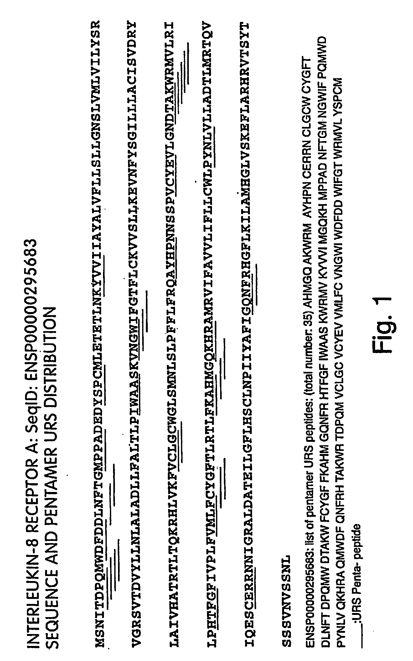
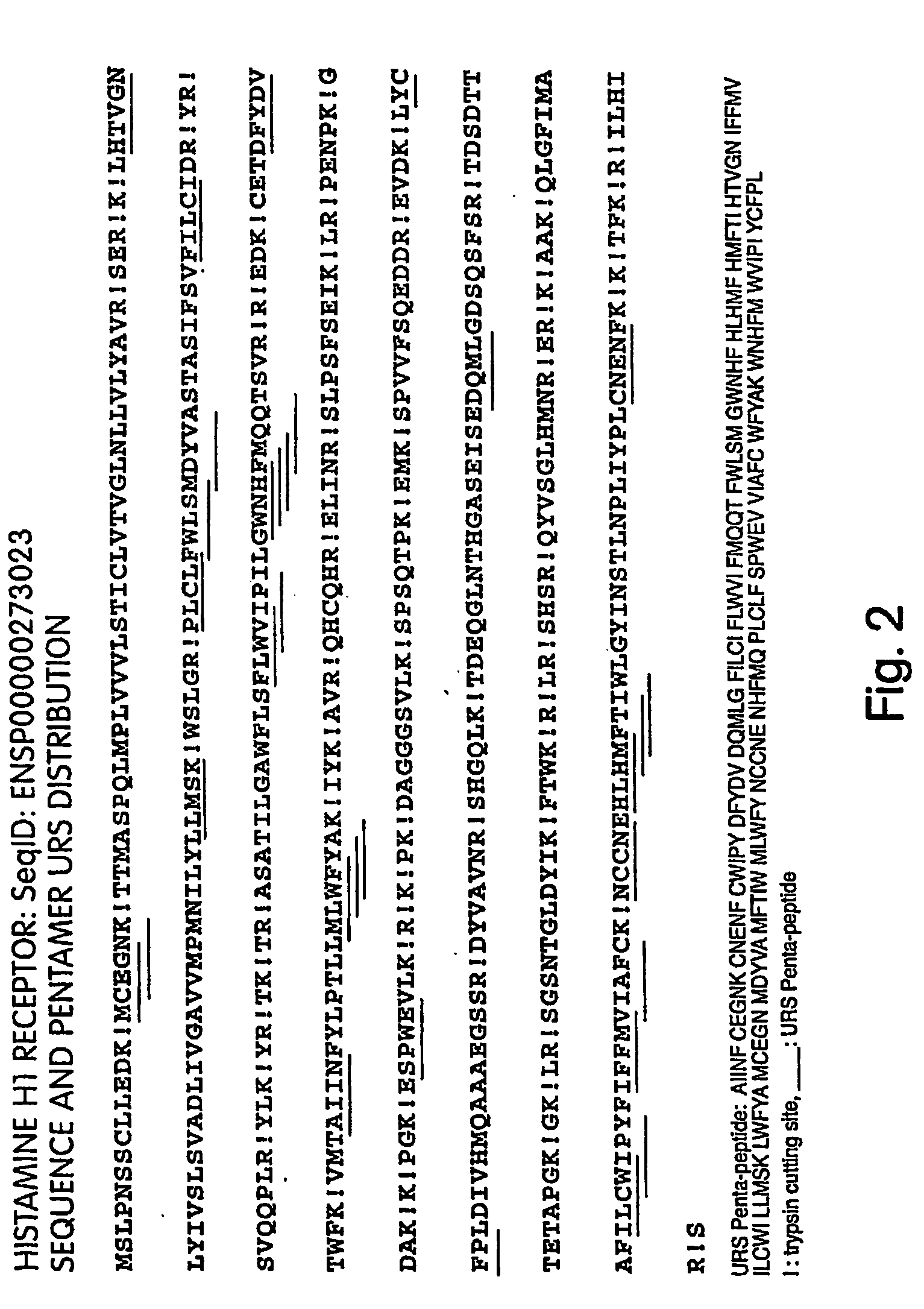
[0275] Based on the foregoing searches, specific URSs were identified for the majority of the human proteome. FIG. 1 depicts the pentamer unique recognition sequences that were identified within the sequence of the Interleukin-8 receptor A. FIG. 2 depicts the pentamer unique recognition sequences that were identified within the Histamine H1 receptor that are not destroyed by trypsin digestion. Further Examples of pentamer unique recognition sequences that were identified within the human proteome are set forth below.

No. ofpentamerSequence ID*URSsPentamer URSsENSP000000002339AMPVS CATQG CFTVW ICFTV MPNAM PHAMP(SEQ ID NOs:1-...


PUM

PropertyMeasurementUnit
Fractionaaaaaaaaaa
Fractionaaaaaaaaaa
Fractionaaaaaaaaaa

Abstract

Disclosed are methods for reliably detecting the presence of proteins in a sample by the use of capture agents that recognize and interact with recognition sequences uniquely characteristic of a set of proteins in the sample. Arrays comprising these capture agents are also provided.

Description

RELATED APPLICATIONS [0001] This application is a divisional application of U.S. Ser. No. 10 / 436,549, filed on May 12, 2003, which claims the benefit of the filing dates of U.S. Provisional Application No. 60 / 379,626, filed on May 10, 2002; U.S. Provisional Application Nos. 60 / 393,137, 60 / 393,233, 60 / 393,235, 60 / 393,211, 60 / 393,223, 60 / 393,280, and 60 / 393,197, all filed on Jul. 1, 2002; U.S. Provisional Application No. 60 / 430,948, filed on Dec. 4, 2002; and U.S. Provisional Application No. 60 / 433,319 filed on Dec. 13, 2002, the entire contents of each of which are incorporated herein by reference.BACKGROUND OF THE INVENTION [0002] Genomic studies are now approaching “industrial” speed and scale, thanks to advances in gene sequencing and the increasing availability of high-throughput methods for studying genes, the proteins they encode, and the pathways in which they are involved. The development of DNA microarrays has enabled massively parallel studies of gene expression as well as ...

Claims


Application Information

Patent Timeline
IPC IPC(8): C12Q1/68G01N33/53G06F19/00C12Q1/48C40B30/04G01N33/68
CPCB82Y5/00B82Y10/00B82Y30/00C12Q1/48G01N33/6851G01N33/68G01N33/6803G01N33/6842C40B30/04Y02A90/10
Inventor LEE, FRANKMENG, XUNCHAN, JOHNZHANG, SHENGSHENGBENKOVIC, STEPHEN
Owner MILLIPORE CORP
Learn More