Devices and methods for intracardiac procedures

a technology for intracardiac surgery and devices, applied in the direction of prosthesis, surgical staples, surgical forceps, etc., can solve the problems of prolonged hospital stay, high degree of trauma, and significant risk of complications, so as to facilitate intervention, reduce trauma, reduce the risk of complications, and improve the effect of patient comfor

Inactive Publication Date: 2006-03-09
EDWARDS LIFESCIENCES LLC
View PDF30 Cites 62 Cited by
  • Summary
  • Abstract
  • Description
  • Claims
  • Application Information

AI Technical Summary

Benefits of technology

[0012] The invention provides devices and methods for performing less-invasive surgical procedures within an organ or vessel, and particularly, within the heart and great vessels of the thoracic cavity. The devices and methods of the invention facilitate intervention within the heart or great vessels without the need for a median sternotomy or other form of gross thoracotomy, substantially reducing trauma, risk of complication, recovery time, and pain for the patient. Using the devices and methods of the invention, surgical procedures may be performed through percutaneous penetrations within intercostal spaces of the patient's rib cage, without cutting, removing, or significantly displacing any of the patient's ribs or sternum. The devices and methods arc particularly well-adapted for heart valve repair and replacement, facilitating visualization within the patient's thoracic cavity, repair or removal of the patient's natural valve, and, if necessary, attachment of a replacement valve in the natural valve position. The invention facilitates valve replacement with any of a variety of commercially-available replacement valves, including mechanical prostheses, bioprostheses, homografts, and allografts.
[0020] The method according to the present invention may be carried out on humans or other mammalian animals. The method is of particular applicability in humans as it allows an alternative approach to open heart surgery and the development of closed cardioscopic surgery. The method according to the invention enables a percutaneous by-pass system to be associated with cardioplegia, venting and cooling of the heart which subverts the need for median sternotomy. This may, in turn, reduce the complications of the surgery.
[0021] The maintenance of the systemic circulation involves establishing a cardiopulmonary by-pass. The blood may be drawn into the by-pass merely by positioning a percutaneous catheter into the right atrium and / or into one or both of the vena cavae through which venous blood may be drawn from the heart into an extracorporeal pump oxygenator. In more preferred embodiments of the invention a single catheter with two inflatable cuffs, or two separate catheters, each with an inflatable cuff are introduced into the vena cavae to occlude them adjacent to their right atrial inlets. This allows isolation of the right atrium and allows blood to be drawn from the vena cavae into the by-pass system. There is also preferably provision for percutaneous communication via one catheter with the right atrium to allow infusion of saline into the right atrium. This infusion has the advantage that it allows the heart to be cooled and improves visual acuity within the right heart allowing direct cardioscopic examination and / or intervention.
[0023] The ascending aorta is preferably occluded by a balloon catheter introduced percutaneously through the femoral artery. This catheter must carry adjacent its tip an inflatable cuff or balloon of sufficient size that upon being inflated it is able to completely occlude the ascending aorta. The length of the balloon should preferably not be so long as to impede the flow of blood or other solution to the coronary arteries or to the brachiocephalic, left carotid or left subclavian arteries. A balloon length of about 40 mm and diameter of about 35 mm is suitable in humans. The balloon is of a cylindrical shape to fully and evenly accommodate the lumen of the ascending aorta. This maximizes the surface area contact with the aorta, and allows for even distribution of occlusive pressure.
[0052] Preferably, the replacement valve is premounted to the holder means and the two are sterilized and packaged together in a sterile pack. In this way, the pack may be opened in the sterile operating room environment with the valve and holder ready for immediate surgical use.
[0064] The system may further include means for attaching the replacement valve to the patient's heart, which comprises, in one embodiment, means for suturing the replacement valve to a valve annulus in the patient's heart. The system preferably includes organizing means for maintaining the sutures in spaced-apart positions outside of the chest cavity after the sutures have been applied to the valve annulus within the heart. The organizing means is preferably fixed to a proximal end of a cannula disposed in a percutaneous intercostal penetration, as described above. In this way, the sutures may be applied to the natural valve annulus within the patient's heart, drawn out of the chest cavity through the cannula lumen, and positioned in spaced-apart positions about the circumference of the proximal end of the cannula. Means may also be provided for maintaining tension on the ends of the sutures after they have been applied to the replacement valve. This facilitates advancing the replacement valve along the sutures, through the lumen in the cannula, and into the chest cavity.

Problems solved by technology

However, these invasive, open-chest procedures produce a high degree of trauma, a significant risk of complications, an extended hospital stay, and a painful recovery period for the patient.
Moreover, while heart valve surgery produces beneficial results for many patients, numerous others who might benefit from such surgery are unable or unwilling to undergo the trauma and risks of current techniques.

Method used


Image

  • Devices and methods for intracardiac procedures
  • Devices and methods for intracardiac procedures
  • Devices and methods for intracardiac procedures

Examples


Embodiment Construction

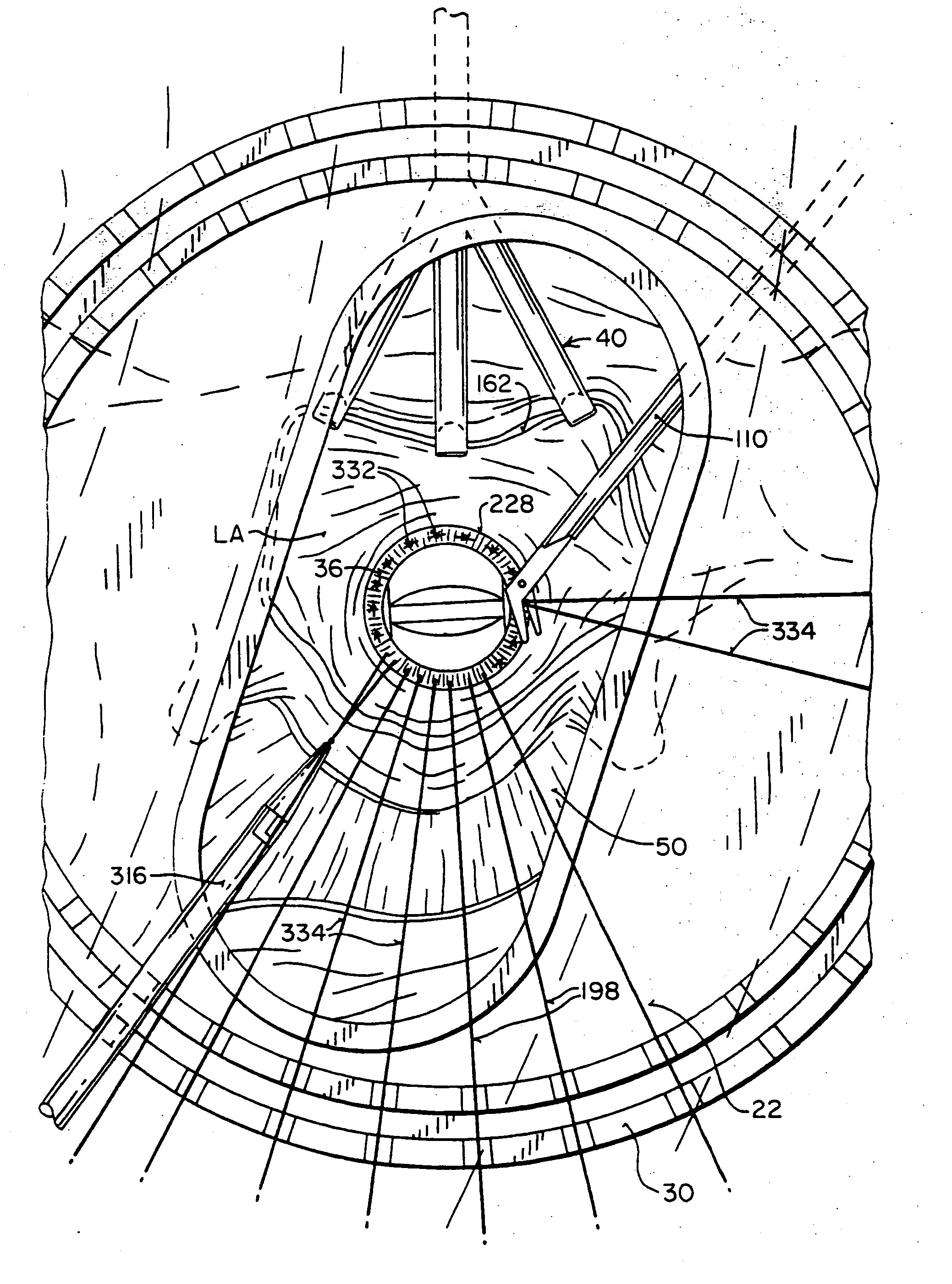
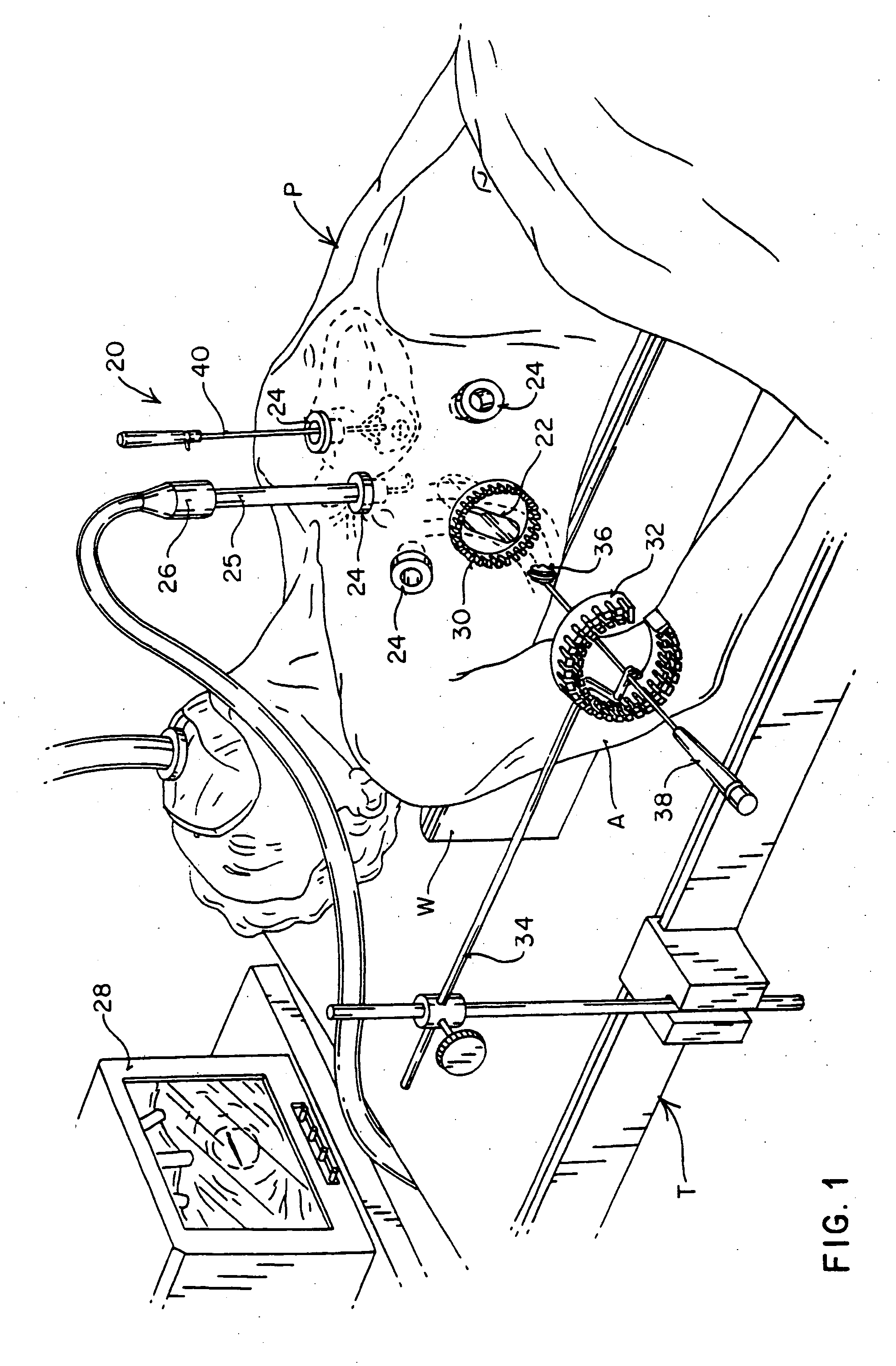
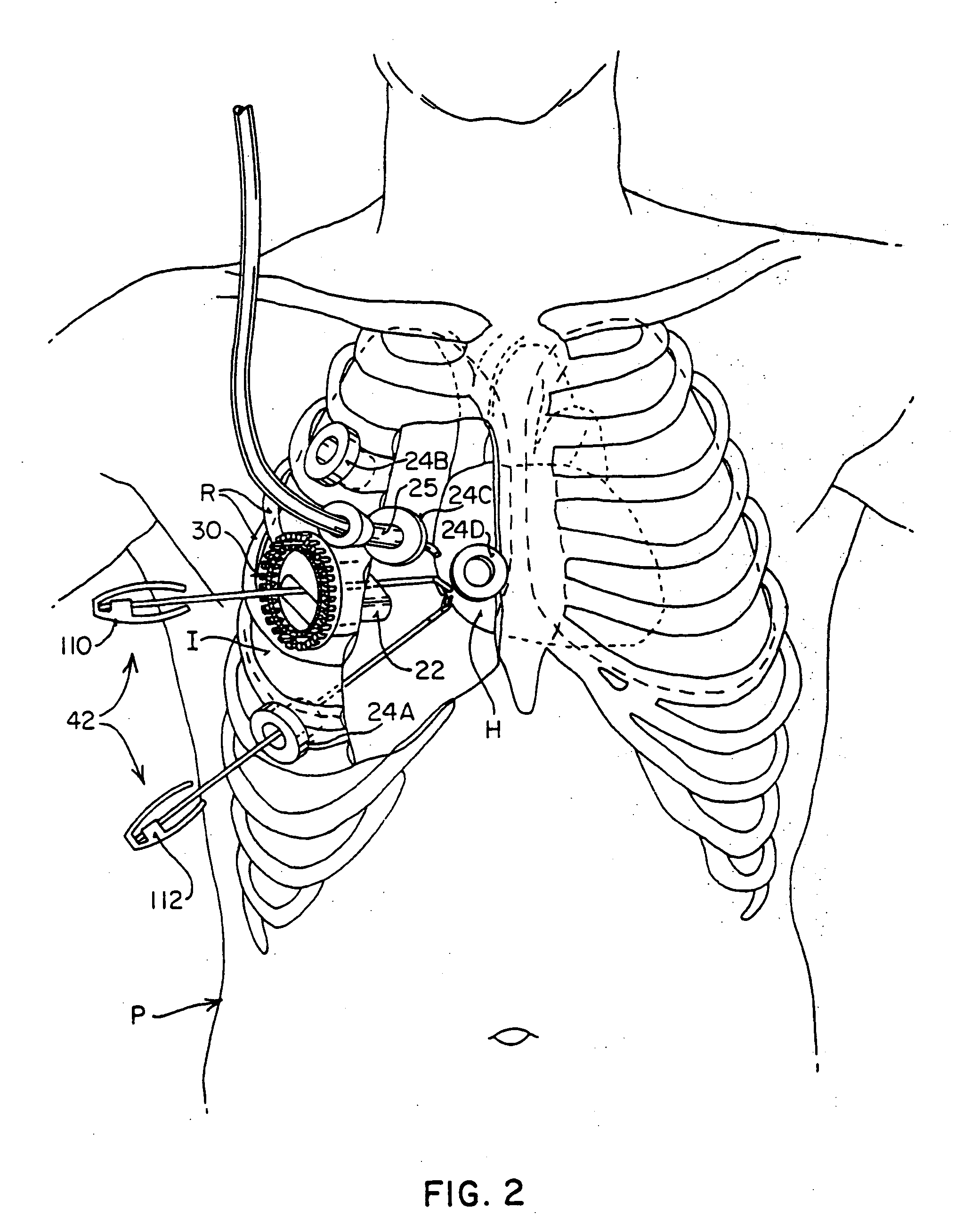
[0109] The invention provides methods and devices for performing surgical interventions within the heart or a great vessel such as the aorta, superior vena cava, inferior vena cava, pulmonary artery, pulmonary vein, coronary arteries, and coronary veins, among other vessels. While the specific embodiments of the invention described herein will refer to mitral valve repair and replacement, it should be understood that the invention will be useful in performing a great variety of surgical procedures, including repair and replacement of aortic, tricuspid, or pulmonary valves, repair of atrial and ventricular septal defects, pulmonary thrombectomy, removal of atrial myxoma, patent foramen ovale closure, treatment of aneurysms, electrophysiological mapping and ablation of the myocardium, myocardial drilling, coronary artery bypass grafting, angioplasty, atherectomy, correction of congenital defects, and other procedures in which interventional devices are introduced into the interior of ...


PUM


Abstract

The invention provides devices and methods for performing less-invasive surgical procedures within an organ or vessel. In an exemplary embodiment, the invention provides a method of closed-chest surgical intervention within an internal cavity of a patient's heart or great vessel. According to the method, the patient's heart is arrested and cardiopulmonary bypass is established. A scope extending through a percutaneous intercostal penetration in the patient's chest is used to view an internal portion of the patient's chest. An internal penetration is formed in a wall of the heart or great vessel using cutting means introduced through a percutaneous penetration in an intercostal space in the patient's chest. An interventional tool is then introduced, usually through a cannula positioned in a percutaneous intercostal penetration. The interventional tool is inserted through the internal penetration in the heart or great vessel to perform a surgical procedure within the internal cavity under visualization by means of the scope. In a preferred embodiment, a cutting tool is introduced into the patient's left atrium from a right portion of the patient's chest to remove the patient's mitral valve. A replacement valve is then introduced through an intercostal space in the right portion of the chest and through the internal penetration in the heart, and the replacement valve is attached in the mitral valve position.

Description

CROSS-REFERENCE TO RELATED APPLICATIONS [0001] This application is a continuation-in-part of commonly-assigned, co-pending application Ser. No. 08 / 163,241, filed Dec. 6, 1993, which is a continuation-in-part of application Ser. No. 08 / 023,778, filed Feb. 2, 1993. This application is also a continuation-in-part of copending application Ser. No. 08 / 159,815, filed Nov. 30, 1993, which is a U.S. counterpart of Australian Patent Application No. PL 6170, filed Dec. 3, 1992. The complete disclosures of all of these applications are hereby incorporated herein by reference.FIELD OF THE INVENTION [0002] This invention relates generally to instruments and techniques for performing less-invasive surgical procedures, and more specifically, to instruments and techniques for less-invasive surgery within the heart and great vessels. BACKGROUND OF THE INVENTION [0003] Various types of surgical procedures are currently performed to investigate, diagnose, and treat diseases of the heart and the great ...

Claims


Application Information

Patent Timeline
Patent Type & Authority Applications(United States)
IPC IPC(8): A61B17/06A61B17/00A61B17/02A61B17/04A61B17/28A61B17/30A61B17/34A61B18/14A61B19/00A61F2/24A61F2/958A61M1/00A61M1/36A61M25/00
CPCA61B17/00234A61B17/0057A61M2210/127A61B17/0218A61B17/0467A61B17/0469A61B17/06061A61B17/064A61B17/068A61B17/12013A61B17/122A61B17/1227A61B17/1285A61B17/29A61B17/2909A61B17/30A61B17/3403A61B18/1442A61B18/1492A61B19/26A61B2017/00243A61B2017/00247A61B2017/00637A61B2017/00663A61B2017/0243A61B2017/047A61B2017/0472A61B2017/0474A61B2017/0475A61B2017/2943A61B2017/306A61B2017/3405A61B2017/3427A61B2017/3492A61B2018/00214A61B2018/00232A61B2018/00261A61B2018/00291A61B2018/00363A61B2018/00392A61B2018/00577A61B2019/464A61F2/2427A61F2/2496A61M1/3664A61M25/0023A61M25/0032A61M25/1011A61M39/0247A61M2025/0003A61M2025/028A61M2025/1052A61M2039/027A61M2039/0279A61M2202/047A61M2205/3344A61M2205/3355A61M2205/366A61M2230/005A61B2090/064A61B90/50A61M1/3623
Inventor STERMAN, WESLEY D.GARRISON, MICHI E.GIFFORD, HANSON S. IIISTEVENS, JOHN H.PETERS, WILLIAM S.
Owner EDWARDS LIFESCIENCES LLC
Why Eureka
Try Eureka