Method and apparatus for ultrasonically increasing the transportation of therapeutic substances through tissue

a technology of ultrasonically increasing and therapeutic substances, applied in the field of implantable ultrasound transducer devices, can solve the problems of systemic methods, undesirable side effects, untargeted organs or tissues, etc., and achieve the effects of enhancing the depth, and enhancing the transportation of therapeutic substances

Inactive Publication Date: 2006-03-16
CYTODOME
View PDF91 Cites 16 Cited by
  • Summary
  • Abstract
  • Description
  • Claims
  • Application Information

AI Technical Summary

Benefits of technology

[0018] The present invention is based, in part, on the recognition that the transportation of therapeutic molecules can be enhanced significantly by an implanted ultrasound device that does not necessarily rely on cavitation as a primary transporting force. Significantly the invention is based on the use of selected ranges of ultrasound energy parameters combined in a manner that will significantly enhance the depth to which therapeutic substances can be transported through tissue at a rate that enables a therapeutically effective dosage to be delivered to targeted cells and tissue. The parameters are selected to enhance substance transportation with a device of a size, configuration and energy output adapted for implantation within the tissue of interest.
[0022] Among the objects of the invention is to enhance the transport of substances through tissue while maintaining the temperature rise of the tissue at a level that will not develop adverse thermal effects. In particular, it is preferred that the ultrasound energy be selected and controlled to avoid an average tissue temperature rise in the effected region that is greater than about two degrees Centigrade.

Problems solved by technology

However, in cases where the targeted clinical disorder is localized, systemic methods may present some disadvantages.
Exposure of untargeted organs or tissues may cause undesirable side effects.
Moreover, in some disorders, such as those involving the neurological system, systemic delivery can fail due to the inability to deliver an adequate quantity of the substance across a biological barrier such as the blood-brain barrier.
However, even when a substance is released locally at the treatment site, that, alone, cannot assure that the substance will diffuse adequately (i.e. (i) deep enough into the tissue; (ii) fast enough; and (iii) at sufficient concentrations) to perform the intended therapeutic function.
The natural biological diffusion process, which is passive and is based on concentration gradient, generally is relatively slow and in many cases may be inadequate to allow a sufficient quantity and concentration of the substances to reach the target tissue in time to achieve the intended therapeutic effect.
Additionally, the rate at which the therapeutic substance is taken up by the cells also may limit the effectiveness of the treatment This is especially true with larger molecules (e.g. genes) which, under natural circumstances, will not be able to be taken up by the cell.
However, because removal of excess tissue about the peripheral margins of the tumor may damage healthy brain cells, the surgeon may be reluctant to excise such peripheral tissue.
Although reliance on a passive, natural diffusion process of a locally placed substance, may be more effective than systemic treatment, it nevertheless presents a number of difficulties, particularly in treating conditions, such as some cancers, in which the rate of cell division or migration is high.
Moreover, many therapeutic substances have short half-lives which adds to the criticality of transporting the therapeutic molecules to the target cells as quickly as possible.
Among the disadvantages of cavitations is that it tends to generate heat within the tissue.
It is believed that the practical utility of ultrasound-induced cavitation to form temporary channels through which therapeutic agents may pass has been limited, as a practical matter, to very short distances, for example, a distance sufficient to pass through the stratus corneum.
The difficulties in applying ultrasound-induced cavitation to enhance drug delivery more deeply into tissue may be a consequence of several factors.
While some tissues can be expected to contain some dissolved gases, the amount available may be insufficient to sustain, or even initiate, cavitation sufficient to generate the microchannels to enhance molecule transportation.
Further, assuming that large enough gas bubbles can be sustained long enough deep in the tissue to enable effective cavitation to occur, such process would be expected to result in a significant increase in tissue temperature consequently resulting in apoptosis.
Unlike the stratus corneum, in which the cavitation process may be facilitated by micro gas bubbles that may either reside in pores on the skin surface or be artificially introduced via a topical gel, other protective membranes of other internal organs as well as internally located tissue may not be readily susceptible of being enriched with microbubbles to sustain cavitation of those tissues.
Perhaps for these reasons, although it has been recognized that it might be desirable to use ultrasound induced cavitation to facilitate drug transport to more deeply located internal tissues and organs, no clinically effective or practical system is believed to have been devised to achieve that objective.
Emission of very high energy level for a short time period will locally elevate tissue temperature leading to cell death.
CED, however, is associated with several limitations, for example, the maximum allowed pressure per tissue volume, the maximum allowed pressurized volume per unit time, the catheter tip position sensitivity and a relatively long treatment time to achieve the desired distribution.
Additionally, CED does not appear to be capable of effecting a drug distribution of more than ten millimeters from the catheter tip over several days.

Method used


Image

  • Method and apparatus for ultrasonically increasing the transportation of therapeutic substances through tissue
  • Method and apparatus for ultrasonically increasing the transportation of therapeutic substances through tissue
  • Method and apparatus for ultrasonically increasing the transportation of therapeutic substances through tissue

Examples


Embodiment Construction

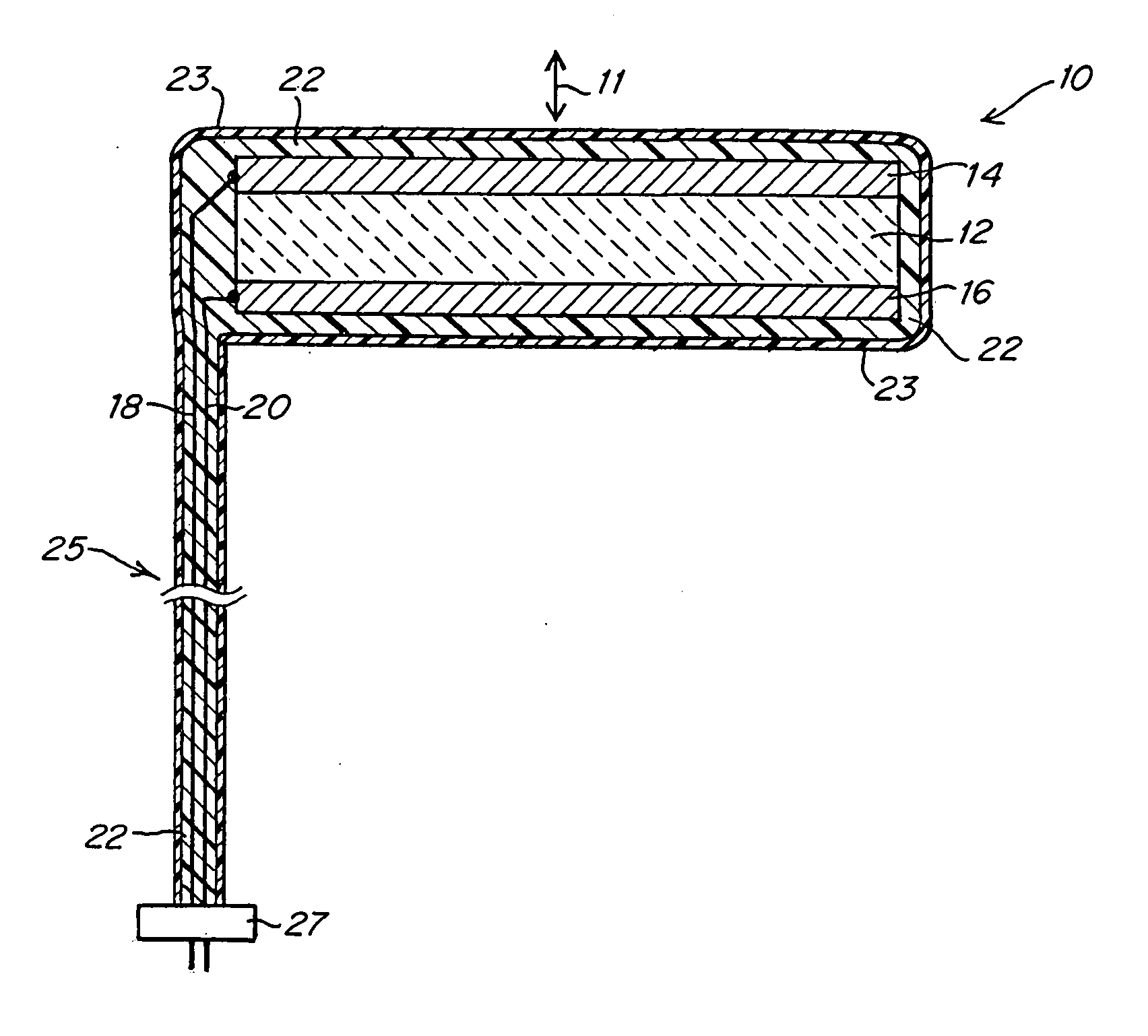
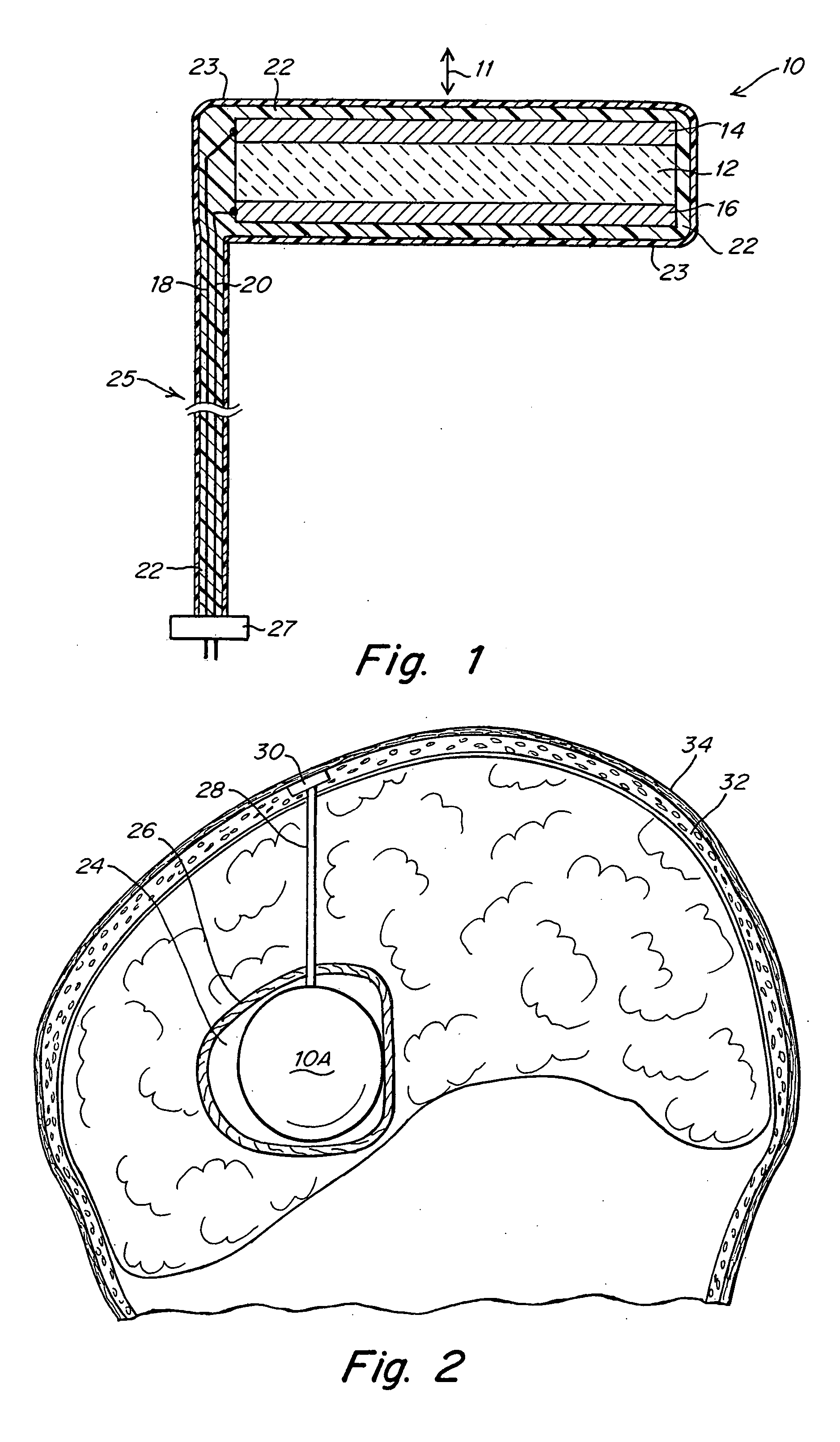
[0033]FIG. 1 illustrates an implantable ultrasound transducer assembly 10 as may be used in practicing the invention. The transducer assembly 10 includes a piezoelectric layer 12, a pair of conductive electrode layers 14, 16 overlying opposite faces of the piezoelectric layer 12 and defining the poles of the transducer, a pair of conductors 18, 20 connected to electrodes 14, 16, an acoustic matching layer 22 and a layer of biocompatible material 23 to encapsulate the components. The conductors 18, 20 are housed in an umbilical cord 25 formed from materials that provide electrical insulation and assure biocompatibility as will be familiar to those skilled in the art of implantable devices. The end of the cord 25 may be connected directly to a controllable power source or may have a connector 27 by which the conductors 18, 20 can be coupled to a source of electrical signals to activate the transducer. The device shown in FIG. 1 will emit ultrasound energy in opposite directions along ...


PUM


Abstract

The transportation of therapeutic substances through internal tissue is enhanced by surgically implanting an ultrasound transducer in immediate proximity to the target tissue and oriented to direct ultrasound having selected characteristics toward the target tissue. The parameters of frequency, mechanical index, pulses per cycle and pulse repetition frequency are selected within defined ranges to cause molecules to be transported at a rate and over a distance substantially greater than by natural diffusion.

Description

This application is a continuation-in-part and claims the benefit of application Ser. No. 10 / 746,311 filed Dec. 24, 2003.FIELD OF THE INVENTION [0001] This invention relates to implantable ultrasound transducer devices and their use to enhance delivery of therapeutic substances to tissue by phonophoresis. BACKGROUND OF THE INVENTION [0002] Most medicinal, pharmacological and other therapeutic substances are delivered systemically by oral, inhalation, injection or intravascular delivery. The substance ultimately reaches the vascular system and is transported to tissue and organs throughout the body. However, in cases where the targeted clinical disorder is localized, systemic methods may present some disadvantages. In order to create a sufficiently high concentration of the substance at the target site, systemic administration requires high dosage in comparison to the amount actually required at the target site. Exposure of untargeted organs or tissues may cause undesirable side effe...

Claims


Application Information

Patent Timeline
Patent Type & Authority Applications(United States)
IPC IPC(8): A61H1/00
CPCA61H23/0245A61M37/0092A61H2201/105
Inventor HEART, GILLTOLKOWSKY, GIDEONBRISKEN, AXEL
Owner CYTODOME
Why Eureka
Try Eureka