
Medical devices to treat or inhibit restenosis

a technology of medical devices and restenosis, applied in the field of medical devices, can solve the problems of precise the exact cellular functions that must be inhibited, and the duration of inhibition needed, so as to achieve the effect of high-efficiency at preventing or inhibiting restenosis

Inactive Publication Date: 2006-03-23
MEDTRONIC VASCULAR INC
View PDF99 Cites 3 Cited by
  • Summary
  • Abstract
  • Description
  • Claims
  • Application Information

AI Technical Summary

Benefits of technology

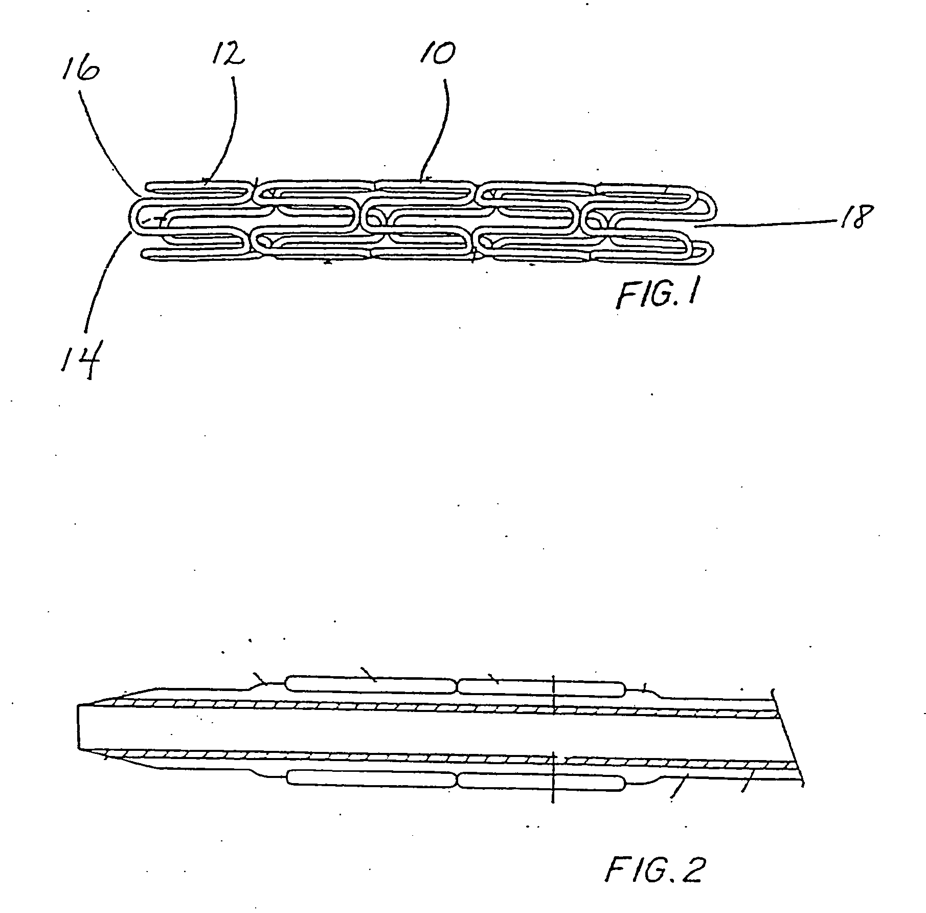
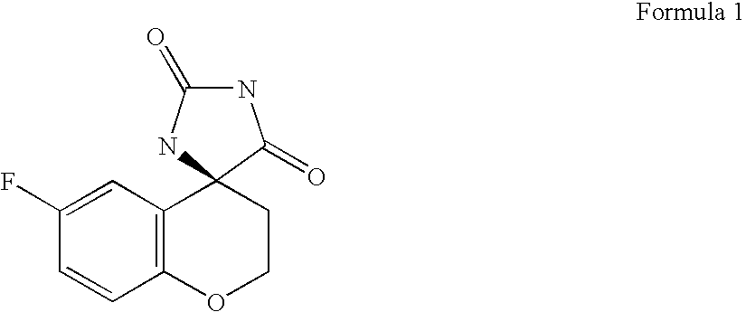
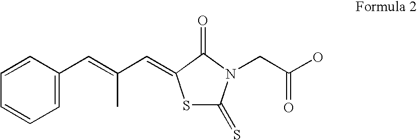
[0011] The present invention provides an in situ drug delivery platform that can be used to administer anti-restenotic tissue levels of aldose reductase (AR) inhibitors, without systemic side effects. It has been found that certain AR inhibitors are highly effective at preventing or inhibiting restenosis when delivered locally to vascular tissue at risk of restenosis. In one embodiment of the present invention the AR inhibitors selected from the group consisting of sorbinil, epalrestat, ponalrestat, methosorbinil, risarestat, imirestat, ALO-1567, quercetin, zopolrestat, AD-5467, NZ-314, M-16209, minalrestat, AS-3201, WP-921, luteolin, tolrestat, EBPC, fidarestat, and the pharmaceutically acceptable derivatives thereof, are particularly effective for this purpose.
[0016] In yet another embodiment of the present invention an anti-restenotic compound-coated intravascular stent can be combined with the systemic delivery of the same or another anti-restenotic compound to achieve a synergistic or additive effect at the medical device placement site. This is particularly beneficial in that non-toxic therapeutic levels of AR inhibitors and other anti-restenotic therapeutics can be combined to achieve dose-specific synergism.

Problems solved by technology

However, balloon catheterization and / or stent deployment can result in vascular injury ultimately leading to VSMC proliferation and neointimal formation within the previously opened artery.
However, many of these drugs, particularly anti-inflammatory and antiproliferative compounds, can be toxic when administered systemically in anti-restenotic-effective amounts.
Furthermore, the exact cellular functions that must be inhibited and the duration of inhibition needed to achieve prolonged vascular patency (greater than six months) are not presently known.
Moreover, it is believed that each drug may require its own treatment duration and delivery rate.
However, side effects including vascular erosion have also been seen.
Vascular erosion can lead to stent instability and further vascular injury.
However, while such treatment may be effective by pharmacological (systemic) administration of AR inhibitors, it would not necessarily be expected that localized administration of small quantities of such AR inhibitors directly to the tissue of the vasculature by elution from a stent would be clinically effective.

Method used


Image

  • Medical devices to treat or inhibit restenosis
  • Medical devices to treat or inhibit restenosis
  • Medical devices to treat or inhibit restenosis

Examples


example 1

Metal Stent Cleaning Procedure

[0102] Medtronic Vascular, Inc. Driver® cobalt alloy coronary stents were placed in a glass beaker and covered with reagent grade or better hexane. The beaker containing the hexane-immersed stents was then placed into an ultrasonic water bath and treated for 15 minutes at a frequency of between approximately 25 to 50 KHz. Next the stents were removed from the hexane and the hexane was discarded. The stents were then immersed in reagent grade or better 2-propanol and vessel containing the stents and the 2-propanol was treated in an ultrasonic water bath as before. Following cleaning the stents with organic solvents, they were thoroughly washed with distilled water and thereafter immersed in 1.0 N sodium hydroxide solution and treated at in an ultrasonic water bath as before. Finally, the stents were removed from the sodium hydroxide, thoroughly rinsed in distilled water and then dried in a vacuum oven overnight at 40° C.

[0103] After cooling the dried s...

example 2

Coating a Clean, Dried Stent Using a Drug / Polymer System

[0104] In the following Example chloroform or tetrahydrofuran is chosen as the solvent of choice. Both the polymer and AR inhibitor are freely soluble in these solvents. Persons having ordinary skill in the art of polymer chemistry can easily pair the appropriate solvent system to the polymer-drug combination and achieve optimum results with no more than routine experimentation.

[0105] 250 mg of NZ-314 is carefully weighed and added to a small neck glass bottle containing 2.8 ml of chloroform or tetrahydrofuran and thoroughly mixed until a clear solution is achieved.

[0106] Next 250 mg of polycaprolactone (PCL) is added to the NZ-314 solution and mixed until the PCL dissolved forming an NZ-314 / polymer solution.

[0107] The cleaned, dried stents are coated using either spraying techniques or dipped into the drug / polymer solution. The stents are coated as necessary to achieve a final coating (drug plus polymer) weight of between ...

example 3

Coating a Clean, Dried Stent Using a Sandwich-Type Coating

[0109] A cleaned, dry stent is first coated with polyvinyl pyrrolidone (PVP) or another suitable polymer followed by a coating of NZ-314. Finally, a second coating of PVP is provided to seal the stent thus creating a PVP-NZ-314-PVP sandwich coated stent.

[0110] The Sandwich Coating Procedure:

[0111] 100 mg of PVP is added to a 50 ml Erlenmeyer flask containing 12.5 ml of chloroform or tetrahydrofuran. The flask was carefully mixed until all of the PVP is dissolved. In a separate clean, dry Erlenmeyer flask 250 mg of NZ-314 is added to 11 ml of the same solvent and mixed until dissolved.

[0112] A clean, dried stent is then sprayed with PVP until a smooth confluent polymer layer was achieved. The stent was then dried in a vacuum oven at 50° C. for 30 minutes.

[0113] Next, successive layers of NZ-314 are applied to the polymer-coated stent. The stent is allowed to dry between each of the successive NZ-314 coats. After the final...


PUM

PropertyMeasurementUnit
Massaaaaaaaaaa
Massaaaaaaaaaa
Dimensionless propertyaaaaaaaaaa

Abstract

Implantable medical devices having anti-restenotic coatings are disclosed. Specifically, implantable medical devices having coatings of aldose reductase (AR) inhibitors are disclosed. Preferred AR inhibitors are enumerated. The anti-restenotic medical devices include stents, catheters, micro-particles, probes and vascular grafts. Intravascular stents are preferred medical devices. The medical devices can be coated using any method known in the art including compounding the AR inhibitor with a biocompatible polymer prior to applying the coating. Moreover, medical devices composed entirely of biocompatible polymer-AR inhibitor blends are disclosed. Additionally, medical devices having a coating comprising at least one AR inhibitor in combination with at least one additional therapeutic agent are also disclosed. Furthermore, related methods of using and making the anti-restenotic implantable devices are also disclosed.

Description

RELATED APPLICATIONS [0001] This application claims the priority of provisional application No. 60 / 611,866, filed Sep. 21, 2004.FIELD OF THE INVENTION [0002] The present invention relates to medical devices and methods of using medical devices to treat or inhibit restenosis. Specifically, the present invention relates to stents that provide in situ controlled release delivery of anti-restenotic compounds. More specifically, the present invention provides intravascular stents that provide anti-restenotic effective amounts of aldose reductase inhibitors directly to tissues at risk for restenosis. BACKGROUND OF THE INVENTION [0003] Cardiovascular disease, specifically atherosclerosis, remains a leading cause of death in developed countries. Atherosclerosis is a multifactorial disease that results in a narrowing, or stenosis, of a vessel lumen. Briefly, pathologic inflammatory responses resulting from vascular endothelium injury causes monocytes and vascular smooth muscle cells (VSMCs) ...

Claims


Application Information

Patent Timeline
IPC IPC(8): A61K31/704A61K31/4188A61F13/00
CPCA61K31/4188A61K31/704A61L27/34A61L27/54A61L29/085A61L2300/606A61L31/10A61L31/16A61L2300/416A61L2300/434A61L29/16
Inventor HEZI-YAMIT, AYALA
Owner MEDTRONIC VASCULAR INC
Learn More