Apparatus and method for lung analysis

a technology of apparatus and lung, applied in the field of apparatus and lung analysis, can solve the problems of requiring equipment which is bulky and expensive to install, requires a large amount of equipment, and is generally expensive to operate, and achieves reliable and reproducible transducer positioning, improved coupling, and improved coupling

Inactive Publication Date: 2006-05-11
PULMOSONIX
View PDF65 Cites 94 Cited by
  • Summary
  • Abstract
  • Description
  • Claims
  • Application Information

AI Technical Summary

Benefits of technology

[0050] In one embodiment, the apparatus further includes a sheath and the transmitter and plurality of receivers are retained on the sheath. In such an embodiment, the sheath can be worn by a subject during use of the apparatus, and individual attachment of the transmitter / receiver(s) to the subjects torso is obviated. This has the advantage of reliable and reproducible transducer positioning and, depending on the nature of the sheath, may improve the coupling between the transducers and the subject's torso.
[0051] In one arrangement, the sheath is a vest worn by the subject. In another arrangement, the sheath is filled with a fluid such as water, saline or gel-like solution and contains hydrophones positioned in the fluid. The hydrophones may provide better coupling, and reduce the influence of the ribs on the detected sound signal. Hydrophones may also provide a much wider frequency response and better noise shielding than other types of receivers. The receiver(s) may be located ventrally, or dorsally. In one embodiment, a plurality of receivers are located on the dorsal spine. Such receivers may be provided in pairs located longitudinally along the dorsal spine. Alternatively, they may be provided in an array centered on the spine. Alternatively / additionally, one or more receivers may be located on the subject's torso and / or below the armpit.

Problems solved by technology

These are generally expensive and involve some degree of risk which is usually associated with the use of x-rays, radioactive materials or gamma-ray emission.
Furthermore, these techniques are generally complicated and require equipment which is bulky and expensive to install and, in most cases, cannot be taken to the bedside to assess biological tissues in patients whose illness prevents them being moved.
However, the process requires sophisticated and sometimes expensive technology and cannot be used in tissues in which there is a substantial quantity of gas, such as the lung.
The vast majority of infants now survive initial acute respiratory illness, but lung injury associated with mechanical ventilation causes many infants to develop ‘chronic lung disease’.
However, trials of new strategies in mechanical ventilation which were expected to reduce barotrauma and / or exposure to oxygen have often had disappointingly little impact on the incidence of chronic lung disease (HIFI Study Group,1989; Bernstein et al, 1996; Baumer, 2000).
Comparison of strategies of conventional mechanical ventilation in animals (Dreyfuss et al, 1985) have indicated that high lung volumes may be more damaging than high intrapulmonary pressures, and has led to the concept of ‘volutrauma’ due to over-inflation of the lung.
However, this technique fails to confer benefit, if the average lung volume is low (HIFI Study Group, 1989), yet it appears to be successful if a normal volume is maintained (McCulloch et al, 1988; Gerstmann et al, 1996).
Even when lung volume is maintained in the “safe window”, changes in the lung condition may manifest due to the general damaged or underdeveloped condition of the lung.
Fluid and blood may accumulate in the lung, posing additional threats to the patient.
However, in the sick newborn, the infant's small size, inability to co-operate and the presence of background noise greatly limits the value of such techniques.
The value of these techniques is further limited by the lack of reproducibility of the sounds and the subjective nature of the analysis which follows.
COPD places enormous economic burden on society.
Medical expenses for COPD patients are extremely high because of frequent visits to the emergency room, extended hospital stays and expensive medications.
The over-distension also markedly reduces the mechanical efficiency of the diaphragm.
Exercise is terminated early because of rapidly rising and unsustainable work of breathing.
The loss of alveolar tissue results in a loss of gas exchange surface area and decreases the number of capillaries available for gas exchange.
It also reduces the elastic recoil of the lung and leads to the collapse of the bronchioles and chronic airflow obstruction.
Thus, lung function is gradually lost through a reduction in gas-exchange area and in the amount of air that reaches the alveoli.
These methods are however somewhat complex and expensive, and are not well suited to the rapid screening of people at risk.
Lung function testing can also be used to identify obstructive airway disease associated with emphysema, but this can only identify the advanced stages of the disease, by which time there has already been widespread and irreversible damage.

Method used


Image

  • Apparatus and method for lung analysis
  • Apparatus and method for lung analysis
  • Apparatus and method for lung analysis

Examples


Embodiment Construction

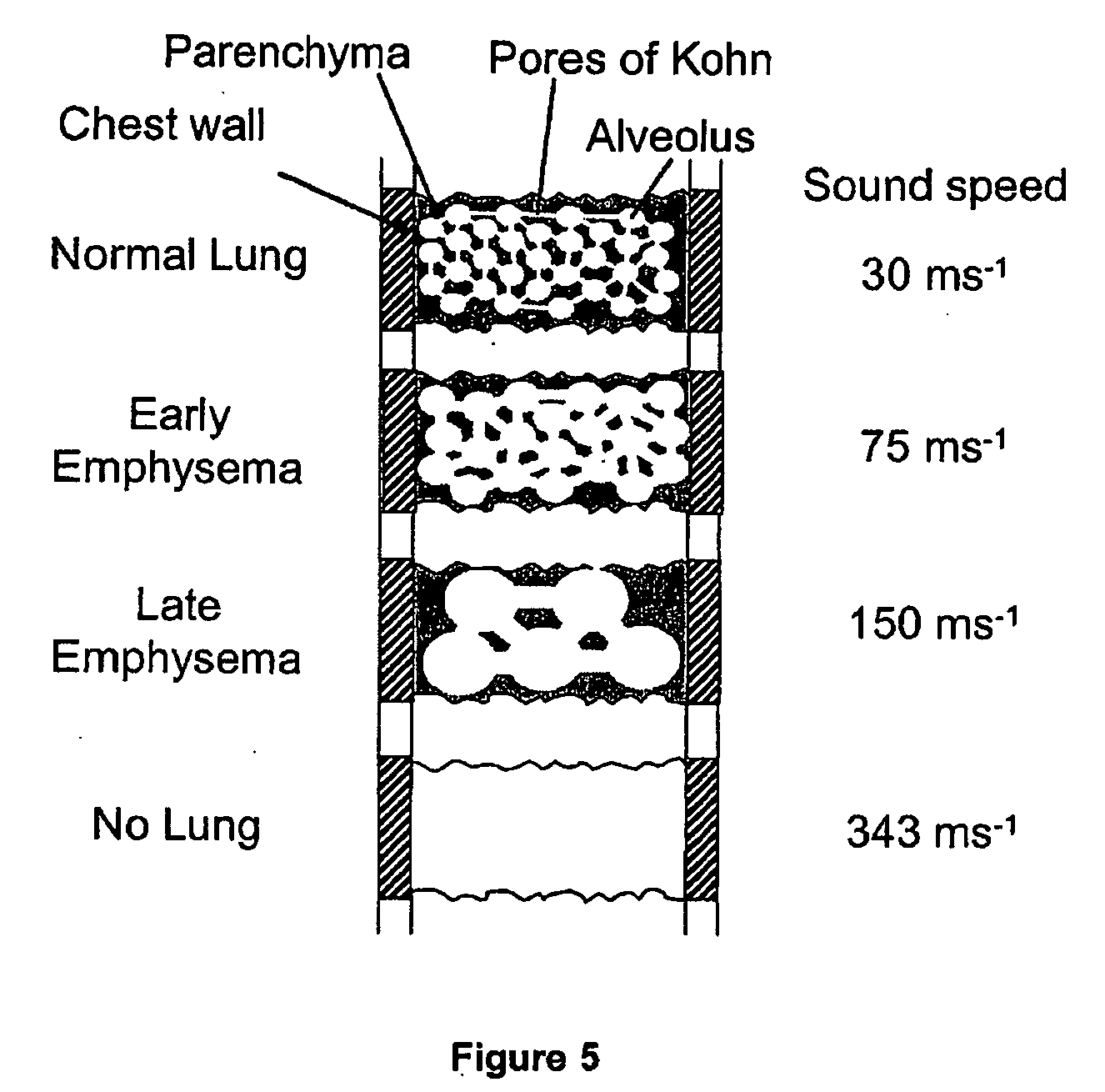
[0065] Characteristics of biological tissues can be determined by measuring the velocity and attenuation of a sound as it propagates through the tissue. This can be achieved by introducing a sound to a particular location or position on the tissue, allowing the sound to propagate through the tissue and measuring the velocity and / or attenuation with which the sound travels from its source to its destination, the destination including a receiver which is spatially separated from the sound source.

[0066] It is particularly desirable that the tissue is porous comprising a composite structure made up of tissue and gas, or has regions of high and low density. Preferably the tissue is of the respiratory system. More preferably the tissue is lung tissue.

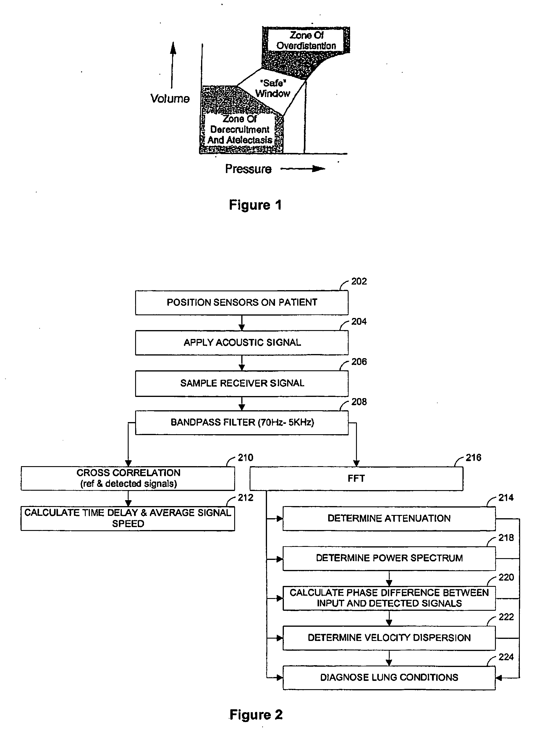
[0067] Referring to FIG. 2, in a method of determining the presence of COPD in a lung, in a step 204, an acoustic signal is applied to the lung. In a step 206, the signal is detected after it has passed through at least part of the lung. In...


PUM


Abstract

An apparatus and method of detecting COPD and in particular, emphysema utilizes a change in acoustic transmission characteristics of a lung due to e.g. the appearance of fenestrae (perforations) in the alveoli of the lung. The use of acoustic signals may provide good sensitivity to the existence of alveolar fenestrae, even for microscopic emphysema, and the appearance and increase in fenestrae may be determined by monitoring acoustic transmission characteristics such as, for example, an increase in acoustic signal velocity and velocity dispersion, and/or a change in attenuation. A transmitter may be located in e.g. the supra-clavicular space and receivers may be mounted on the chest. Measurements may be correlated between pairs of receivers to determine acoustic transmission profiles.

Description

[0001] The present application is a continuation-in-part of U.S. Ser. No. 10 / 272,494 entitled “Method and Apparatus for Determining Conditions of Biological Tissues” filed on 15 Oct. 2002, which is a continuation of International Patent Application No. PCT / AU01 / 00465 entitled “Method and Apparatus for Determining Conditions of Biological Tissues” filed on 20 Apr. 2001. The present application also claims priority from Australian provisional patent application No. 2004902932 entitled “Apparatus and Method for Lung Analysis” filed on 2 Jun. 2004. The contents of each of these applications are incorporated by reference in their entirety in the present application.[0002] The present invention relates to a method of determining characteristics of biological tissues in humans and animals. In particular, it relates to determining the characteristics of tissues such as the lungs and airways by introducing a sound to the tissue, and measuring one or more characteristics of the sound. The inv...

Claims


Application Information

Patent Timeline
Patent Type & Authority Applications(United States)
IPC IPC(8): A61N1/39A61B5/085
CPCA61B5/08A61B5/085A61B5/7257A61B8/08A61B2562/0204A61B2562/046G01S15/88
Inventor WILKINSON, MALCOLM HOWARDRAMSDEN, CLIVE ANDREWBERGER, PHILIP JOHN
Owner PULMOSONIX
Why Eureka
Try Eureka