Biomolecule transduction motif Mph-1-BTM and the use thereof

a biomolecule and transduction motif technology, applied in the field of mph1 biomolecule transduction motif, can solve the problems of side effects on other cells, insufficient efficacy to be applied clinically, and restricted research to develop drugs

Inactive Publication Date: 2006-07-06
FORHUMANTECH CO LTD
View PDF6 Cites 7 Cited by
  • Summary
  • Abstract
  • Description
  • Claims
  • Application Information

AI Technical Summary

Problems solved by technology

The fact that only small-size molecules can permeate through the membrane of living cells at very low rates has restricted the research to develop drugs to cure, prevent or diagnose diseases using macromolecules including, for example, proteins and nucleic acids.
Furthermore, these methods can deliver macromolecules to only a few of the target cells, and its efficiency is not sufficient enough to be applied clinically.
Also, most of these methods cause side effects on the other cells.
However, the effects of biologically active macromolecules delivered into the cytosol and their general application have not been clearly verified yet.
However, concerning beneficial-effects and side effects, it has numerous problems to be resolved.
In particular, regarding liposomes, since the side effects against cells and the cytotoxicity are very serious, their application was limited to basic research.
As for retroviruses, it has a problem in that it cannot infect undeviding cells.
Adenovirus or adeno-associated virus vector also has a very limited clinical application.
Furthermore, these two types of viral vectors may elicit immune responses against the other viral proteins, so its treating efficiency has many doubts.
The proteins, which were synthesized in bacteria, however, were known to be inefficient in folding structures and functions in comparison to the naturally folded proteins in vivo.
However, these methods require further studies and full understanding on many molecular cell biological intermediate steps, and their transduction efficiencies are very low.
And they are not cost effective.
The first is that PTDs ruin the plasma membrane, and transmit the molecules across it.
However, experiments with artificially combined amino acids with 12 arginines and 12 lysines suggested that the hypothesis that the existence of lysine and arginine at specific positions induced the formation of new channels might be wrong (Rothbard J. B., et al., Nature Med.
In addition, considering the facts that only the proteins, which were bound covalently or non-covalently to PTDs, were transferred through the cell membrane, the hypothesis, that PTDs ruin the plasma membrane and translocate the molecules across it, was not acceptable.

Method used


Image

  • Biomolecule transduction motif Mph-1-BTM and the use thereof
  • Biomolecule transduction motif Mph-1-BTM and the use thereof
  • Biomolecule transduction motif Mph-1-BTM and the use thereof

Examples


example 1

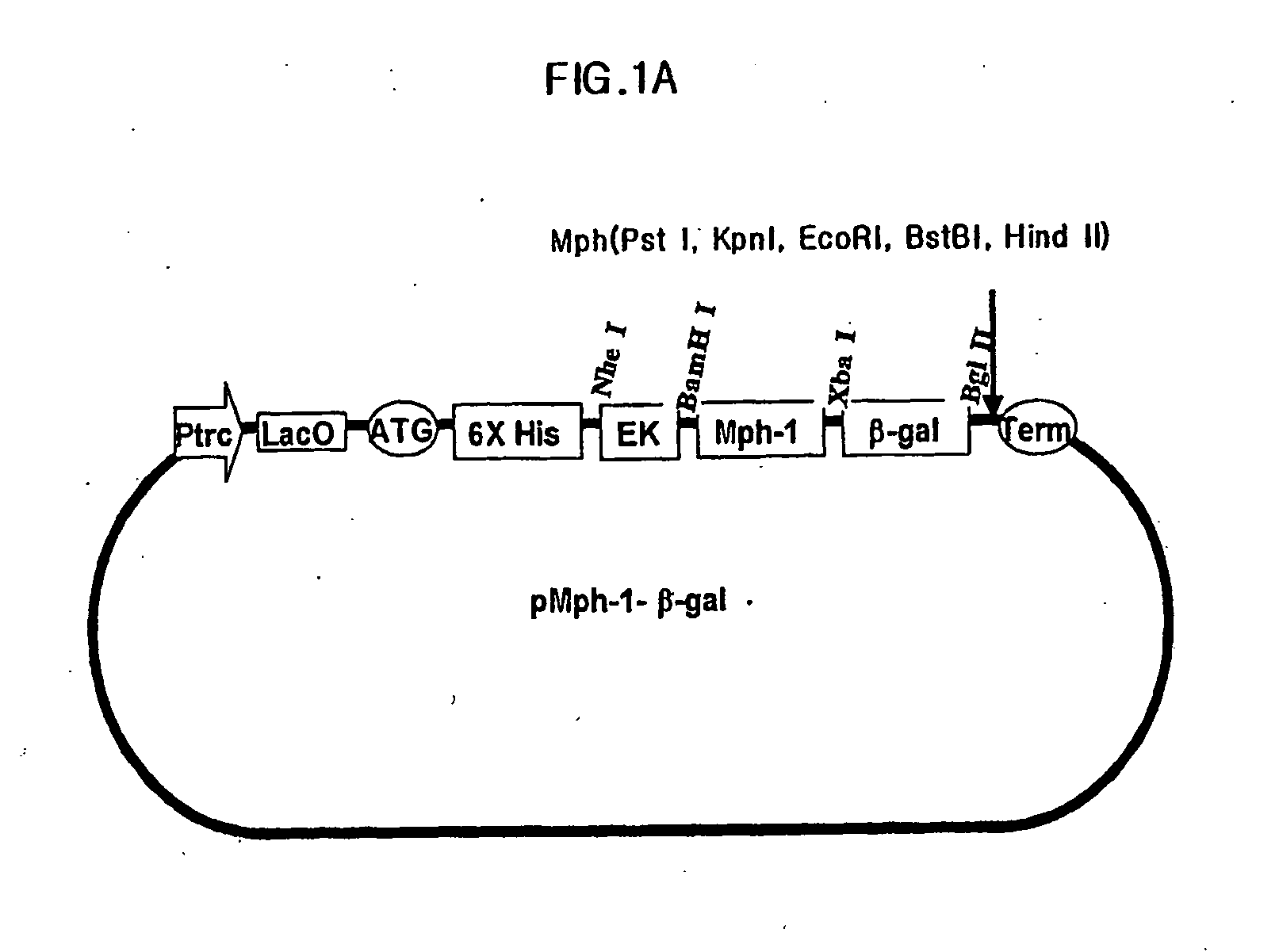
Preparation of Expression Vector Including Mph-1-BTM of Biomolecule Transduction Peptide

[0067] Nucleic acid sequence encoding peptide corresponding to amino acids from 858th of Tyrosine to 868th of Arginine from N-terminus mouse transcription factor of Mph-1 (GeneBank Code: U63386) was combined with the nucleic acid sequence encoding reporter protein of β-galactosidase. In order to do this, firstly, a primer of SEQ ID NO:2 containing the nucleic acid sequence encoding peptide corresponding to amino acids from 858th of Tyrosine to 868th of Arginine from N-terminus of Mph-1 and BamHI site for cloning, and a primer of SEQ ID NO:3 containing nucleic acid sequence of 3′ terminus of β-galactosidase and restriction enzyme of Bgl II site for cloning were synthesized. Then, PCR was carried out using pIND / lacZ vector (Invitrogen corp.), as a template, with pfu turbo DNA polymerase (Stratagene, cat.# 600252-51). After digesting the PCR products with restriction enzymes of BamHI and Bgl II, th...

example 2

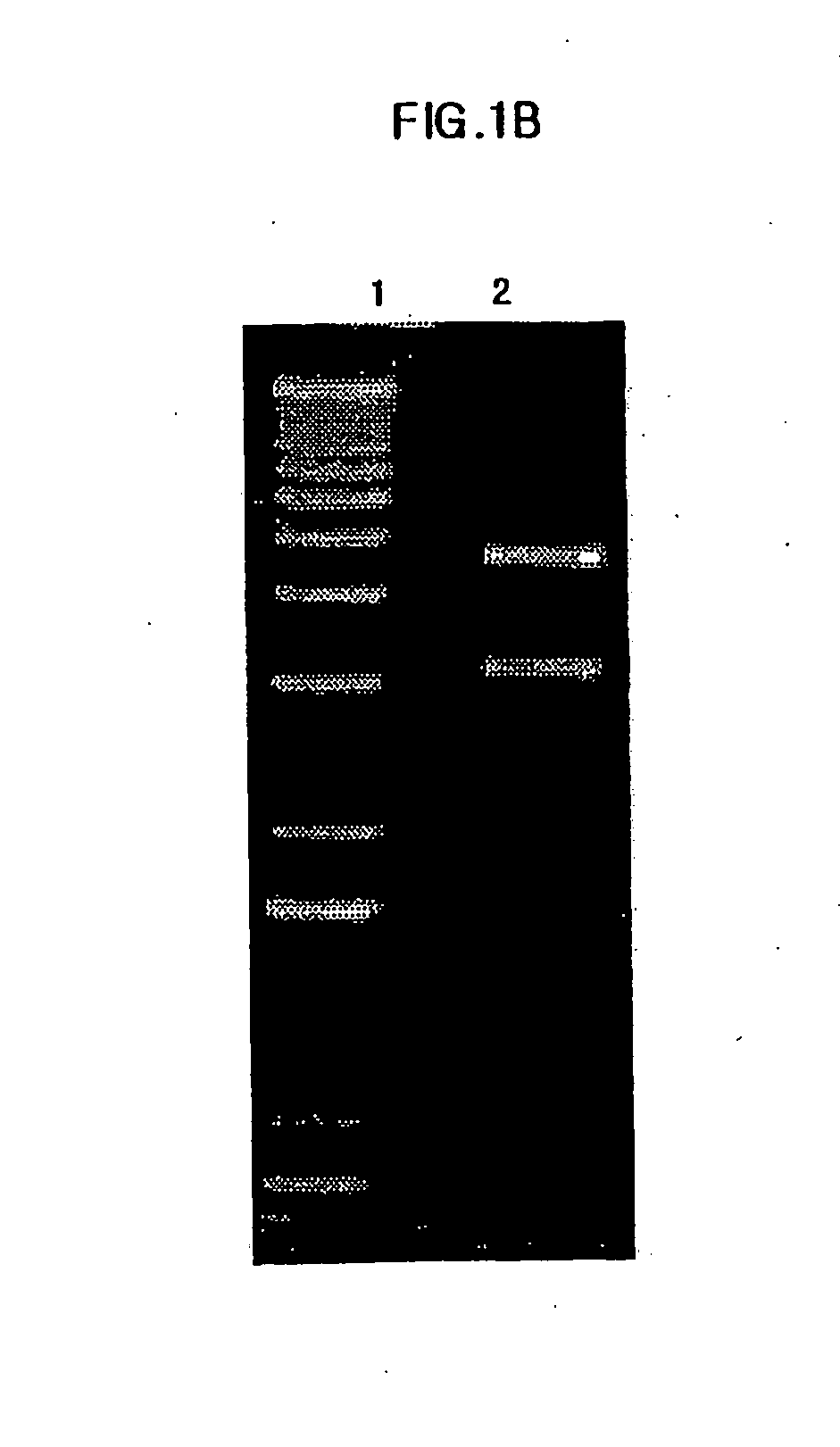
Preparation of E. Coli Transformant, and Expression and Isolation of Fusion Protein Therefrom

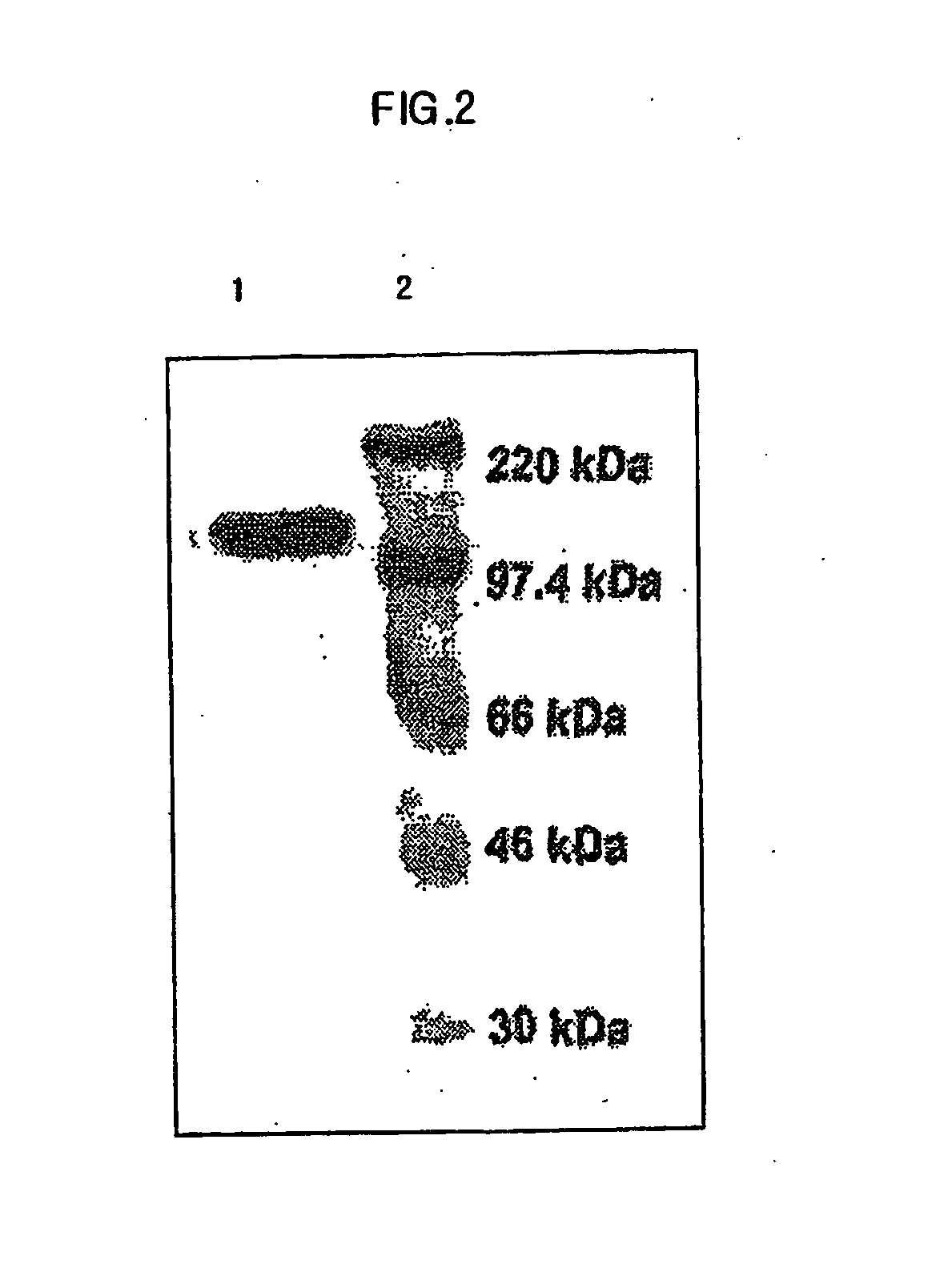
[0068] DH5a (ATCC No. 53863) was transformed using pMph-1-β-gal of the Example 1 by heat shock transformation. 2 ml of the transformants were transferred to 100 ml of LB medium, and pre-incubated for 12 hrs at 37° C. with agitation. Then, the pre-incubated transformants were inoculated to 1000 ml of LB medium (10 g of pancreatic digests of casein, 5 g of yeast extract, 10 g of sodium chloride) and were incubated for 4 hrs at 37° C. Following the incubation, 1 mM of IPTG (Isopropyl β-D-thiogalactosipyranoside, GibcoBRL cat.# 15529-019) was added in order to induce the expression of lac operon, and then incubated again for 8 hrs to elicit expression of the fusion protein. Next, the cell culture solution was centrifuged at 6,000 rpm for 20 min at 4° C. After removing the supernatant, resultant pellet was dissolved in 10 ml of buffer solution 1 (50 mM of NaH2PO4, 300 mM of NaCl, 10 mM of Imidaz...

example 3

Transduction of Fusion Protein Across Cell Membrane

[0069] HUVEC (ATCC:CRL-1730) (2×10⁵), HeLa (ATCC:CCL-2) (2×10⁵), and 293 (ATCC:CRL-1573) (2×10⁵), which were incubated in 10% FBS (Fetal Bovine Serum) of DMEM, were respectively transferred to Lab-tek II chamber slides. Jurkat cells (ATCC: CRL-10915) (2×10⁵) incubated in 10% FBS RPMI were transferred to poly-L-lysin coated slide. Each of the slides was treated with the purified 0.5 μM of Mph-1-β-galactosidase for 30 min at 37° C. in 5% CO2 incubator. And the supernatants were removed from the slides. The incubation mixtures were washed using ice-cold PBS four times and then were solidified with 2% formaldehyde solution for 10 min. After removing the solidified solutions, the mixtures were washed again using ice-cold PBS four times. Subsequently, the respective mixtures were treated with 700 μl of β-galactosidase staining solution (Roche. Co.) for 45 min at 37° C. in 5% CO2 incubator. Following the staining, the mixtures were washed...


PUM

PropertyMeasurementUnit
pHaaaaaaaaaa
Fluorescentaaaaaaaaaa
chemicalaaaaaaaaaa

Abstract

This invention relates to a novel Biomolecule Transduction Motif (BTM), Mph-1 peptide, which has the potential to transduce many biological response modifiers effectively into the cytoplasm, intracellular organelles or nucleus of prokaryotic or eukaryotic cells in vivo and in vitro, and the related technological methods using Mph-1 BTM. This Mph-1 BTM can be used in the development of new recombinant protein vaccines or DNA / RNA vaccines, gene and protein therapy, production of pharmacologically or medicinally useful proteins, or pharmaco-medicinal drug therapy.

Description

CROSS REFERENCE TO RELATED APPLICATIONS [0001] This application is a Continuation-in-Part (CIP) of U.S. patent application Ser. No. 10 / 501,665, which is the National Stage of International Application No. PCT / KR03 / 00122, filed on Jan. 20, 2003, which claims priority from Korean Patent Application No. 10-2002-0003183, filed on Jan. 19, 2002.BACKGROUND OF THE INVENTION [0002] 1. Field of the Invention [0003] This invention relates to Mph-1 Biomolecule Transduction Motif (BTM), a novel intracellular biomolecule transduction peptide, which delivers biologically active, functional or / and regulatory molecules in vivo and in vitro into the cytosol, organelles or nucleus of prokaryotic and eukaryotic cells, and the utilization of the same. [0004] 2. Background Art [0005] Generally, living cells are not permeable to macromolecules such as proteins or nucleic acids. The fact that only small-size molecules can permeate through the membrane of living cells at very low rates has restricted the r...

Claims


Application Information

Patent Timeline
Patent Type & Authority Applications(United States)
IPC IPC(8): C12N9/24C07K14/56C07K14/54C07K14/52C07K14/435C12N15/09A61K38/00A61P37/06C07K7/06C07K14/47C07K16/28C07K19/00C12P21/02
CPCC07K7/06C07K2319/01A61P37/06
Inventor LEE, SANG-KYOULEE, SEUNG-KYOU
Owner FORHUMANTECH CO LTD
Why Eureka
Try Eureka