

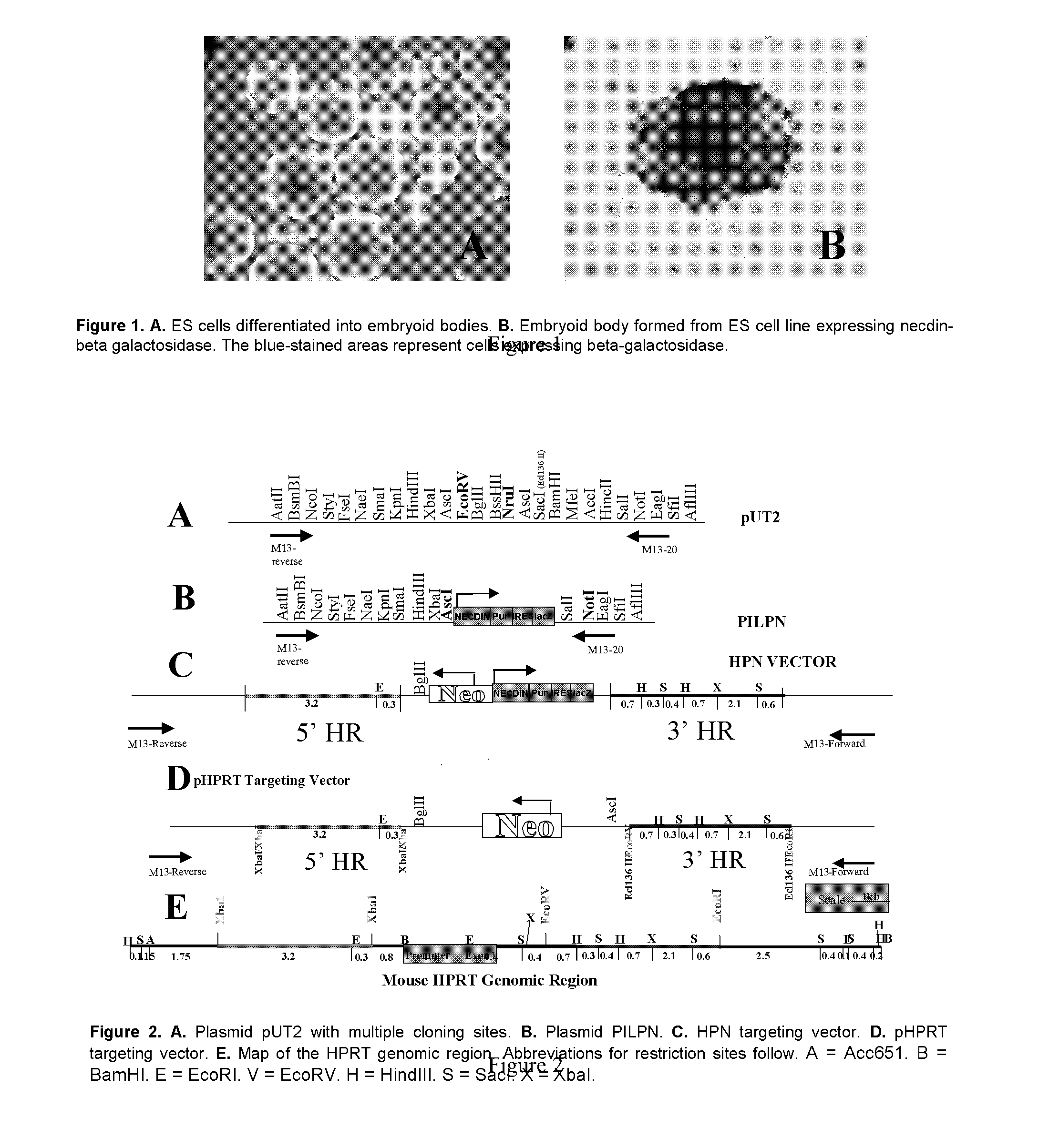
Embryoid Body - Based Screen
- Summary
- Abstract
- Description
- Claims
- Application Information
AI Technical Summary
Benefits of technology
Problems solved by technology
Method used
Image

Examples
example 1
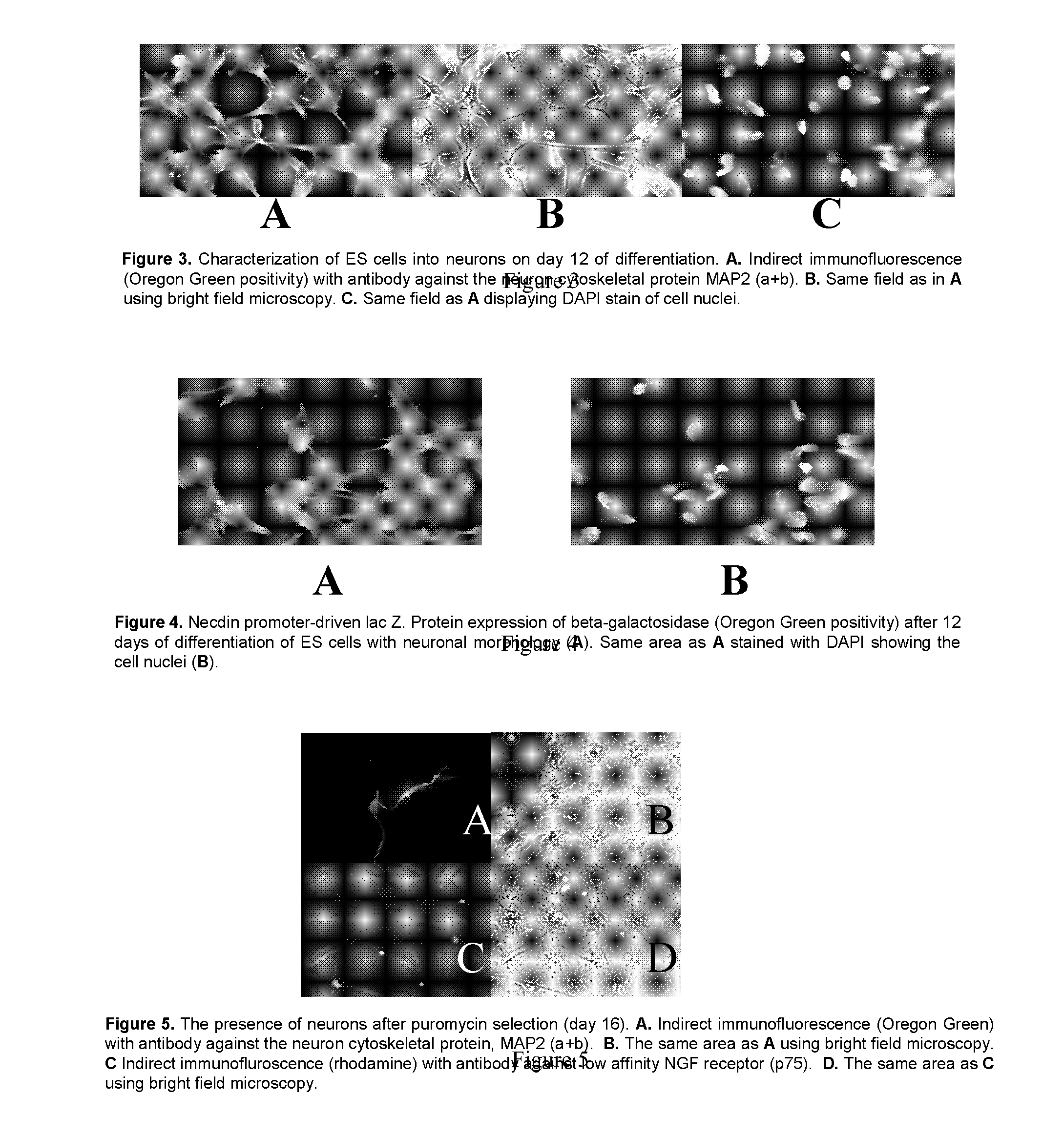
Construction of Vectors
Construction of HPRT Targeting Vector
[0106]FIG. 2E is a map of the mouse HPRT genomic locus. The promoter and exon 1 of the HPRT gene is in the central region and designated by the green box. The pUT1 vector backbone was used to construct the targeting vector. To make the targeting vector (FIGS. 2D and 2E), the 5′ and 3′ homologous regions flanking the 5′ region of the promoter (red line) and the 3′ region of exon 1 (blue line) were used. Transcription of the HPRT gene requires the promoter region (green box). The targeting vector was designed such that homologous recombination of this vector at the HPRT locus eliminates the promoter-exon 1 region (green box) thereby completely eliminating HPRT gene expression. The strategy utilized for construction of the HPRT targeting vector follows. Plasmid pUT1 was used as a backbone to engineer the targeting vector. The source of the UPRT gene was a BAC clone identified by screening a BAC library made from 129 / SvJ mous...
example 2
Development of ES Cell Lines
Transfection of ES Cells with the HPRT Targeting Vector
[0108] ES cells were electroporated (180 V, 500 uF) with 10 μg linearized HPRT targeting vector (FIG. 2D). After electroporation, ES cells were cultured on a fibroblast feeder layer at 37° C. and 7.5% CO2 in standard ES cell culture media of Dulbecco's minimal essential medium with high glucose, L-glutamine (DMEM: Invitrogen, Carlsbad, Calif.) plus 1 mM sodium pyruvate, 10% fetal calf serum, 100 μM β-mercaptoethanol and 1000 U / ml LIF (ESGRO®, Chemicon). After 24 h, G418 selection was performed by G418 (400 μg / ml) addition and the cells were allowed to grow for 5 days. Only cells transfected with the HPRT targeting vector survived in the presence of G418. After 5 days, 30 μM 6-TG was added to the standard ES cell culture media and cells were grown for 7-10 days. Only cells with a mutated HPRT gene grew in 6-TG.
Selection of Cells Containing the Targeted IPRT Gene
[0109] To identify that the mutation ...
example 3
Effect of Small Molecules on ES Cell Differentiation
[0111] An ES cell line (ES-6; Preliminary Studies Section C.2.) containing a stably integrated necdin-puromycin-IRES-β-galactosidase cassette was allowed to form EB. Briefly, 1000 cells in 200 μl of media without LIF were plated into 96-well PCR plates. On the third day, the EB were transferred to 96-well tissue culture plates in which each well was coated with laminin. The medium was changed on the fourth day and appropriate small molecule compounds were added with each compound being assessed in triplicate. Six wells received no treatment and six wells received treatment with vehicle control (DMSO or ethanol diluted in similar manner as small molecule compounds). The cells were incubated with compounds for 48 hr, compounds removed by washing with PBS and cells allowed to grow and differentiate in media for 4 days. Cells were lysed on the fifth day and the lysates were analyzed for β-galactosidase expression. FIG. 9A shows the ef...
PUM
 Abstract
Description
Claims
Application Information