Virtual flow cytometry on immunostained tissue-tissue cytometer

a flow cytometer and immunostained tissue technology, applied in the field of automated light microscopic image analysis, can solve the problems of limited population statistic analysis, system cannot perform chromogen-based brightfield cell analysis, and cannot be useful

Inactive Publication Date: 2007-01-25
CUALING HERNANI D
View PDF2 Cites 58 Cited by
  • Summary
  • Abstract
  • Description
  • Claims
  • Application Information

AI Technical Summary

Benefits of technology

[0041] c) Use of single cell image analysis result instead of the prior art pixel and area-based image analysis. The object advantage of the invention overcomes these difficulties by using single cell population statistic detection to detect the whole cells, membrane cytoplasm nucleus and all, irregardless of color mixtures inside the cells. The frequency distribution of variable staining single cells is displayed for the user. Optimal evaluation of these results are then rapidly visible and accessible for approval or system modification based on the actual visualized original tissue and cell distribution in the colored image.
[0042] d) Use of a single cell graphic plot only made possible by the single cell image analysis technic to display the features of the collections of cells in tissue with an objective display of feature distribution data, verifiable and modifiable by the user.
[0043] e) Use of a novel thresholding algorithm that dynamically adjust for staining and sectioning variabilities by using the novel epsilon parameter.
[0044] f) Use of a novel thresholding algorithm that dynamically adjust for a wide range of light intensity of the transmitted light microscope by using the novel epsilon parameter.
[0045] g) Use of the combination of the above to provide a new and improved apparatus and method that converts tissue immunohistochemistry results to a data and display, commonly ascribed to a prior art flow cytometry apparatus.
[0046] Slide based Tissue Cytometry as being described herein will aid in slide based diagnosis in providing a “flow cytometry”-like function to histochemically or immunohistochemically stained cells fixed on a microscopic slide.

Problems solved by technology

The subject is locally adaptable in machine vision field and may not be useful in detecting immunostained cells in tissue, wherein these cells are in a contiguous distribution with a gradation and mixture of bleeding colors, as is often the situation with immunohistochemical stains of cells using brown chromogen and blue counterstain.
The system will not perform chromogen based brightfield cell analysis.
The Chromavision ACIS, with some of their patents described above, could do many slide based analysis but has limited the population statistic analysis to getting pixels that are positively stained in hot spots areas and over all the area of the image frame.
Since these cells are so numerous, and since they tend to clump, counting each individual nucleated cell would add an excessive processing burden, at the expense of speed, and would not necessarily provide an accurate count due to clumping.
It has been using the pixel mask technology and because of similar accuracy issues has not implemented its population statistic reporting.
Imaging flow cytometry U.S. Pat. No. 6,251,615 will not allow visualization of routinely immunohistochemically stained cells in brightfield microscopy but uses fluorochrome reactive antigens and fluorescent microscopy displayed cells.
It has been found, however, that present prior art apparatus and methods fail to meet the demand for a low cost, efficient, customizable imaging microscope that is capable of extracting or overlapping, concurrent data acquisition and analysis over color image obtained by brightfield light.
A problem found in alternative systems is that they are capable of imaging a set of pixels representing the stained object over all the other digital objects in the image frame which is not an accurate representation since some microscopic images contain background stromal tissue or other cells other than the relevant class.
Another problem with available systems is the need for special filters, reliance on machine obtained hot spots and non-biased approaches, and complexity in the system optics is required, increasing the complexity to user and system.

Method used


Image

  • Virtual flow cytometry on immunostained tissue-tissue cytometer
  • Virtual flow cytometry on immunostained tissue-tissue cytometer
  • Virtual flow cytometry on immunostained tissue-tissue cytometer

Examples


Embodiment Construction

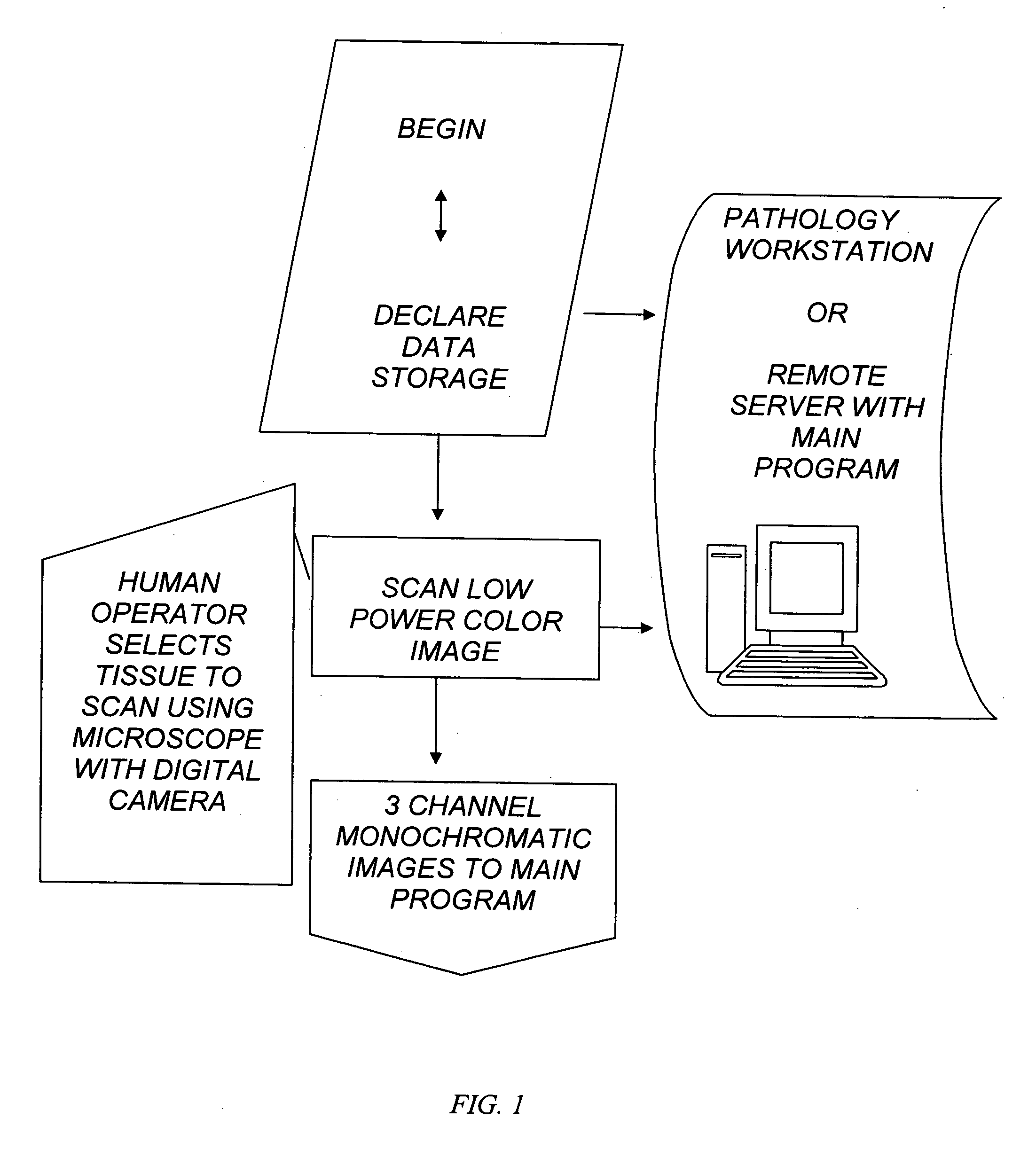
[0085]FIG. 1 is a block diagram of the interface of the system. The system includes a human operator or an automated slide delivery system, to place and select the tissue to scan for low power color image. The image is scanned of 3 channel RGB monochromatic planes which are sent to the main program. The main program and its data storage are in preferably a pathology workstation with monitor display or alternatively located in a remote server.

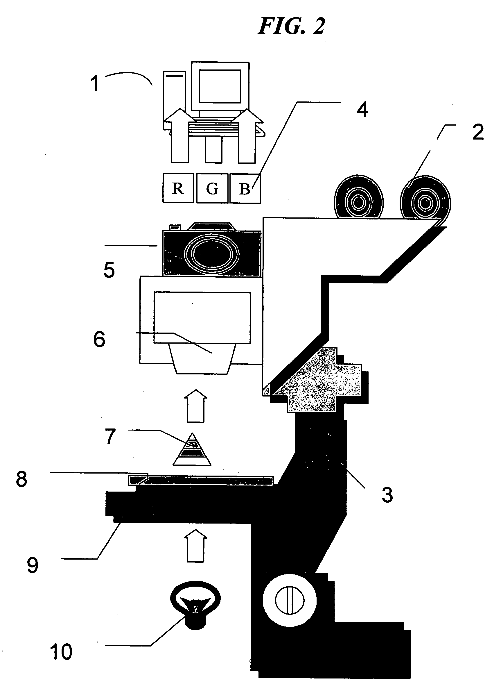
[0086] The general purpose computer 1, preferably a personal computer (PC) FIG. 2 controls the operation of the image processor board preferably a Pentium with PCI bus advanced chip, running Windows 9X or greater or a PowerPC with PCI bus running OS 8.5 or greater and able to run executable programs. The frame memory of the image processor is memory mapped to the PC which performs most of the complicated computations that the image processor cannot handle. This includes searching and other operations requiring random access of the frame memory....


PUM

PropertyMeasurementUnit
set cell diameteraaaaaaaaaa
optical densityaaaaaaaaaa
sizeaaaaaaaaaa

Abstract

The invention provides an automated method of single cell image analysis which determines cell population statistic, applicable in the field of pathology, disease or cancer diagnosis, in a greatly improved manner over manual or prior art scoring techniques. By combining the scientific advantages of computerized automation and the invented method, as well as the greatly increased speed with which population can be evaluated, the invention is a major improvement over methods currently available. The single cells are identified and displayed in an easy to read format on the computer monitor, printer output or other display means, with cell parameter such as cell size and staining distribution at a glance. These output data is an objective transformation of the subjective visible image that the pathologist or scientist relies upon for diagnosis, prognosis, or monitoring therapeutic perturbations. Using our novel proposed technology, we combine the advantages provided by the clinical standard tool of flow cytometry in quantifying single cells and also retain the advantages of microscopy in retaining the capability of visualizing the immunoreactive cells. Unlike flow cytometry however, the invention uses commonly available formalin fixed immunostained tissue and not fresh viable cells. To accomplish this aim, we resort to new and improved advanced image analysis using a unique, useful, and adaptive process as described herein. The method uses multi-stage thresholding and segmentation algorithm based on multiple color channels in RGB and HS I spaces and uses auto-thresholding on red and blue channels in RGB to get the raw working image of all cells, then refines the working image with thresholding on hue and intensity channels in HS I using an adaptive parameter epsilon in entropy mode, and further separates different groups of cells within the same class, by auto-thresholding within the working image region. The Immunohistochemistry Flow cytometry (IHCFLOW) combination results in a new paradigm that is both useful, novel, and provides objective tangible result from a complex color image of tissue.

Description

CLAIM OF PRIORITY [0001] This application claims the benefit of priority, under 35 USC.sctn.119 (e) (1), of provisional application No. 60 / 701,774, filed Jul. 25, 2005. The entirety of that provisional application is incorporated herein by reference.FEDERALLY SPONSORED RESEARCH [0002] Not applicable. REFERENCE TO A COMPUTER FLOW CHART [0003] Program flow chart in the drawings TECHNICAL FIELD [0004] The invention relates generally to a system for automated light microscopic image analysis, specifically to computerized methods of converting immunohistochemistry color image to objective, clinically useful, statistical single cell data now commonly associated with flow cytometry- but herein performed on paraffin embedded tissue sections. BACKGROUND OF INVENTION—INTRODUCTION [0005] The optical microscope in the diagnostic and biomedical laboratory is routinely used by pathologists and research scientist to make diagnosis and perform experiments. These users perform these functions by vis...

Claims


Application Information

Patent Timeline
Patent Type & Authority Applications(United States)
IPC IPC(8): G01N33/567G06F19/00C12M3/00
CPCG01N33/57492G01N33/56966
Inventor CUALING, HERNANIZHONG, ERIC E.
Owner CUALING HERNANI D
Why Eureka
Try Eureka