Platelet-derived growth factor D, DNA coding therefor, and uses thereof

a growth factor and platelet technology, applied in the field of platelet-derived growth factor d, can solve the problems of cardiovascular failure, fluid accumulation in the pericardial cavity, and embyonic lethality around midgestation, and achieve the effect of efficient high-volume screening

Inactive Publication Date: 2007-02-22
LUDWIG INST FOR CANCER RES +1
View PDF1 Cites 0 Cited by
  • Summary
  • Abstract
  • Description
  • Claims
  • Application Information

AI Technical Summary

Benefits of technology

[0044] Polypeptides comprising conservative substitutions, insertions, or deletions, but which still retain a biological activity of PDGF-D are within the scope of the invention. Persons skilled in the art will be well aware of methods which can readily be used to generate such polypeptides, for example the use of site-directed mutagenesis, or specific enzymatic cleavage and ligation. The skilled person will also be aware that peptidomimetic compounds or compounds in which one or more amino acid residues are replaced by a non-naturally occurring amino acid or an amino acid analog may retain the required aspects of the biological activity of PDGF-D. Such compounds can readily be made and tested for their ability to show the biological activity of PDGF-D by routine activity assay procedures such as the fibroblast proliferation assay and are also within the scope of the invention.
[0051] A variation on this contemplation would be to insert a proteolytic site between a CUB-removed monomer of VEGF, VEGF-B, VEGF-C, VEGF-D, PDGF-C, PDGF-A, PDGF-B, PDGF-C, PDGF-D or PlGF and the mutant CUB domain which is dimerized to a CUB-removed monomer of VEGF, VEGF-B, VEGF-C, VEGF-D, PDGF-A, PDGF-B, PDGF-C, PDGF-D or PlGF. Addition of the specific protease(s) for this proteolytic site would cleave the CUB domain and thereby release an activated dimer that can then bind to its corresponding receptor. In this way, a controlled release of an activated dimer is made possible.
[0060] PDGF-D may also be relevant to a variety of lung conditions. PDGF-D assays could be used in the diagnosis of various lung disorders. PDGF-D could also be used in the treatment of lung disorders to improve blood circulation in the lung and / or gaseous exchange between the lungs and the blood stream. Similarly, PDGF-D could be used to improve blood circulation to the heart and O2 gas permeability in cases of cardiac insufficiency. In a like manner, PDGF-D could be used to improve blood flow and gaseous exchange in chronic obstructive airway diseases.
[0065] Use of this screen system provides a means to determine compounds that may alter the biological function of PDGF-D. This screening method may be adapted to large-scale, automated procedures such as a PANDEX® (Baxter-Dade Diagnostics) system, allowing for efficient high-volume screening of potential therapeutic agents.

Problems solved by technology

Disruption of the VEGFR genes results in aberrant development of the vasculature leading to embryonic lethality around midgestation.
Inactivation of VEGFR-3 results in cardiovascular failure due to abnormal organization of the large vessels (Dumont et al.
Targeted inactivation of both copies of the VEGFR-3 gene in mice resulted in defective blood vessel formation characterized by abnormally organized large vessels with defective lumens, leading to fluid accumulation in the pericardial cavity and cardiovascular failure at post-coital day 9.5.
However, the role of VEGFR-3 in the development of the lymphatic vasculature could not be studied in these mice because the embryos died before the lymphatic system emerged.

Method used


Image

  • Platelet-derived growth factor D, DNA coding therefor, and uses thereof
  • Platelet-derived growth factor D, DNA coding therefor, and uses thereof
  • Platelet-derived growth factor D, DNA coding therefor, and uses thereof

Examples


example 1

Expression of Human PDGF-D in Baculovirus Infected Sf9 Cells

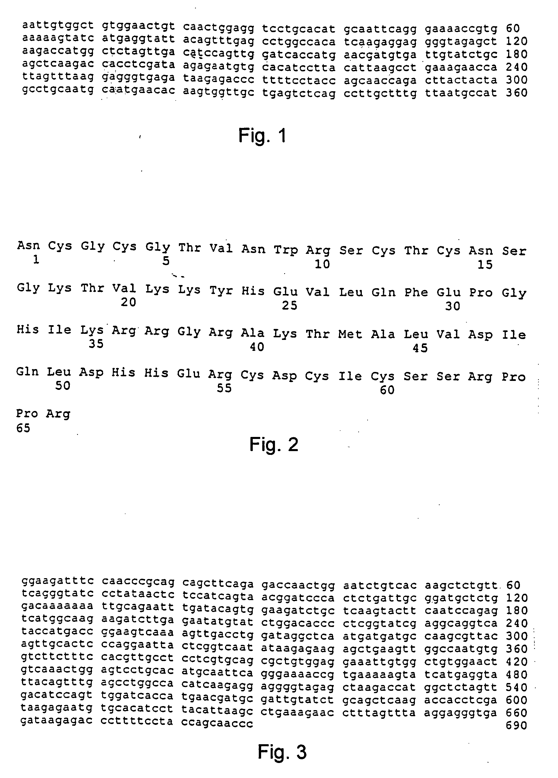
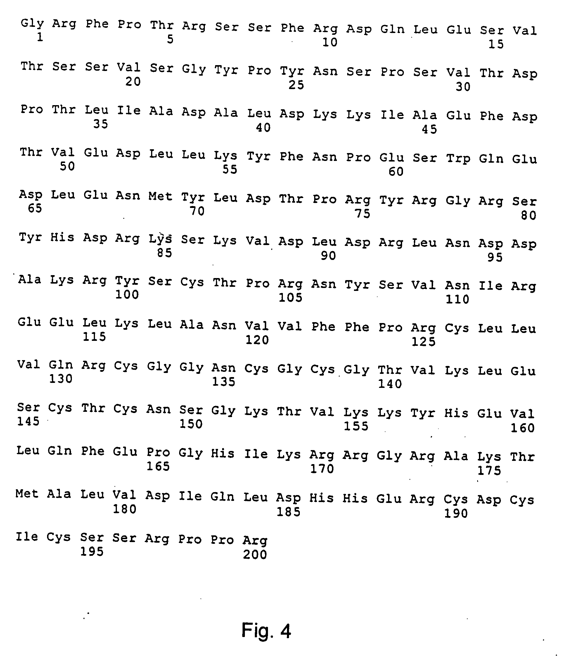
[0129] The portion of the cDNA encoding amino acid residues 24-370 of SEQ ID NO:8 was amplified by PCR using Taq DNA polymerase (Biolabs). The forward primer used was 5′GATATCTAGAAGCAACCCCGCAGAGC 3′ (SEQ ID NO:33). This primer includes a XbaI site (underlined) for in frame cloning. The reverse primer used was 5′ GCTCGAATTCTAAATGGTGATGGTGATGATG TCGAGGTGGTCTTGA 3′ (SEQ ID NO:34). This primer includes an EcoRI site (underlined) and sequences coding for a C-terminal 6×His tag preceded by an enterokinase site. The PCR product was digested with XbaI and EcoRI and subsequently cloned into the baculovirus expression vector, pAcGP67A. Verification of the correct sequence of the cloned PCR product was done by nucleotide sequencing. The expression vectors were then co-transfected with BaculoGold linearized baculovirus DNA into Sf9 insect cells according to the manufactures protocol (Pharmingen). Recombined baculovirus were amplified ...

example 2

Generation of Antibodies to Human PDGF-D

[0132] Rabbit antisera against full-length PDGF-DD and against a synthetic peptide derived from the PDGF-D sequence (residues 254-272, amino acid sequence RKSKVDLDRLNDDAKRYSC of SEQ ID NO:36) were generated. These peptides were each conjugated to the carrier protein keyhole limpet hemocyanin (KLH, Calbiochem) using N-succinimidyl 3-(2-pyridyldithio)propionate (SPDP) (Pharmacia Inc.) according to the instructions of the supplier. 200-300 micrograms of the conjugates in phosphate buffered saline (PBS) were separately emulsified in Freunds Complete Adjuvant and injected subcutaneously at multiple sites in rabbits. The rabbits were boostered subcutaneously at biweekly intervals with the same amount of the conjugates emulsified in Freunds Incomplete Adjuvant. Blood was drawn and collected from the rabbits. The sera were prepared using standard procedures known to those skilled in the art. The antibodies to full-length PDGF-DD were affinity-purifie...

example 3

Expression of PDGF-D Transcripts

[0134] To investigate the tissue expression of PDGF-D in several human tissues, a Northern blot was done using a commercial Multiple Tissue Northern blot (MTN, Clontech). The blots were hybridized at according to the instructions from the supplier using ExpressHyb solution at 68° C. for one hour (high stringency conditions), and probed sequentially with a 32P-labeled 327 bp PCR-generated probe from the human fetal lung cDNA library (see description above) and full-length PDGF-B cDNA. The blots were subsequently washed at 50° C. in 2×SSC with 0.05% SDS for 30 minutes and at 50° C. in 0.1×SSC with 0.1% SDS for an additional 40 minutes. The blots were then put on film and exposed at −70° C. As shown in FIG. 15, upper panel, the highest expression of a major 4.4 kilobase (kb) transcript occurred in heart, pancreas and ovary while lower expression levels were noted in several other tissues including placenta, liver, kidney, prostate, testis, small intesti...


PUM

PropertyMeasurementUnit
pHaaaaaaaaaa
temperatureaaaaaaaaaa
pHaaaaaaaaaa

Abstract

PDGF-D, a new member of the PDGF / VEGF family of polypeptide growth factors, is described, as well as nucleotide sequences encoding, methods for producing, pharmaceutical compositions containing this new growth factor, and its antibodies and other antagonists. Also disclosed are transfected and transformed host cells expressing PDGF-D, and uses thereof in medical and diagnostic applications.

Description

CROSS REFERENCE TO RELATED APPLICATIONS [0001] This application is a division of application Ser. No. 10 / 086,623, filed Mar. 4, 2002, which is a continuation-in-part of U.S. application Ser. No. 09 / 691,200, filed Oct. 19, 2000, which is a continuation-in-part of U.S. application Ser. No. 09 / 438,046, filed Nov. 10, 1999 and claims the benefit of U.S. Provisional Application No. 60 / 107,852, filed Nov. 10, 1998; U.S. Provisional Application No. 60 / 113,997, filed Dec. 28, 1998; U.S. Provisional Application No. 60 / 150,604, filed Aug. 26, 1999; U.S. Provisional Application No. 60 / 157,108, filed Oct. 4, 1999; and U.S. Provisional Application No. 60 / 157,756, filed Oct. 5, 1999, the disclosures of which are hereby incorporated by reference in their entireties into the present application.FIELD OF THE INVENTION [0002] This invention relates to growth factors for cells expressing receptors to a novel growth factor that include endothelial cells, connective tissue cells (such as fibroblasts) my...

Claims


Application Information

Patent Timeline
Patent Type & Authority Applications(United States)
IPC IPC(8): C12Q1/68C07H21/04C12P19/34A61K38/00C07K14/49
CPCA61K38/00C07K14/49C12Q1/6886C12Q2600/158
Inventor ERIKSSON, ULFAASE, KARINLI, XURIPONTEN, ANNICAUUTELA, MARKOALITALO, KARIOESTMAN, ARNEHELDIN, CARL-HENRIK
Owner LUDWIG INST FOR CANCER RES
Why Eureka
Try Eureka