
Method of in situ detection of proteins using aptamers

Inactive Publication Date: 2007-06-14
HOSPITAL FOR SICK CHILDREN
View PDF6 Cites 10 Cited by
  • Summary
  • Abstract
  • Description
  • Claims
  • Application Information

AI Technical Summary

Benefits of technology

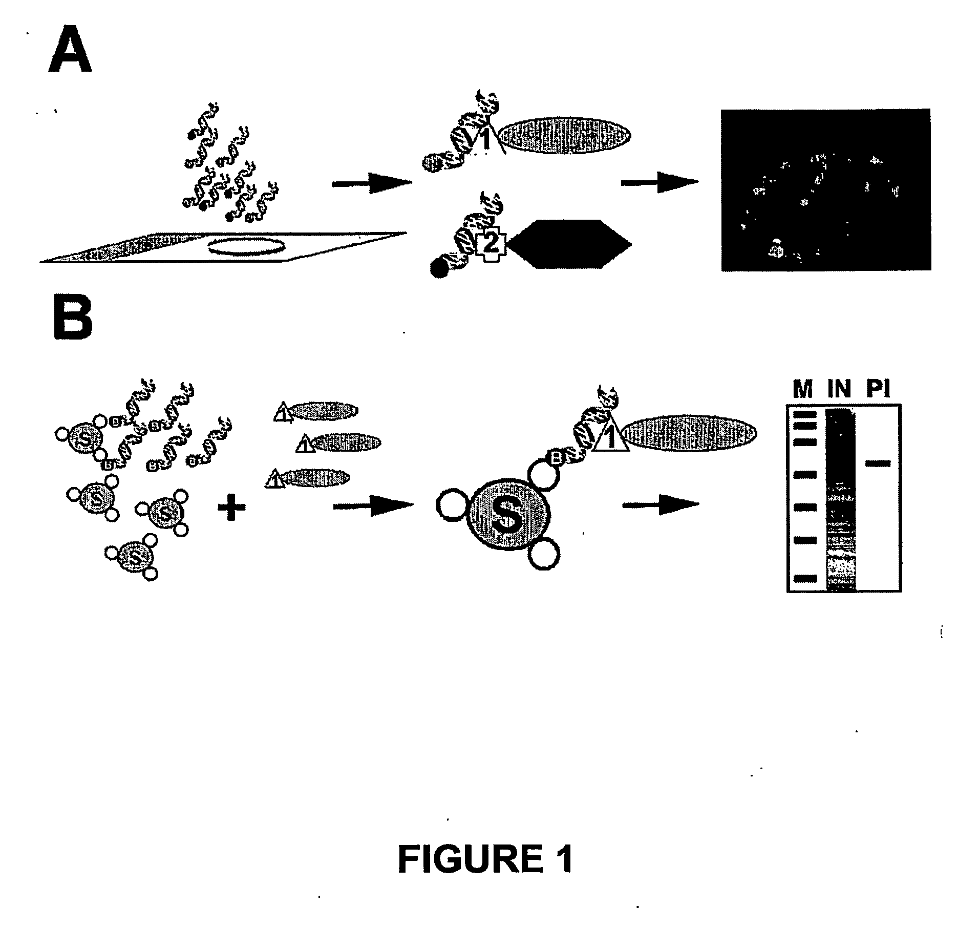
[0012] The present invention is a method of detecting proteins in situ wherein a peptide tag is used that can bind a double stranded nucleic acid aptamer with high affinity. The peptide tag is expressed as a fusion protein encoding both the tag and a known protein, which protein is desired to detect / localize in situ. The peptide tag portion of the fusion protein is recognized / binds to its cognate nucleic acid aptamer which itself is functionalized so that it is readily detectable by a variety of methods. The nucleic acid aptamer sequence thus detects proteins in situ which has not previously been contemplated. The method of the invention provides for the detection and localization of a protein in situ with specificity within sub-cellular and sub-nuclear structures at high resolution. The method also allows for the multiplex visualization of sub-cellular and sub-nuclear complexes of proteins within a cell. The method can also be used for protein purification for both proteomic and therapeutic applications. Further, the method allows for detection of different proteins and protein complexes in the same cell without cross-hybridization of the detecting nucleic acid aptamers. Lastly, only nanomolar quantities of DNA aptamer are required in the present method. Detected proteins can then be further isolated and purified.

Problems solved by technology

While both antibodies and FPs have been attractive reagents for cell biologists because of their versatility and ease of use, several limitations with these systems exist.
One limitation is the small repertoire of bright and spectrally distinct proteins or fluorophores that are available.
In addition, a specific combination of excitation filter, emission filter and dichroic mirror is required to visualise the fluorescence emission from a particular fluorophore or combination of fluorphores during epifluorescence microscopy, with the result that detection of three different proteins is the practical limit.
Furthermore, the detection of three or more proteins simultaneously in situ using antibodies requires that each antibody be produced in a different and immunologically distinct animal so that secondary antibodies conjugated to each fluorophore do not cross-hybridise, resulting in false positive detection.
These methods are only applicable to the imaging of chromatin and thus do not represent a general imaging technology for proteins.
However, the proposed method proved unsuccessful.

Method used


Image

  • Method of in situ detection of proteins using aptamers
  • Method of in situ detection of proteins using aptamers
  • Method of in situ detection of proteins using aptamers

Examples


example 1

Vector Construction and Aptamer Preparation

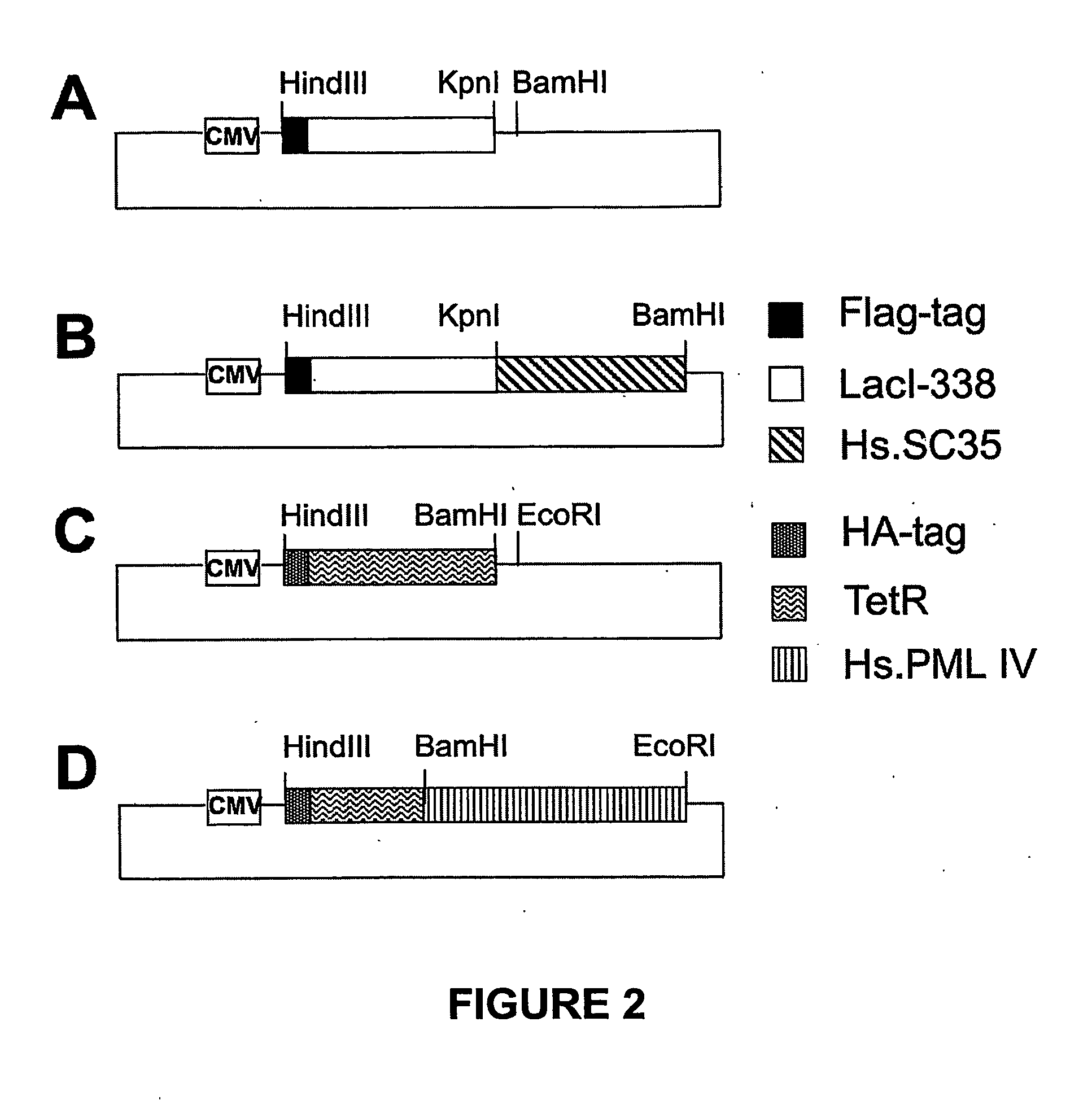
[0078] The mammalian expression vector pGD-Flag-Lac338 and its derivative pGD-Flag-Lac-SC35 were constructed as follows (FIGS. 1A and B). Plasmid pcDNA3.1 / His-C (Invitrogen) was cut with HindIII and Asp718 and ligated to an Asp718 / HindIII cut PCR product (Flag-Lac338) to produce pGD-Flag-Lac338. The Flag-Lac338 PCR product encodes the amino acid sequence of the Flag epitope (MDYKDDDDK) fused to the first 338 amino acids of the Lac repressor (LacI) and was amplified from the vector p3′SS-GFP-Lac-NLS (Robinett et al., 1996) using the following primers:

(Sequence ID No. 1)LACFLAG-1 (tgacgtaagcttaggatggactataaagacgatgacgataaaccagtaacgttatacga);and(Sequence ID No. 2)LAC3R-338 (ctataaggtaccgccccctccacttccaccgcccccagaggcggtttgcgtattgggcgcca).

[0079] To generate pGD-Flag-Lac-SC35, both pGD-Flag-Lac338 and the vector pBSK-SC35 were first cut with Asp718, blunt ended with Klenow in the same buffer, followed by phenol / chlorophorm extraction and preci...

example 2

Cell Culture and Transfection

[0083] SK-N-SH cells were cultured according to the American Type Culture Collection (ATCC) guidelines for each cell line. Cells were split the day before transfection and 2×10⁵ cells were seeded at 105 cells / ml onto 18 mm square coverslips in 8 or 6 well plates. The following day cells were transfected with 1-2 μg of pGD-Flag-Lac338 and pGD-TET-PML DNA alone or combined per well using Lipofectamine 2000 (Invitrogen™) as suggested by the manufacturer.

example 3

Aptamer Hybridisation, Immunofluorescence and Microscopic Imaging of LacI and TetR-Tagged Proteins

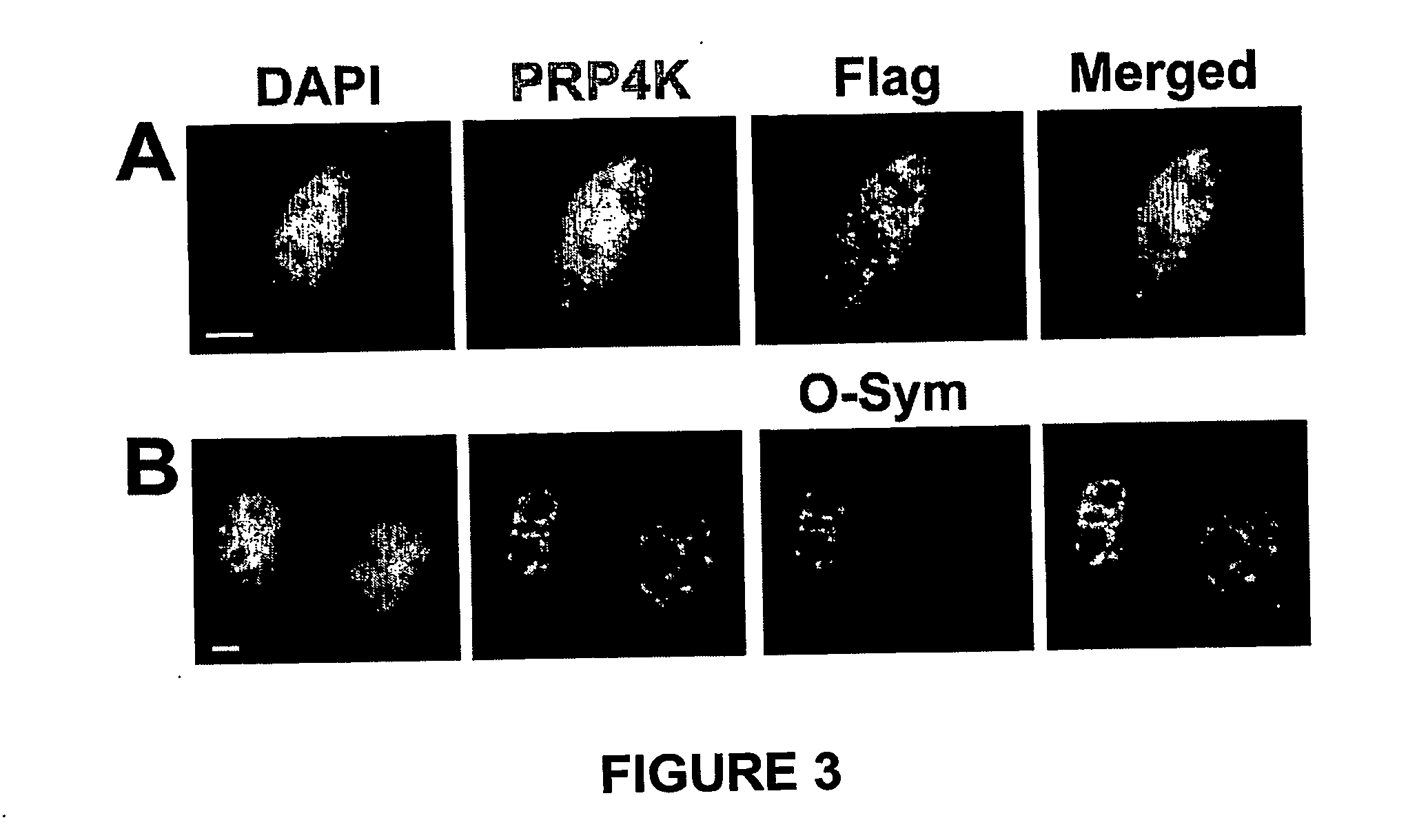
[0084] Twenty-four to 36 hours post transfection cells were fixed in 1% paraformaldehyde in PBS for 5 min. followed by permeabilisation in 0.5% Triton X-100 for 5 min. at RT. Following several washes in PBS, cells were then blocked for 20 min. at RT with O-Sym Binding / Blocking (OSB) buffer (10 mM Tris-HCl pH 7.5, 0.1 mM EDTA, 150 mM KCl, 600 μg / ml sheared Herring sperm DNA, 200 μg / ml BSA). Directly following the blocking step, cells were hybridised for 1-2 hours at 37° C. with the O-Sym aptamer (labelled with either Cy3, biotin or both) alone or combined with Tet-O aptamer (labelled with either Cy5, biotin or both) in OSB buffer at a concentration of 50-100 nM. After hybridisation with the O-Sym or Tet-O aptamer, coverslips were either washed with 3×PBS and mounted in anti-fade reagent for immediate immunofluorescence detection or were further processed for immunofluorescent localisati...


PUM

PropertyMeasurementUnit
Nuclear structureaaaaaaaaaa
Affinityaaaaaaaaaa
Paramagnetismaaaaaaaaaa

Abstract

The present invention is a method for the detection and localization of proteins in situ. The method comprises contacting a peptide tag labelled protein with a nucleic acid aptamer that recognizes the peptide tag. The nucleic acid aptamer is functionalized such that is detectable by a variety of methods. The method allows for the detection of one or more proteins and / or protein complexes in sub-cellular and sub-nuclear structures with high specificity and also allows for the purification of such identified proteins.

Description

FIELD OF THE INVENTION [0001] The invention relates to the detection and localization of proteins. More specifically, the present invention is directed to a method for the in situ detection and localization of one or more proteins or protein complexes with specificity within cellular sub-structures at high resolution. The method is herein referred to as Aptamer In Situ Detection, (Aptamer-ID™) or Aptamer protein isolation (Aptamer-PI™). BACKGROUND OF THE INVENTION [0002] Cellular proteins are often detected using conventional antibody techniques such as with the use of antibodies directed against the proteins themselves or to peptide tags fused to these proteins (e.g. Flag-tag or HA-tag, Terpe, 2003). The antibodies can be functionalised with fluorescent molecules or gold particles in order that the protein of interest be recognised by light (LM) and electron microscopy (EM). To visualise the localisation and dynamics of specific proteins in living cells, natural and engineered fluo...

Claims


Application Information

Patent Timeline
IPC IPC(8): C12Q1/70C12Q1/68G01N33/68
CPCB82Y5/00B82Y10/00G01N33/68G01N33/6803G01N33/6842G01N33/6848
Inventor DELLAIRE, GRAHAMBAZETT-JONES, DAVID P.
Owner HOSPITAL FOR SICK CHILDREN
Learn More