Compositions and methods for promoting attachment of cells of endothelial cell lineage to medical devices

a technology of endothelial cells and cell lines, which is applied in the field of compositions and methods for promoting the attachment of cells of endothelial cell lines to an intravascular device, can solve the problems of unacceptably high thrombosis rates limited arterial bypass procedures, and the association of small caliber grafts with these devices, so as to promote the endothelialization of the vascular device, promote the spread of cells

Inactive Publication Date: 2007-07-19
AFFINERGY INC
View PDF24 Cites 15 Cited by
  • Summary
  • Abstract
  • Description
  • Claims
  • Application Information

AI Technical Summary

Benefits of technology

[0015] Alternatively, provided is a method for promoting endothelialization of a vascular device so that to a selected metallic surface of the device, once that surface is coated and the device implanted, promoted is attachment of cells of endothelial cell lineage. The method comprises the steps of: (a) contacting a biofunctional coating composition described herein to at least one metallic surface of a vascular device to be endothelialized, so that the biofunctional coating composition binds to the at least one metallic surface, in forming a coated metallic surface on the vascular device; wherein the biofunctional coating composition comprises at least one surface-binding domain having an amino acid sequence consisting essentially of SEQ ID NOs:1-8, coupled to at least one endothelial-cell binding domain having an amino acid sequence consisting essentially of SEQ ID NOs:9-46; and (b) implanting the device into an individual (human or non-human) in need of the device; wherein cells of endothelial cell lineage (produced by the individual) contact, attach and adhere to the coated surface of the device (primarily mediated by the cells binding to the at least one endothelial binding domain of the biofunctional coating composition), in promoting spread of cells of endothelial cell lineage over the coated metallic surface of the device, and in promoting endothelialization of the vascular device. Promoting endothelialization on the implanted device may further promote one or more of healing of tissue or vasculature adjacent to the implanted device, promote incorporation (integration) of the implanted device into the adjacent tissue, and reduce occurrence of thrombosis as related to the implanted device.

Problems solved by technology

Arterial bypass procedures are limited by the availability of a vascular conduit, such as internal mammary artery or saphenous vein.
Unfortunately, synthetic conduits made from polytetrafluoroethylene (PTFE) or polyethylene terephthalate (PET) suffer from unacceptably high rates of thrombosis in small caliber grafts due to their lack of an adherent, quiescent endothelium.
However numerous problems have been reported to be associated with these devices, including thrombosis, neointima formation, and restenosis.
However, spontaneous endothelialization of endovascular grafts is rare in human patients, perhaps because the graft materials are engineered to resist molecular adhesion and coagulation, and endothelial progenitor cells have no ability to adhere, survive, and proliferate on such materials.

Method used


Image

  • Compositions and methods for promoting attachment of cells of endothelial cell lineage to medical devices

Examples


example 1

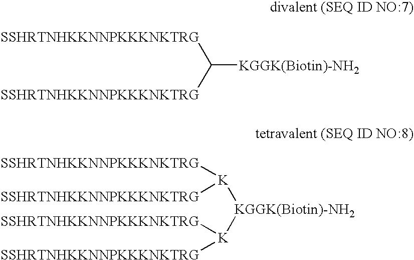
[0046] Illustrated in this example are various methods for producing a surface-binding domain and a endothelial-binding domain for the biofunctional coating compositions according to the present invention. Many of the peptides comprising the binding domains in the biofunctional coating composition according to the present invention (i.e., a surface-binding domain and an endothelial-binding domain) were developed using phage display technology.

[0047] Phage display technology is well-known in the art, and can be used to identify additional peptides for use as binding domains in the interfacial binding materials according to the present invention. In general, using phage display, a library of diverse peptides can be presented to a target substrate, and peptides that specifically bind to the substrate can be selected for use as binding domains. Multiple serial rounds of selection, called “panning,” may be used. As is known in the art, any one of a variety of libraries and panning metho...

example 2

[0050] This example illustrates the discovery and characterization of surface-binding domains comprising peptides having binding specificity for a metallic surface of a medical device, such as a stainless steel surface of a stent.

A. Phage Screening and Selections.

[0051] As a specific illustrative example, nonspecific binding sites in wells containing stainless steel stent material in polystyrene microtiter plates were blocked with a buffer containing 1% bovine serum albumin for 2 hours at room temperature. The wells and stainless steel stent material were then washed three times with PBS-T. The plate was incubated for 1 hour at room temperature with shaking at 50 rpm. Each of 17 different phage display libraries was diluted in PBS+1% BSA and was added at a concentration of 1010 pfu / ml in a total volume of 250 μl. After a 1 hour incubation at room temperature with shaking at 50 rpm, 70 μl of bovine serum was added, and then the plates were incubated at 37° C. with shaking for 1 ho...

example 3

[0061] This example illustrates the discovery and characterization of endothelial-binding domains comprising peptides having binding specificity for cells of endothelial cell lineage.

A. Phage Screening and Selections.

[0062] Phage libraries were pooled into four groups and screened for peptides that bind to human umbilical vein endothelial cells (HUVECs). The four pools were first pre-cleared on non-target cells (e.g., cells other than cells of endothelial cell lineage). For each pool, 10 μL of phage (1010 of phage) was added to 1×10⁶ cells of each of the following cell types: HEK-293 cells, aortic vascular smooth muscle cells (AoSMC), and platelets. The phage and cells were incubated for 1 hour at room temperature. Cells and attached phage were pelleted by centrifugation. The phage remaining in the supernatant were used for subsequent selections on HUVECs. Selections on HUVECS were performed using methods known in the art, including by one or more of biopanning with the cells inc...


PUM

PropertyMeasurementUnit
diameteraaaaaaaaaa
molecular weightaaaaaaaaaa

Abstract

The present invention provides compositions and methods for an improved coating for medical devices. Provided is a biofunctional coating composition comprising at least one binding domain that has binding specificity for a metallic surface material of a medical device, and at least one binding domain that has binding specificity for cells of endothelial cell lineage. Methods for coating a metallic surface of a medical device, and for manufacturing of a medical device, comprise contacting the metallic surface to be coated with the biofunctional coating material in an amount effective to form a coating, and may further comprise contacting the coated surface with cells of endothelial cell lineage to bind the cells of endothelial cell lineage to the coated surface.

Description

CROSS REFERENCE TO RELATED APPLICATIONS [0001] This is a nonprovisional application which claims priority benefit of U.S. Provisional Application No. 60 / 758,029, filed 11 Jan. 2006; which is hereby incorporated by reference herein.FIELD OF THE INVENTION [0002] The present invention relates to compositions and methods for promoting the attachment of cells of endothelial cell lineage to an intravascular device. BACKGROUND OF THE INVENTION [0003] Atherosclerosis causes stenosis and occlusion of arteries. Stenting and bypass surgery are often used to treat severe disease in small caliber arteries (defined as less than 6 mm in diameter). Arterial bypass procedures are limited by the availability of a vascular conduit, such as internal mammary artery or saphenous vein. Unfortunately, synthetic conduits made from polytetrafluoroethylene (PTFE) or polyethylene terephthalate (PET) suffer from unacceptably high rates of thrombosis in small caliber grafts due to their lack of an adherent, quie...

Claims


Application Information

Patent Timeline
Patent Type & Authority Applications(United States)
IPC IPC(8): A61F2/02C07K14/705C07H21/04C12P21/06
CPCA61K38/00A61L27/34A61L31/10C07K2319/00C07K7/08C07K14/705C07K7/06
Inventor HAMILTON, PAUL THEODOREKENAN, DANIEL JAMESSOLAN, AMY KATHERINE
Owner AFFINERGY INC
Why Eureka
Try Eureka