
DNA amplification of a single cell

a single cell, dna technology, applied in the field of dna amplification, to achieve the effect of improving accuracy and reliable amplification of dna

Inactive Publication Date: 2004-01-06
AMGEN RES (MUNICH) GMBH
View PDF3 Cites 89 Cited by
  • Summary
  • Abstract
  • Description
  • Claims
  • Application Information

AI Technical Summary

Benefits of technology

The method of the present invention for the first time allows the amplification of the entire genome of a single cell even from unextracted DNA samples. This enables e.g. the genomic analysis of individual isolated disseminated tumor cells, applying comparative genomic hybridization (CGH) to single cells. Therefore, this method provides, e.g., the means to identify the individual genetic changes in a single cell that might promote dissemination and ectopic growth of disseminated tumor cells with metastatic potential. The genomic profile of such single disseminated cells could provide useful information on whether certain clonal genotypes are associated with disseminative events.
Furthermore, the method of the present invention for the first time allows for the reproducible application of CGH to individual cells, whereas other protocols for "whole genome amplification" such as PEP, DOP-PCR and Alu-PCR do not provide homogenous staining patterns by CGH wherein the origin of test-DNA is a single cell, single chromosomes or parts thereof. This reproducible application of CGH on individual single cells can be explained, inter alia, by the fact that a non-degenerate primer is used that drastically reduces the complexity of primer binding sites of oligonucleotides hitherto used in the DOP and PEP techniques. These previously numerous different sites require equally numerous different specific PCR conditions impossible to achieve during the same reaction. Additionally, since the amplification method of the present invention does not depend on repetitive sequences within the genome (like IRS-PCR), it is possible to reliably amplify the genomic DNA of single cells from species where such repetitive sequences are less frequent or even non-existent.
In an additional preferred embodiment, said first and said second primer of the method of the present invention are hybridized to each other separately from said DNA fragments and are added to said DNA fragments after they hybridized to each other. The addition of the hybridized primers to said DNA fragments is effected prior to step (ca) or step (cb). Such a pre-hybridization of primers leads, inter alia, to a higher hybridization efficiency to said DNA fragments and interfering to chromosomal DNA can be avoided.
The inventive method described herein can be applied to DNA of different sources, such as solid tumor DNA isolated from frozen sections and / or cryosections and / or paraffin embedded, formalin fixed specimens. For decades these tissue sections have been stored mainly for histopathological diagnosis. Single cells or small samples, comprising a limited amount of cells, from histopathological tissue can be screened for specific genetic changes and compared with other areas from the same tissue that may exhibit distinctly different histopathological features or, for control purposes, with areas of apparently normal tissue. Global screening of copy number sequence changes within a tumor genome from archival tissue material could increase the knowledge about cytogenetic alterations in solid tumors significantly. A direct comparison of these cytogenetic data with histological and histochemical results and clinical follow up data would become possible.
In yet a more preferred embodiment of the method of the present invention steps (a) to (e) are carried out in one reaction vessel. This has the advantage that a potential template loss is avoided and, moreover, an additional opening and closing of the reaction vessel, which may involve contamination and is troublesome, is avoided.
Another important application of the presented method is prenatal diagnosis using embryonic or fetal cells in maternal blood. The ability to use embryonic or fetal cells enriched from maternal blood of pregnant women for prenatal diagnosis of chromosomal abnormalities has been a long-sought goal for those pursuing a non-invasive alternative to current methods, such as amniocentesis or chorionic villus sampling. The localization and identification of novel disease genes allows for mutation analysis or linkage studies on fetuses at risk for single gene disorders or chromosomal abnormality etc. The method of the present invention improves the accuracy as well as applicability of methods for the diagnosis of preimplantation genetic disorders or for the diagnosis on fetal cells isolated from maternal blood, whereby analyses can be performed on a single cell level, thus abolishing the need for preceding enrichment of cells. As demonstrated in the appended examples, the method of the present invention provides for the reliable amplification of DNA from a single fetal or embryonic cell isolated from maternal blood, inter alia, from the umbilical vein blood.

Problems solved by technology

Thus, the technical problem consists in providing means and methods which comply with the needs as described above and which eliminate the above-mentioned disadvantages.

Method used


Image

  • DNA amplification of a single cell
  • DNA amplification of a single cell
  • DNA amplification of a single cell

Examples


example 2

Amplification of DNA of a Single Cell

In order to avoid template loss, all preparatory steps were performed in one tube. For DNA isolation, restriction enzyme digest, primer ligation and PCR-amplification all buffers and conditions were adjusted for optimal performance to guarantee highest reliability and reproducibility. Generally, high concentrations of proteinase K, Msel, T4 DNA ligase and Taq polymerase gave the best results.

A single cell (e.g. peripheral blood lymphocyte or bone marrow stoma cell) in 1 .mu.l pick buffer (50 mM Tris / HCl, pH 8.3, 75 mM KCl 3mM MgCl.sub.2, 137 mM NaCl) was added to 2 .mu.l Proteinase K digestion buffer (10 mM Tris-Acetate (pH 7.5), 10 mM Mg-Acetate, 50 mM K-Acetate (0.2 .mu.l of Pharmacia One-Phor-AII-Buffer-Plus), 0.67% TWEEN (Sigma), 0.67% Igepal (Sigma), 0.67 mg / ml Proteinase K) and incubated for 10h at 42.degree. C. in a PCR machine with heated lid. Proteinase K was inactivated at 80.degree. C. for 10 minutes. After inactivation of Proteinase K...

example 3

CGH With Amplified DNA From Peripheral Blood Lymphocytes

DNA isolated from a single unfixed and paraformaldehyde (PFA)-fixed peripheral blood lymphocyte was amplified as described in Example 2 and competitively hybridized with 1 .mu.g nick-translated, unamplified placenta DNA. Labeling was most efficient using ThermoSequenase in combination with a ratio of dTTP / bio-dUTP of 7 / 1. Reamplification was performed in 30 .mu.l using 0.5 .mu.l LigMse-21 primer (100 .mu.M), 1 .mu.l dNTP (dATP, dCTP, dGTP, 10 mM each, 8.6 mM dTTP) and 1.3 .mu.l biotin-16-dUTP (1 mM, Bboehringer Mannheim), 13 U ThermoSequenase (USB) in 1.times.ThermoSequenase buffer and 0.5 .mu.l of the primary PCR. In total 25 cycles were programmed with the temperatures set to 94.degree. C. (1 min.), 65.degree. C. (30 sec.), 72.degree. C. (2 min.) for 1 cycle; 94.degree. C. (40 sec.), 65.degree. C. (30 sec.), 72.degree. C. (1 min. 30 sec.) for 94.degree. C. (40 sec.), 65.degree. C. (30 sec.), 72.degree. C. (2 min.) for 9 cycle...

example 4

Detection of Chromosomal Changes in a Patient With Down's Syndrome

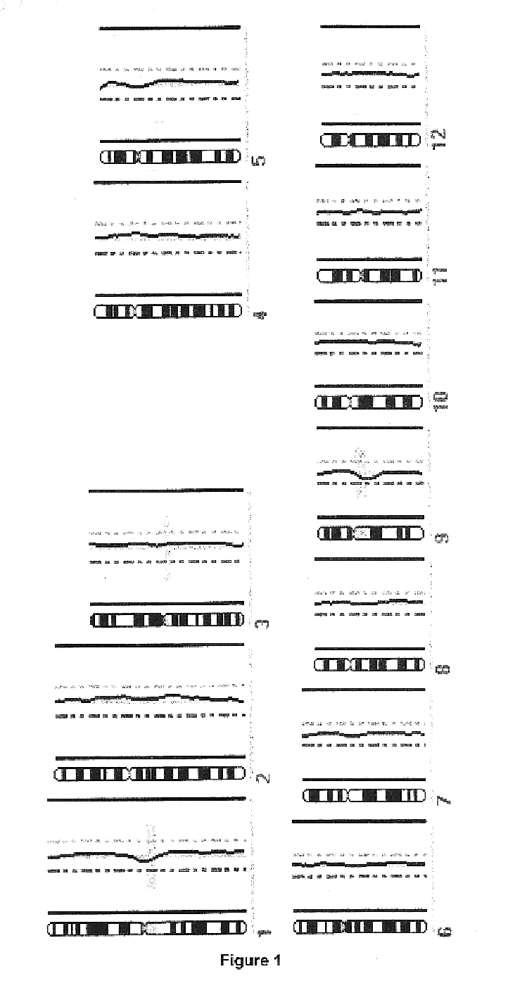
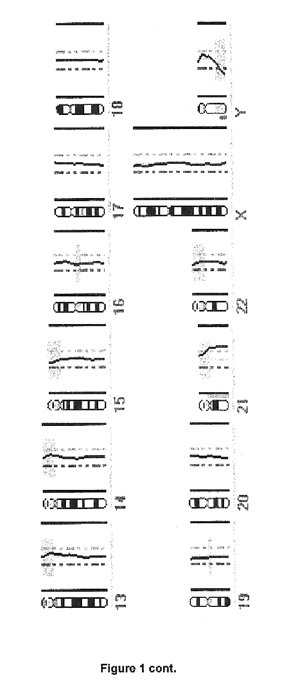
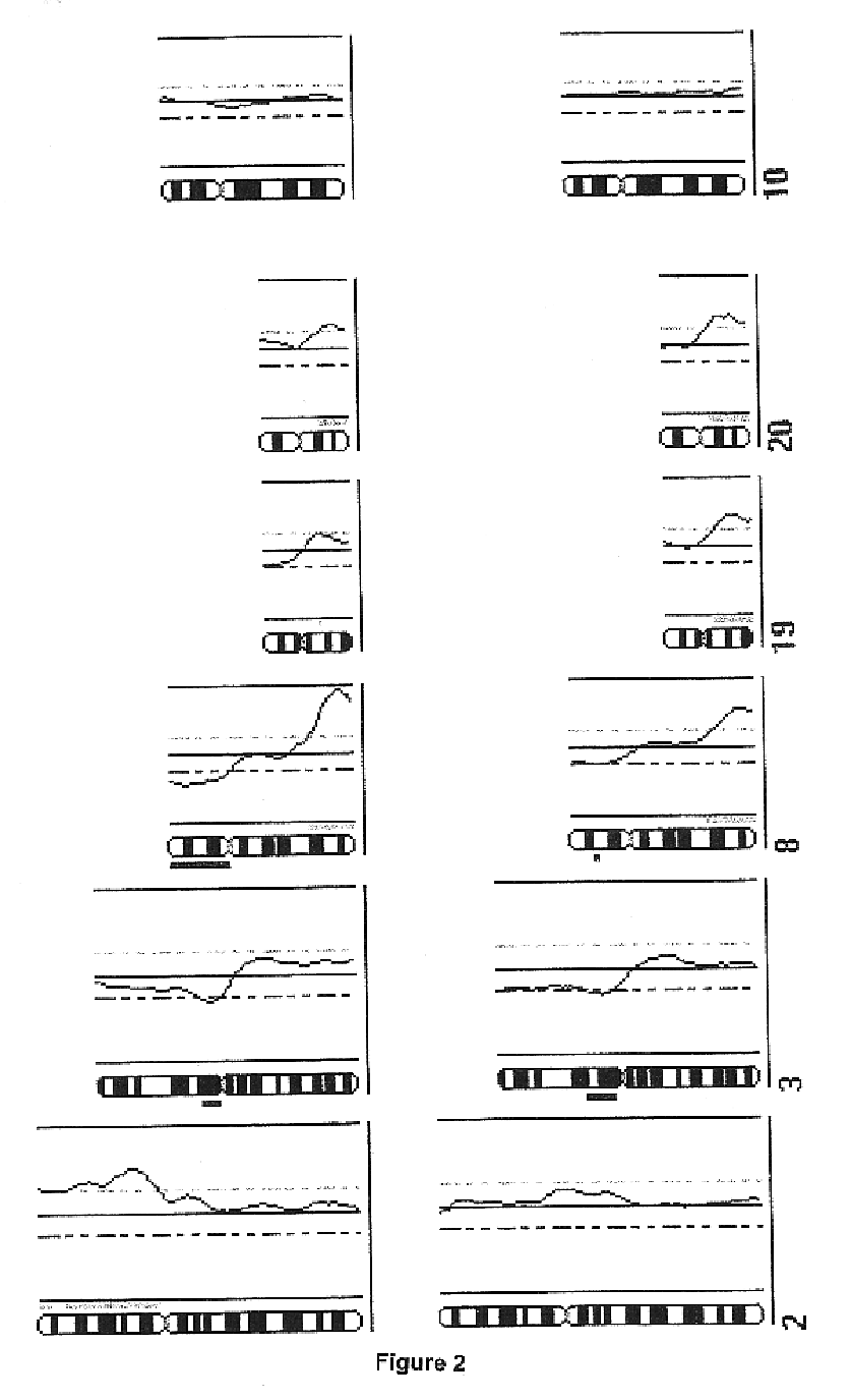
In order to determine whether known chromosomal changes could be detected by the single cell analysis, DNA of a single leukocyte from a blood sample of a patient with Down's syndrome and its DNA was isolated and amplified as described in Example 2. The CGH-profile showed a gain of chromosome 21 as single detectable abnormality (FIG. 1).


PUM

PropertyMeasurementUnit
temperatureaaaaaaaaaa
pHaaaaaaaaaa

Abstract

The present invention relates to a novel method for the amplification of DNA, this method being particularly useful for the amplification of the DNA or the whole genome of a single cell, chromosomes or fragments thereof. Described is also the use of the method in DNA analysis for medical, forensic, diagnostic or scientific purposes, like comparative genomic hybridization (CGH)-, fluorescence in situ hybridization (FISH)-, polymerase chain reaction (PCR)-, single strand conformation polymorphism (SSCP)-, DNA sequence-, "loss of heterozygosity" (LOH)-, fingerprint- and / or restriction fragment length polymorphism (RFLP)-analysis.

Description

This application is a national phase application of PCT / EP99 / 06912 filed Sept. 17, 1999, which claims priority to EP 98 11 7745.4 filed Sept. 19, 1998 and EP 98 11 7799.1 filed Sept. 18, 1998, and which are incorporated in their entirety by reference.1. Field of the InventionThe present invention relates to a novel method for the amplification of DNA, this method being particularly useful for the amplification of the DNA or the whole genome of a single cell, chromosomes or fragments thereof. The present invention further relates to the application of the method in DNA analysis for medical, forensic, diagnostic or scientific purposes, like comparative genomic hybridization (CGH), fluorescence in situ hybridization (FISH), polymerase chain reaction (PCR), single strand conformation polymorphism analysis (SSCP), DNA sequence analysis, "loss of heterozygosity" analysis (LOH), fingerprint analysis, and / or restriction fragment length polymorphism analysis (RFLP).2. Description of the Rela...

Claims


Application Information

Patent Timeline
Patent Type & Authority Patents(United States)
IPC IPC(8): C12Q1/68C12N15/09G01N33/566G01N33/53
CPCC12Q1/6809C12Q1/6855
Inventor KLEIN, CHRISTOPHSCHMIDT-KITTLER, OLEG
Owner AMGEN RES (MUNICH) GMBH
Learn More