Protein crystal structure
a technology of protein crystal structure and crystal structure, applied in the direction of crystal growth process, enzymes, biochemistry apparatus and processes, etc., can solve the problem of unclear function of pre-set and post-set domains
- Summary
- Abstract
- Description
- Claims
- Application Information
AI Technical Summary
Benefits of technology
Problems solved by technology
Method used
Image
Examples
example 1.1
Protein Constructs
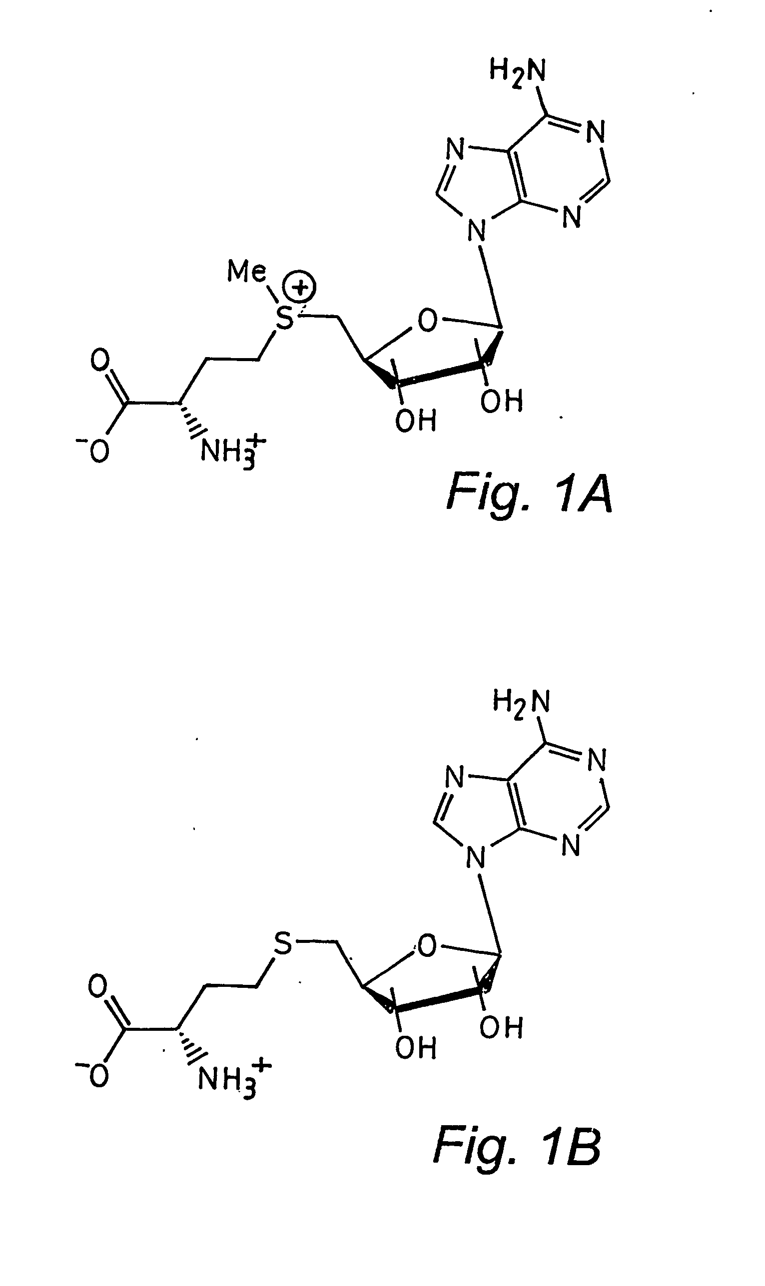
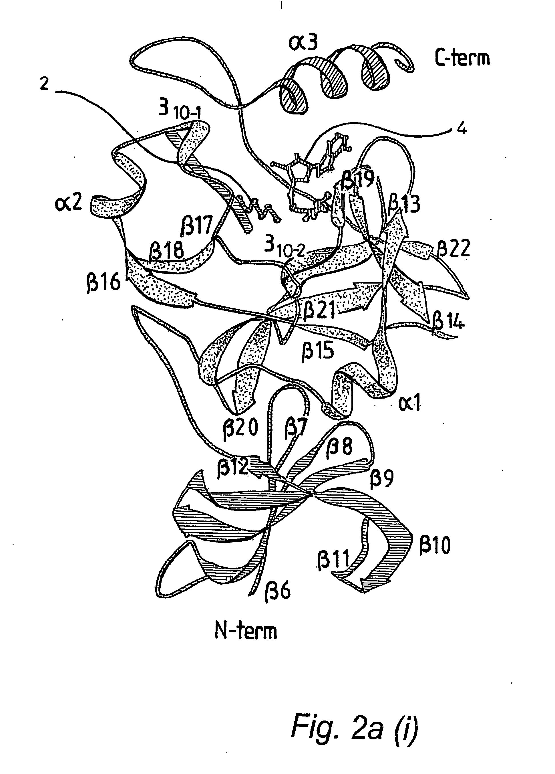
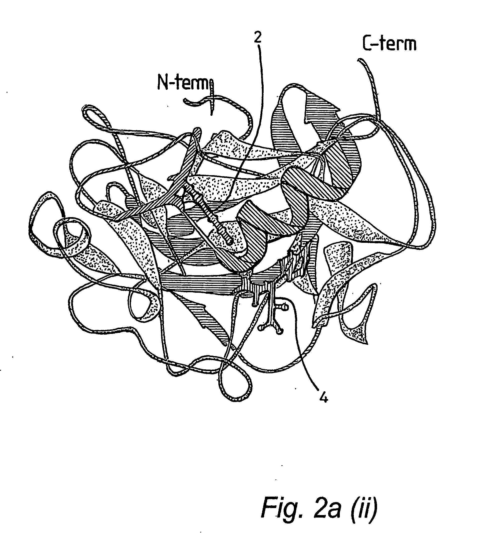
[0066] A truncated form of the SET 7 / 9 HMT protein lacking the first 51 residues of the N terminal portion, referred to as ΔSET7 / 9 (residues 52-366), was expressed as a GST-fusion in pGEX 6P1 in E. coli BL21. The GST was removed by overnight treatment with PreScission™ Protease (Amersham) prior to gel filtration. Preparation of ΔSETT7 / 9 in D2O for nmr studies resulted in a series of N-terminal degradation products which were still catalytically active. Subsequently a further truncated form of the protein, ΔΔSET7 / 9 (residues 108-366), was prepared as above and found to be stable for growth in D2O and was consequently used for further nmr and crystallography experiments. Site-directed alanine mutations were introduced using the Stratagene Quikchange Mutagenesis kit, mutations were confirmed by DNA sequencing and electrospray mass spectrometry. Synthetic peptides were prepared using conventional in vitro techniques. AdoHcy was obtained from Fluka, Switzerland.
example 1.2
HMTase Activity Measurements
[0067] The methyltransferase activity of SET7 / 9 and the various mutant constructs described in the text were determined in a reaction volume of 20 μl containing 3 μM AdoMet supplemented with [methyl-3H] AdoMet (4 μCi) (Amersham Biosciences, UK) and 750 μM purified methylase in reaction buffer (50 mM Tris pH 8.5, 100 mm NaCl, 1 mM EDTA, 1 mM DTT) with 50 μM histone peptide (see below). Following incubation at 37° C. for 60 min the reaction was vacuum blotted onto membrane (Hybond-C, Amersham Biosciences, UK) washed and activity measured by scintillation counting.
example 1.3
Analytical Analysis of Histone Methylation
[0068] The histone methyltransferase assay for analytical purposes was carried out under slightly different conditions than those described in Example 1.2. For analytical purposes the reaction was performed at 37° C. in 50 mM Tris-HCl, pH 8.0, 100 mM NaCl, 1 mM DTT, with 300 μM AdoMet (Fluka, Switzerland), 100 μM H3 20mer peptide (ARTKQTARKSTGGKAPRKQY), and with 1.5 μM enzyme. At desired time intervals an aliquot of the reaction was removed and quenched in 8 M urea and acidified with glacial acetic acid. The reaction products were separated by reverse phase HPLC (Jasco (UK) Ltd) on a Zorbax 300SB-C18 column (Rockland Technologies, Inc. USA) using a gradient from 0 to 40% acetonitrile in the presence of 0.05% trifluoroacetic acid at 55° C. Fractions from the peptide peak were analysed using a Reflex III MALDI time-of-flight mass spectrometer (Bruker Daltonik, GmbH, Germany) to obtain positive ion mass spectra.
PUM
| Property | Measurement | Unit |
|---|---|---|
| structure | aaaaa | aaaaa |
| hydrophobic | aaaaa | aaaaa |
| hydrophobic interactions | aaaaa | aaaaa |
Abstract
Description
Claims
Application Information
 - R&D
- Intellectual Property
- Life Sciences
- Materials
- Tech Scout
- Unparalleled Data Quality
- Higher Quality Content
- 60% Fewer Hallucinations
Browse by: Latest US Patents, China's latest patents, Technical Efficacy Thesaurus, Application Domain, Technology Topic, Popular Technical Reports.
© 2025 PatSnap. All rights reserved.Legal|Privacy policy|Modern Slavery Act Transparency Statement|Sitemap|About US| Contact US: help@patsnap.com


