
Biosensors comprising heat sealable spacer materials

a technology of heat sealing spacers and biosensors, applied in the field of biosensors, can solve the problems of difficult to obtain electrode patterns with small resolution and smooth edges, and achieve the effect of excellent edge quality

Inactive Publication Date: 2007-10-18
NIPRO DIAGNOSTICS INC
View PDF7 Cites 87 Cited by
  • Summary
  • Abstract
  • Description
  • Claims
  • Application Information

AI Technical Summary

Benefits of technology

[0004] In addition to improved accuracy, spatial resolution of the electrode is important because the smaller the surface area of the electrode, the smaller the sample volume required. This is desirable with, for example, glucose monitoring for diabetics, where the patient must test his or her blood glucose multiple times a day. Smaller blood volume requirements allow the patient to obtain blood from areas with lower capillary densities than the fingers, such as the upper arm and forearm, which are less painful to lance.
[0006] Coupled with the need to better define the anode area, is a desire to simplify manufacturing steps of the new generation of biosensors in order to provide a more robust process, high production yields and high quality sensors. New materials are being explored that could be beneficial in attaining this goal.
[0007] To solve the foregoing problems, the Inventors have developed a unique method of defining the anode area of a biosensor by utilizing a heat sealable spacer material to accurately define one or more edges of the anode instead of a dielectric layer. The Inventors have found that this method is particularly useful when used with a laser ablation technique. With the laser ablation technique, an electroactive material, such as gold is sputtered in a thin film onto a substrate. A laser then traces across the substrate and ablates the electroactive material, leaving an electrode pattern on the substrate. This technique produces electrodes with better resolution and smoother edges than with screen printing. In addition to greatly improving the accuracy and reproducibility of the anode area, the method of fabricating the biosensor is simpler than current process as it no longer requires depositing a separate dielectric layer. SUMMARY OF INVENTION
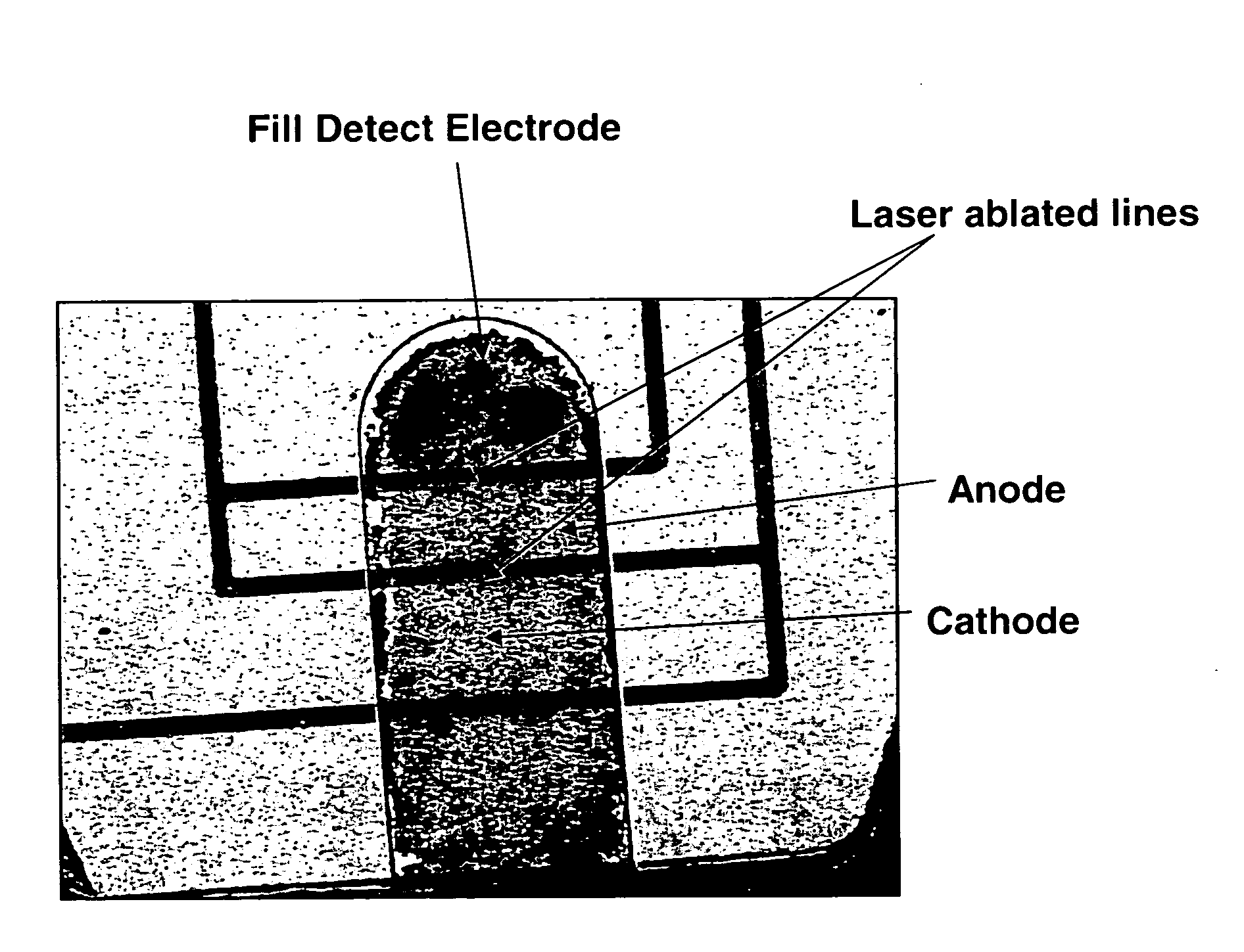
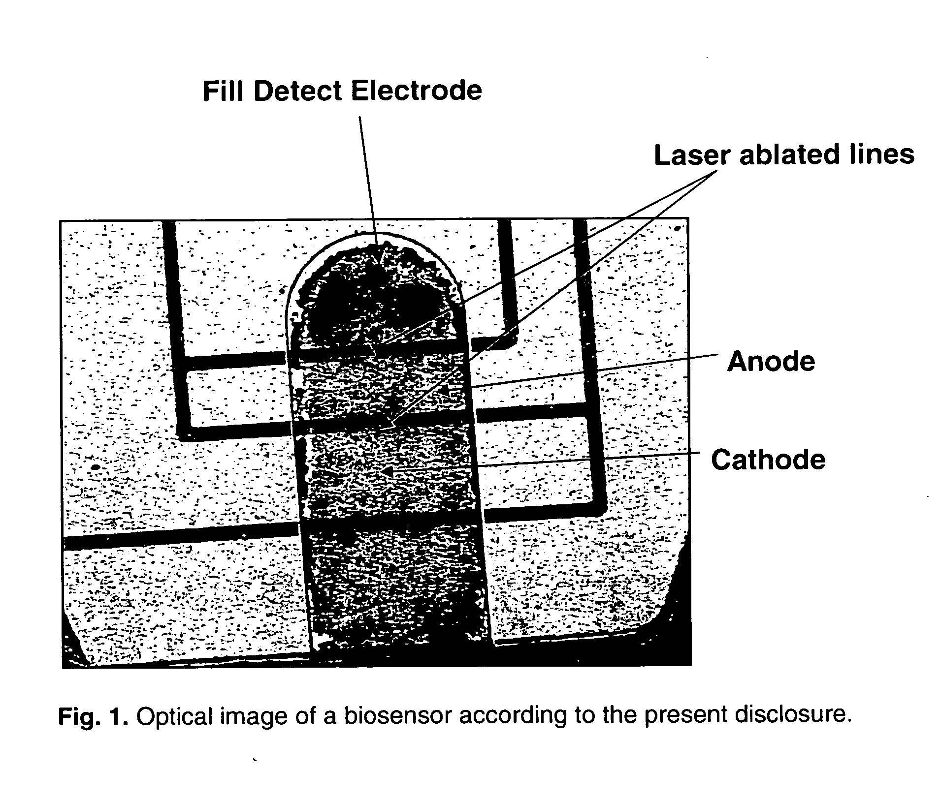
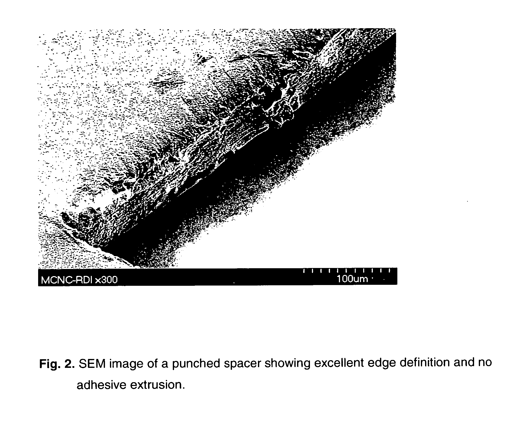
[0009] Whatever the composition of the spacer material, it typically has at least one opening punched through it, and covers at least a portion of the working electrode, such as the anode. The punched opening defines at least one edge of the anode, and typically two opposing edges. The remaining two opposing edges are typically defined by ablated laser lines, and thus also have excellent edge quality.

Problems solved by technology

Because screen printing involves extruding the paste through the screen onto the substrate, it is difficult to obtain electrode patterns with small resolution and smooth edges.

Method used


Image

  • Biosensors comprising heat sealable spacer materials
  • Biosensors comprising heat sealable spacer materials
  • Biosensors comprising heat sealable spacer materials

Examples


example 1

[0082] A thin film of gold (30 nm) was sputtered onto a plastic film substrate (PET). The gold layer was then laser ablated using a focused beam approach, in which Galvo mirrors were used to direct the laser beam to ablate the material according to a desired electrode pattern. The remaining gold layer was formed into desired patterns for an electrode array, which included an anode, cathode, and two fill detect electrodes.

[0083] Next, the second layer or spacer layer of the biosensor was formed by first punching out sample cavities in a polyester film having a heat seal coating. The polyester film used for the spacer was a commercially available PET film (3M Scotchpak™ MA370M), which had a total thickness of 3.7 mils, including the heat seal coating of 0.8 mils.

[0084] The punched spacer material was laminated onto laser ablated electrode substrate to form assembled biosensors having an anode, cathode and two fill detect electrodes. As shown in FIG. 1, the anode area was defined on ...

example 2

[0087] Once the sensors were assembled according to Example 1, chemistry was dispensed into the sample cavities using micropipetting. Blood volume required to fill the sample cavity of this biosensor was 0.25 ul when a 100 μm thick spacer layer was used. Table 1 below shows the relative percentages by weight of the various ingredients dispensed into the sample cavities.

TABLE 1IngredientWeight PercentPhosphate, Monobasic (Buffer) 0.64%Phosphate, Dibasic (Buffer) 0.92%Silwet L-7206 (Spreading Agent)0.051%Triton-X 100 (Spreading Agent)0.051%Methocel F4M (Binder)20.00%Sucrose (Enzyme Stabilizer) 5.00%Hexammine Ruthenium (III) Chloride (Mediator) 5.88%PQQ Dependent Glucose Dehydrogenase (Enzyme)10.00%18 mega ohm deionized waterBalance

[0088] The chemistry solution was then dried and a cover was applied over the sample cavities to form capillary gaps into which blood sample could be drawn. Blood testing data was taken on the finished samples, with sample sizes ranging from 40-60 per bloo...


PUM

PropertyMeasurementUnit
Temperatureaaaaaaaaaa
Fractionaaaaaaaaaa

Abstract

Disclosed herein is a biosensor for measuring analyte in a fluid that comprises a substrate layer having disposed thereon at least one each of an electrode, cathode, anode, and a novel spacer material. The spacer material according to the present disclosure comprises a heat sealable organic layer that covers at least a portion of the anode and defines at least one edge of the anode, wherein the spacer material has at least one hole punched through it and defines a cavity or well for accepting chemistry. Also disclosed is a method of making such biosensors.

Description

[0001] The present disclosure relates to biosensors for measuring an analyte in a bodily fluid, such as blood, wherein the biosensor comprises a heat sealable, organic spacer material that particularly defines at least one edge of a working electrode disposed on the biosensor. The present disclosure also relates to methods of making the biosensor and methods of measuring analytes in bodily fluid using the biosensor. [0002] Electrochemical sensors have long been used to detect and / or measure the presence of analytes in a fluid sample. In the most basic sense, electrochemical sensors comprise a reagent mixture containing at least an electron transfer agent (also referred to as an “electron mediator”) and an analyte specific bio-catalytic protein, and one or more electrodes. Such sensors rely on electron transfer between the electron mediator and the electrode surfaces and function by measuring electrochemical redox reactions. When used in an electrochemical biosensor system or device,...

Claims


Application Information

Patent Timeline
IPC IPC(8): G01N33/487
CPCG01N27/3272
Inventor POPOVICH, NATASHA D.SLOMSKI, DENNIS
Owner NIPRO DIAGNOSTICS INC
Learn More