Method of Monitoring a Microorganism That Causes Infectious Disease of a Laboratory Animal

a laboratory animal and microorganism technology, applied in the field of monitoring a microorganism, can solve the problems of antigen antibody reaction, take a long time for the test procedure, etc., and achieve the effects of low contamination of the facility, high throughput, and easy and rapid condu

Inactive Publication Date: 2007-12-13
NAGAMUNE TERUYUKI +1
View PDF0 Cites 18 Cited by
  • Summary
  • Abstract
  • Description
  • Claims
  • Application Information

AI Technical Summary

Benefits of technology

[0018] (4) A population can be tested with high throughput.
[0019] In short, the method of this invention using a micro flow channel chip is conducted in a completely closed system, and the operation using this system can be conducted easily and rapidly. Therefore, it is also advantageous in that the risk of infection to a human body by an infectious microorganism and contamination of facility is low, thus a microorganism that causes infectious disease of a laboratory animal can be monitored in safety.

Problems solved by technology

According to conventional methods used to monitor an infectious disease of a laboratory animal, massive blood is needed for one test (in the case of mouse, only 100 μl corresponds to 1 / 10 of total blood of the individual), therefore, there are some problems to be solved as follows, (1) The microbiological condition of a parent population has to be estimated from the result of a random inspection of the parent population, and no other method can not be adopted.
(3) It take a long time for the test procedure and antigen antibody reaction.

Method used


Image

  • Method of Monitoring a Microorganism That Causes Infectious Disease of a Laboratory Animal
  • Method of Monitoring a Microorganism That Causes Infectious Disease of a Laboratory Animal
  • Method of Monitoring a Microorganism That Causes Infectious Disease of a Laboratory Animal

Examples


example 1

[0052] In the following experiments, PBS (Na2HPO4 0.61 g, KH2PO4 0.19 g, NaCl 8.00 g, KCl 0.20 g, MilliQ (Millipore) IL), PBST (0.05% Tween20-PBS), and a washing solution (2% skim milk-PBST), and a blocking solution (2% skim milk-PBST) were used, and they were composed of the compositions described in brackets. Surface of the substrate, where antigen antibody reaction was conducted, was coated with Indium-Tin Oxide (ITO) and a glass substrate (manufactured by Tatsunami glass, 26×76 mm) introduced with aldehyde group was used on the coated substrate.

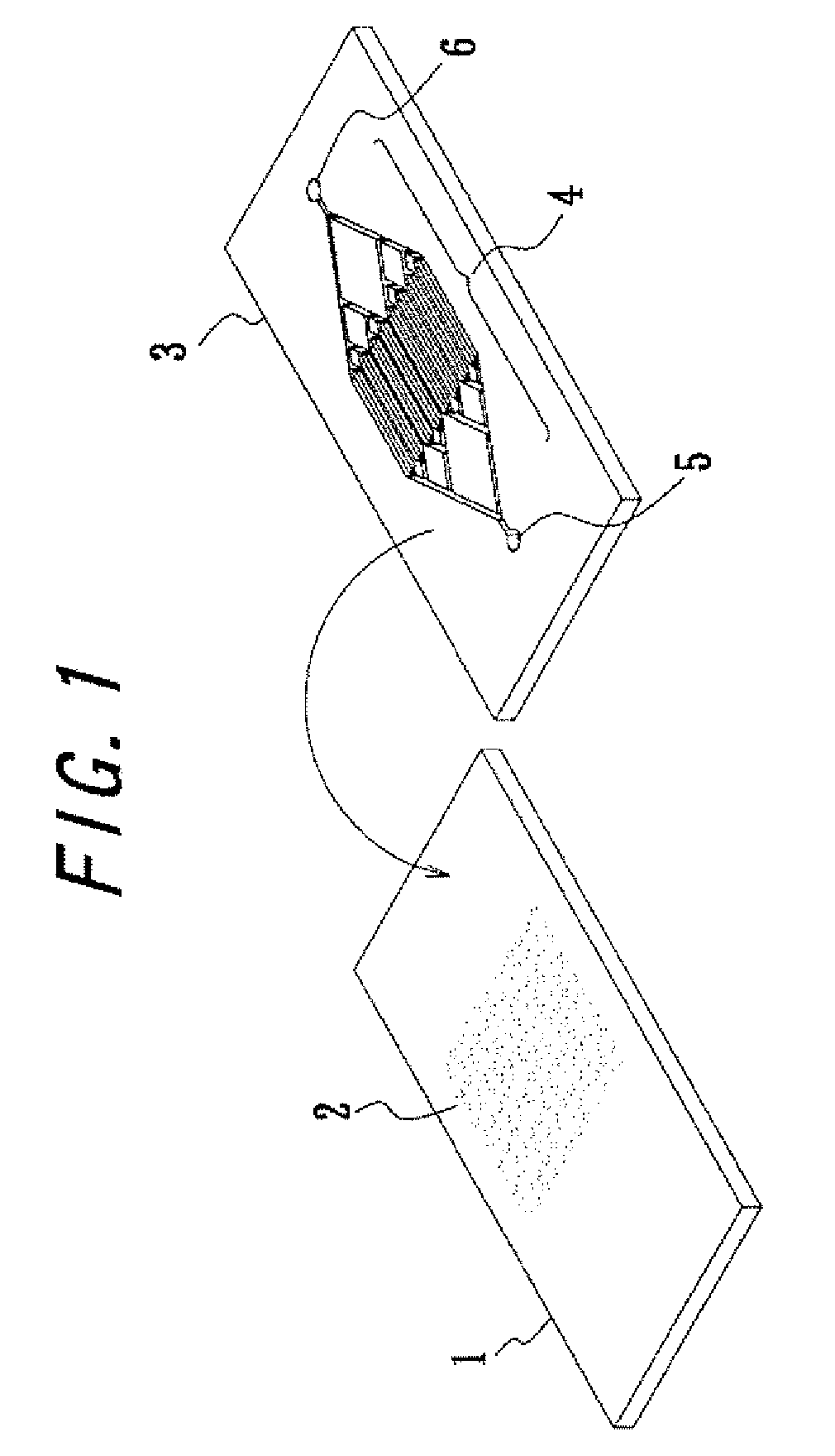
[0053] At first, about 0.45 μg of mycoplasma (MP) antigen (DENKA SEIKEN Co., Ltd.) was sprayed on the substrate using an electrospray deposition device (manufactured by Fuence). At conducting the spray, a glass mask having a slit (width 200 μm, length 12 mm) was used. By using the mask, antigens were deposited on the substrate in the shape of thin linear figure. The substrate was set with a flow channel made of polydimethylsiloxane havin...

example 2

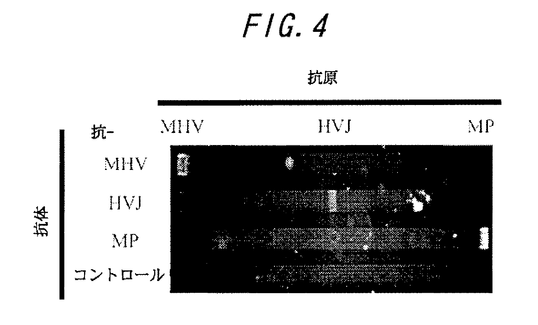
[0059] Cross reactivity was determined to examine on the cross contamination. In the same manner as example 1, antigens of pneumonia virus of Mice (MHV) (DENKA SEIKEN Co., Ltd.), Sendai virus (HVJ), and mycoplasma (MP) were sprayed by an electrospray deposition device. Thereafter anti-pneumonia virus of Mice (MHV) antibody, anti-Sendai virus (HVJ) antibody, and anti-mycoplasma (MP) antibody (DENKA SEIKEN Co., Ltd.) derived from mouse were passed through each flow channels as the primary antibodies, and the antigen antibody reactions were conducted. Then the amounts of antigens bound on the substrate were detected using anti-mouse antibody labeled with Alexa Fluor 488 as the secondary antibody. At the same time, a flow channel without flowing a primary antibody was set as a control. The results are shown in FIG. 4. According to the results, the non-specific binding of the secondary antibody was not observed in the control. On the other hand, it was revealed that respective antibodies...

example 3

[0060] Experiments on the actual samples were conducted using serum collected from mouse, The micro flow channel chip was sprayed and immobilized with the antigens of pneumonia virus of Mice (MHV) (DENKA SEIKEN Co., Ltd.), Sendai virus (HVJ), and mycoplasma (MP) using an electrospray deposition device. The test sample to be subjected to microorganism monitoring was diluted tenfold and 10 μl of the diluted sample was passed through the flow channels, then a labeled anti-mouse antibody was subjected for detection. As the result, antibody against pneumonia virus of Mice was detected in the test sample. Therefore it is assumed that the mouse is infected or has an infected record by pneumonia virus of Mice.


PUM

PropertyMeasurementUnit
volumeaaaaaaaaaa
lengthaaaaaaaaaa
widthaaaaaaaaaa

Abstract

This invention provides a method to monitor a microorganism that causes infectious disease of a laboratory animal by using a micro flow channel chip immobilized with a molecular to be tested such as an antigen or an antibody of the microorganism that causes infectious disease, the method comprises flowing serum or body fluid taken from the laboratory animal through the minute flow channel of the micro flow channel chip and detecting the antigen antibody reaction on the chip. The method of this invention enabled medical inspection of an infectious disease of a laboratory animal and microorganism monitoring of a laboratory animal, by using minute amount of animal serum or body fluid in a closed system rapidly and sensitively.

Description

BACKGROUND OF THE INVENTION [0001] 1. Field of the Invention [0002] This invention relates to a method of monitoring a microorganism that causes infectious disease of a laboratory animal using a micro flow channel chip. According to the method of the invention, pathogenic microorganism that causes infectious disease of a laboratory animal can be detected quickly and sensitively in a closed system, using a trace amount of animal serum or body fluid. [0003] 2. Related Art [0004] When an experimenter is conducting experiments by handling animals, there is a danger that a pathogen harmful for human may be lurking in the experimental animals and the experimenter may be infected by the pathogen. It is possible that an experimental animal may die except for experimental handling because of the pathogen existing in the experimental animal. Moreover, it is possible that an experimental animal may be in the latency period of a pathogenic disease. In these eases, the reliability of the animal ...

Claims


Application Information

Patent Timeline
Patent Type & Authority Applications(United States)
IPC IPC(8): C12Q1/70G01N33/53C12Q1/04G01N33/543G01N33/569G01N37/00
CPCC12Q1/04G01N33/569G01N33/54366
Inventor NAGAMUNE, TERUYUKITAJIMA, AKIHIKOYAMAGATA, YUTAKAAOKI, HIROYOSHI
Owner NAGAMUNE TERUYUKI
Why Eureka
Try Eureka