Detection of nucleic acid differences using combined endonuclease cleavage and ligation reactions

a nucleic acid and endonuclease technology, applied in the field of nucleic acid differences, can solve the problems of inability to accurately discriminate, lack of sensitivity to be an efficient technique, and inability to identify additional low frequency mutations with clinical significance, etc., to achieve the effect of broad application and increase the throughput capacity

Inactive Publication Date: 2008-01-03
CORNELL RES FOUNDATION INC
View PDF5 Cites 2 Cited by
  • Summary
  • Abstract
  • Description
  • Claims
  • Application Information

AI Technical Summary

Benefits of technology

[0020] The present invention has proven effective in identifying unknown frameshift, nonsense, and missense mutations as demonstrated in tests screening for various mutations in six genes associated with the development of inherited or sporadic cancers. Furthermore, paired germline DNA can be evaluated side-by-side with tumor DNA, allowing this invention to even distinguish missense mutations from nearby or adjacent inherited polymorphisms. The assay also has broad applicability with respect to types of sequence variations that can be detected, and can identify 98% of the typical mutations or polymorphisms found in the human genome. In addition, it is capable of scanning for mutations in a region as long as 1.7 kb or greater. The present invention can detect mutations in a high background of normal sequence. The present invention can detect a mutation or polymorphism in a 1:20 dilution with wild-type DNA and can even detect mutations / polymorphisms in cases as low as 1:50 dilution with wild-type DNA. This high sensitivity makes the present invention amenable to pooled samples and, therefore, significantly increasing its throughput capabilities.

Problems solved by technology
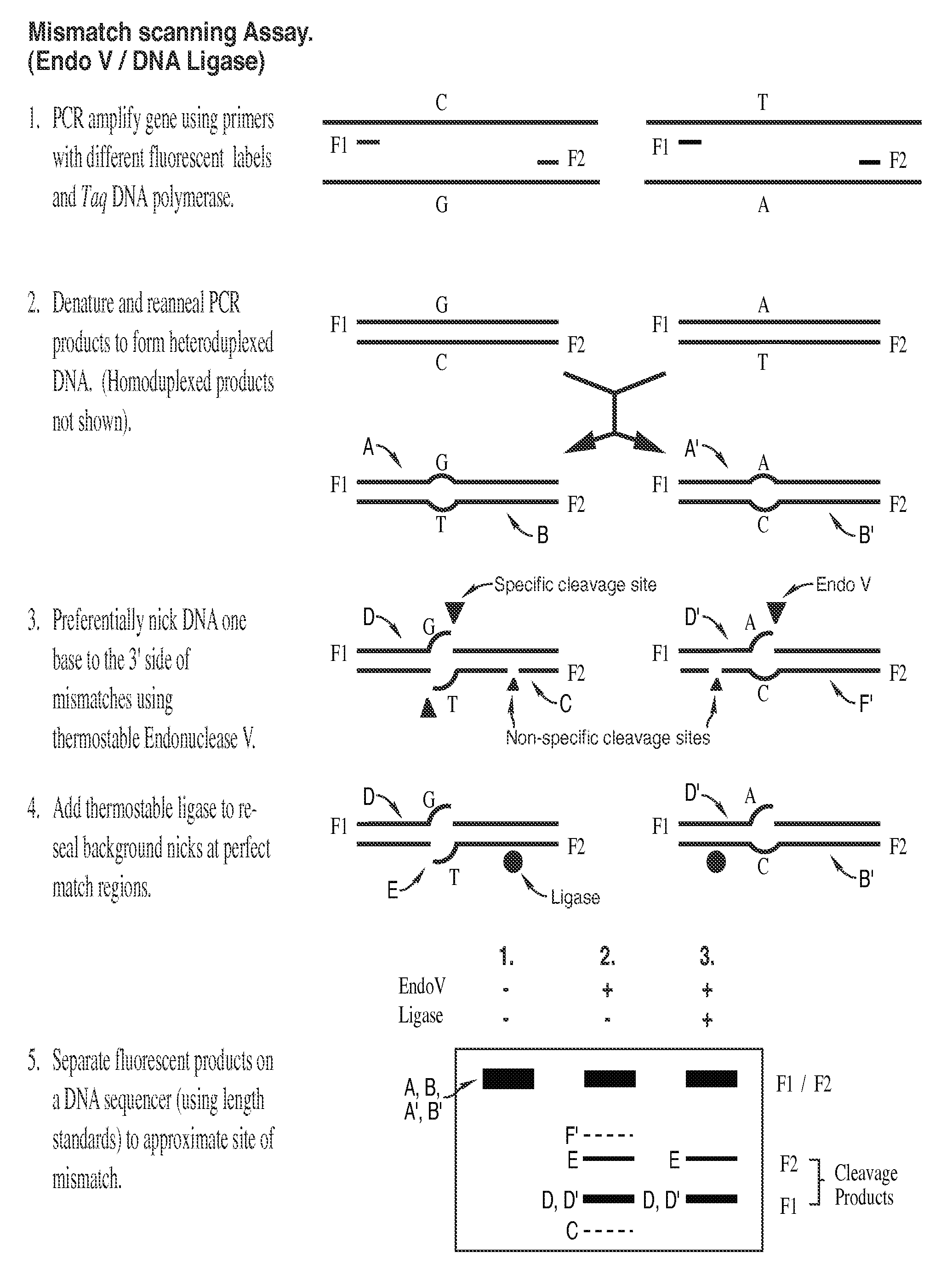
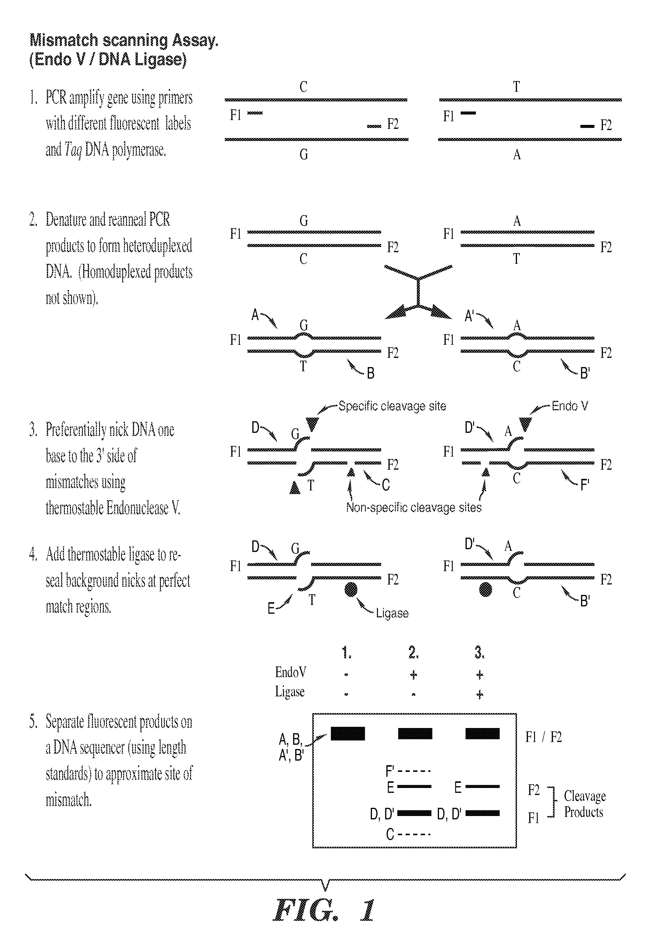
Identifying unknown mutations in clinical samples presents similar difficulties as screening for known mutations, as well as some novel complications.
Unfortunately, present methods of identifying additional low frequency mutations with clinical significance are restricted in their applications.
Most methods to date lack either the accuracy to discriminate or the sensitivity to be an efficient technique.
This method has low sensitivity which prevents an accurate analysis of pooled samples, and, in regard to solid tumor samples, the technique is vulnerable to stromal contamination.
In blinded studies, direct sequencing was unable to identify 20% of K-ras mutations in microdissected tumor samples.
These results reflect the limits of sensitivity associated with direct sequencing and further demonstrate the difficulty of automating this approach.
Despite the high scanning capacity levels, this approach has some characteristics that limits its utility.
VDA is unable to detect all mutations and has particular difficulty in detecting frameshift mutations, for example in the BRCA2 and p53 genes.
Primer extension arrays also fail to detect slippage of mononucleotide repeat sequences.
One consequence of direct hybridization that may account for these inaccurate results involves the disruption of secondary structure.
Since a secondary structure is usually energetically unfavorable with respect to fragment / array hybridizations, the variant fragment may bind the perfect match complement on the array with higher binding affinity than the true perfect match fragment.
Such illegitimate hybridizations would produce a false positive signal.
Direct hybridization of mutation-containing PCR fragments to sequences on the array has the additional difficulty of simultaneously assaying sequence tracts with localized regions of both high G+C and A+T content.
Certain mutations within these tracts can significantly decrease the Tm and thus lead to false negative signals.
Although these techniques contain some very desirable characteristics, none of them are complete with respect to both previously-discussed throughput and sensitivity.
The rest of these techniques tend to be rapid and can detect low level mutations, but they cannot distinguish missense from silent polymorphisms.
In addition, since these methods do not locate the position of the mutation, they are less compatible with follow up techniques such as direct sequencing.
This is currently the most robust approach for finding mutations which generate truncated proteins in large genes; however, it does not detect missense mutations or polymorphisms.
These enzymatic cleavage approaches identify the approximate position of most polymorphisms; however, these enzymes often nick matched DNA causing a high background noise.
This high background tends to limit their usefulness with respect to solid tumor studies.

Method used


Image

  • Detection of nucleic acid differences using combined endonuclease cleavage and ligation reactions
  • Detection of nucleic acid differences using combined endonuclease cleavage and ligation reactions
  • Detection of nucleic acid differences using combined endonuclease cleavage and ligation reactions

Examples


example 1

Reagents, Media, and Strains

[0084] All routine chemical reagents were purchased from Sigma Chemicals (St. Louis, Mo.) or Fisher Scientific (Fair Lawn, N.J.). deoxynucleotide, BSA, and ATP were purchased from Boehringer-Mannheim (Indianapolis, Ind.). Deoxyoligonucleotides were ordered from Integrated DNA Technologies Inc. (Coralville, Iowa). HiTrap SP columns were purchased from Amersham-Pharmacia Biotech (Piscataway, N.J.).

[0085] Restriction enzymes, T4 DNA ligase and DNA polymerase I (Klenow fragment) were purchased from NewEngland Biolab (Beverly, Mass.). DNA sequencing kits, PCR kits, and GENESCAN-500 (TAMRA) Size Standard were purchased from Applied Biosystems Division of Perkin-Elmer Corporation (Foster City, Calif.). Pfu DNA polymerase, PCR buffer and TaqPlus Precision PCR kit were purchased from Stratagene (La Jolla, Calif.). Protein assay kit was obtained from Bio-Rad (Hercules, Calif.).

[0086] FB medium (one liter) consisted of 25 gram Bacto tryptone, 7.5 gram yeast extra...

example 2

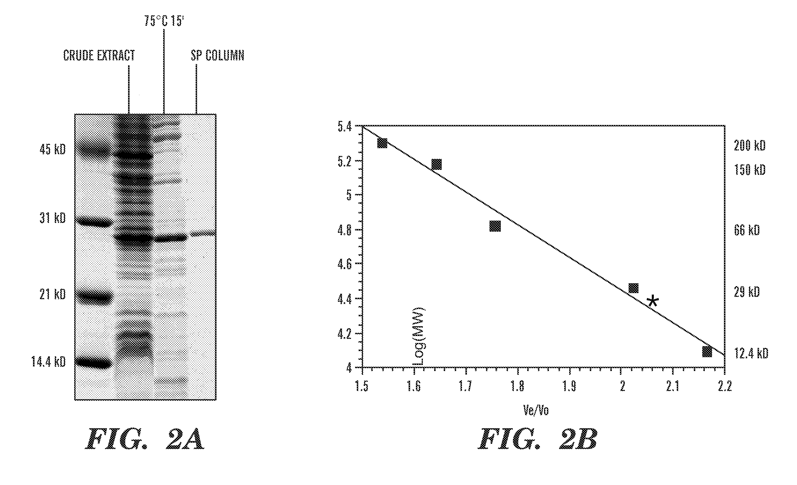
Plasmid Construction, Cloning, Expression, and Purification of Thermotoga maritima Endonuclease V

[0089] Through BLAST searches (Altschul, S. F., et al., J. Mol. Biol., 215(3):403-10 (1990), which is hereby incorporated by reference), a putative open reading frame of 225 amino acid has been identified in the Thermotoga maritima genome that shows 34% sequence identity to the E. coli endonuclease V gene. To prove that this, Tma ORF indeed encodes an endonuclease V, it was cloned and overexpressed in E. coli.

[0090] The putative endonuclease V gene (nfi) from Thermotoga maritima was amplified by PCR using forward primer EV.Tma.01A (5′ GGA GGG AAT CATATG GAT TAC AGG CAG CTT CAC A 3′ (SEQ. ID. No. 3), the NdeI site is underlined) and reverse primer EV.Tma.02R (5′ GCG CCT GGA TCCACT AGT TCA GAA AAG GCC TTT TTT GAG CCG T 3′ (SEQ. ID. No. 4), the SpeI and BamHI sites are underlined). The PCR reaction mixture (100 μl) consisted of 50 ng of Thermotoga maritima genomic DNA, 10 μM of forward p...

example 3

Oligonucleotide Substrates Preparation

[0096]E. coli endonuclease V demonstrates high activity with double-stranded DNA strands containing deoxyinosine-deoxyinosine or deoxyinosine-base mismatch. Yao, M. and Kow, Y. W., J. Biol. Chem., 269(50):31390-96 (1994), which is hereby incorporated by reference. This general characteristic of endoV was used in order to functionally identify the purified Tma endoV enzyme. A double-stranded oligonucleotide containing a base mismatch was designed as a substrate to monitor for cleavage activity by the purified enzyme. FIG. 3A shows a simple assay system using two differentially labeled fluorescent oligonucleotides. The top strand is labeled with 6-FAM and the bottom strand is TET labeled. The mismatch position of the deoxyinosine nucleotide is off-center so that nicked products do not comigrate on a denaturing polyacrylamide gel. The differential double labeling allows the nicking events on both strands to be easily observed and distinguished on ...


PUM

PropertyMeasurementUnit
pHaaaaaaaaaa
Tmaaaaaaaaaa
temperatureaaaaaaaaaa

Abstract

The present invention is a method for detecting DNA sequence differences including single nucleotide mutations or polymorphisms, one or more nucleotide insertions, and one or more nucleotide deletions. Labeled heteroduplex PCR fragments containing base mismatches are prepared. Endonuclease cleaves the heteroduplex PCR fragments both at the position containing the variation (one or more mismatched bases) and to a lesser extent, at non-variant (perfectly matched) positions. Ligation of the cleavage products with a DNA ligase corrects non-variant cleavages and thus substantially reduces background. This is then followed by a detection step in which the reaction products are detected, and the position of the sequence variations are determined.

Description

[0001] This application is a divisional of U.S. patent application Ser. No. 09 / 998,481, filed Nov. 30, 2001, which claims benefit of U.S. Provisional Patent Application Ser. No. 60 / 250,435, filed Dec. 1, 2000, which are hereby incorporated by reference in their entirety.[0002] The present invention arose from research sponsored by the National Institutes of Health under Grant Nos. ROI-CA 65930 and ROI-CA 81467. The United States Government may have certain rights.FIELD OF THE INVENTION [0003] The present invention relates to detecting nucleic acid differences using combined endonuclease (“endo”) cleavage and ligation reactions. BACKGROUND OF THE INVENTION Detection of DNA Sequence Variation [0004] There is a great need in both basic and clinical research to identify DNA sequence variations with high efficiency and accuracy. The current techniques for detection of such variation can be divided into two groups: 1) detection of known mutations or polymorphisms and 2) detection of unkn...

Claims


Application Information

Patent Timeline
Patent Type & Authority Applications(United States)
IPC IPC(8): C12Q1/68C12N9/14G01N33/53C12N9/16C12N15/09C12Q1/683G01N33/566
CPCC12Q1/683C12Q2531/113C12Q2521/501C12Q2521/301
Inventor BARANY, FRANCISCAO, WEIGUOHUANG, JIANMINLU, JING
Owner CORNELL RES FOUNDATION INC
Why Eureka
Try Eureka