Extracardiac Blood Flow Amplification Device

a technology of blood flow amplification and extracardiac vein, which is applied in the direction of blood vessels, prostheses, therapy, etc., can solve the problems of poor blood circulation in the leg arteries, poor renal failure or hypertension, poor blood circulation in the leg, etc., and achieves the effect of increasing the blood flow through the blood vessel, reducing the pressure in the second chamber, and improving the blood pressure of the aortic blood

Inactive Publication Date: 2008-09-25
RAINBOW MEDICAL LTD
View PDF47 Cites 6 Cited by
  • Summary
  • Abstract
  • Description
  • Claims
  • Application Information

AI Technical Summary

Benefits of technology

[0048]During a systolic phase of operation of the implanted device, the increased systolic pressure of the arterial blood is communicated to the pressure-transmitting fluid. This increased pressure is applied to the second surface, expanding the second chamber, and applying force to the common surface, thereby storing potential energy in the source of the elastically-derived force. Motion of the common surface expands the first chamber, opening the valve and drawing blood into the first chamber and the base chamber. During a subsequent diastolic phase of operation, the decreased diastolic pressure of the arterial blood is communicated to the pressure-transmitting fluid, resulting in lower pressure in the second chamber. The common surface converts the stored potential energy to kinetic energy, applying a force to both the first and second surfaces, thereby contracting the chambers. The valve closes, and the first chamber expels the blood downstream. The device thus increases blood flow through the blood vessel.
[0049]In some embodiments of the present invention, a diastolic heart failure treatment system comprises a blood flow regulation device. The device is adapted to be coupled in pressure communication with an artery of a subject, typically a descending thoracic aorta, and in fluid communication with a left atrium of the subject. The device is configured to utilize the increased blood pressure of aortic blood during systole to suck atrial blood from the left atrium into the device. The sucking of atrial blood from the left atrium lowers the blood pressure in the left atrium, thereby increasing blood flow from the pulmonary veins into the left atrium. This increased blood flow reduces backing up of blood in the lungs, and typically serves to treat diastolic heart failure.

Problems solved by technology

Renal failure or hypertension is caused by insufficient blood flow to the kidneys.
Claudication results from poor circulation of blood in the leg arteries, typically caused by atherosclerosis as a result of smoking, diabetes, or high cholesterol.
In some cases, particularly in diabetes, poor blood circulation in the leg results in amputation.
Retinal vessel occlusion (retinal artery or vein occlusion) often results in vision loss.
Decreased cardiac output to the systemic circulation typically increases venous blood pressure, which often leads to blood backing up in the lungs.
Low cardiac output also results in decreased blood perfusion to organs, such as the liver, kidney, brain, and heart itself.
Over time, the effects of heart failure contribute to a worsening of the condition.
Reduced blood supply to the heart causes less effective contraction of the heart.
To compensate, the heart attempts to increase output by increasing muscle strength, which leads to myocardial hypertrophy (enlargement of the heart with thickening and stiffening of the heart wall).
These conditions in turn lead to reduced cardiac output, resulting in a vicious cycle.
Systolic heart failure is characterized by a deficiency in systolic heart function, which causes insufficient expulsion of blood during systole.
Diastolic heart failure is characterized by a deficiency in diastolic heart function, which causes insufficient ventricular filling during diastole.

Method used


Image

  • Extracardiac Blood Flow Amplification Device
  • Extracardiac Blood Flow Amplification Device
  • Extracardiac Blood Flow Amplification Device

Examples


Embodiment Construction

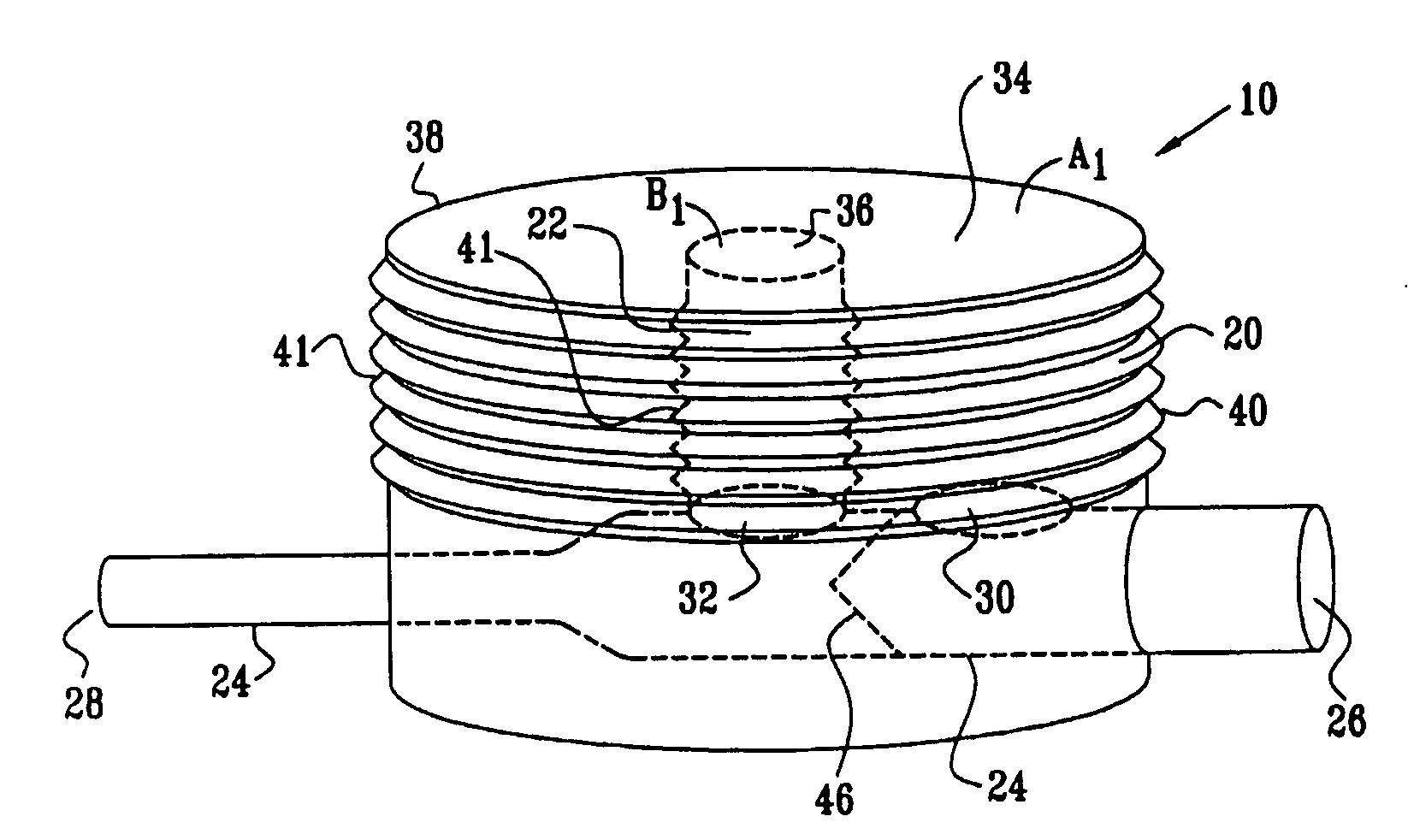
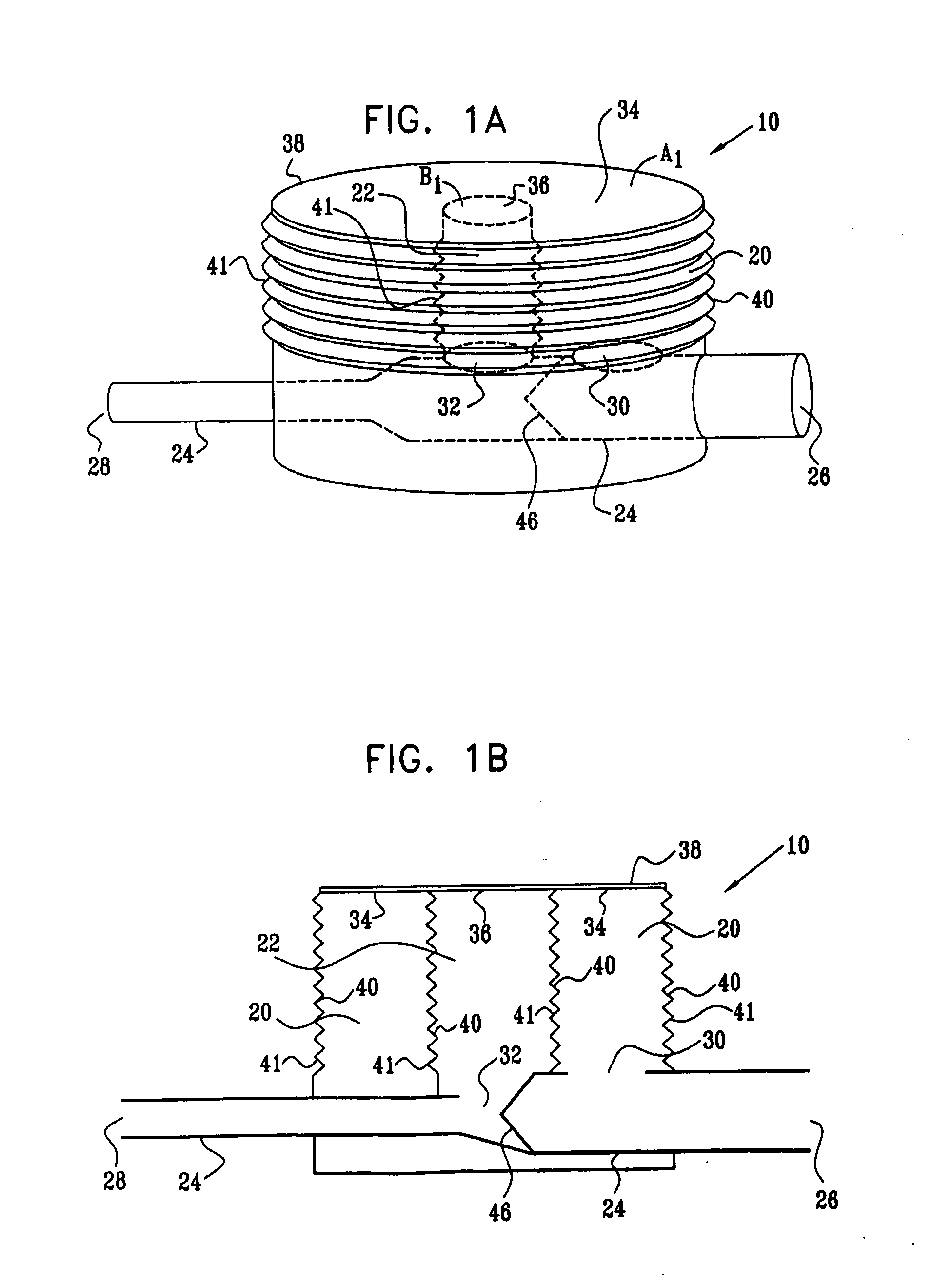
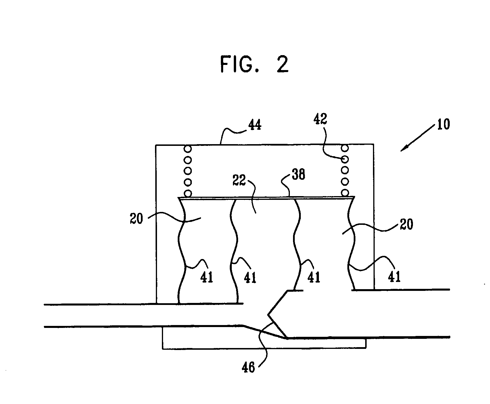
[0159]FIGS. 1A and 1B are a pictorial view and a schematic cross-sectional view, respectively, of an extracardiac fully-implantable blood flow amplification device 10, in accordance with an embodiment of the present invention. Device 10 comprises a first flexible chamber 20, a second flexible chamber 22, and a lumen 24. The lumen is adapted to be coupled to an artery (not shown) of a subject, by coupling a proximal inflow end 26 of the lumen to a first site of the artery, and a distal outflow end 28 of the lumen to a second site of the artery, the second site distal to the first site with respect to blood circulation. Typically, but not necessarily, a diameter of outflow end 28 is less than a diameter of inflow end 26. For example, the diameter of outflow end 28 may be between about 50% and about 80% of the diameter of inflow end 26.

[0160]In the embodiment shown in FIGS. 1A and 1B, the chambers are generally cylindrically shaped, and second chamber 22 is shown surrounded by first ch...


PUM


Abstract

Apparatus (10) is provided including a first chamber (20) and a second chamber (22), adapted to be in fluid communication with a first volume and a second volume of oxygenated blood of a subject, respectively, the first chamber (20) and the second chamber (22) having a first surface (34) and a second surface (36), respectively. The apparatus (10) further includes a third surface (38) adapted to apply an elastically-derived force, at least a first portion of the third surface (38) in mechanical communication with the first surface (34), and at least a second portion of the third surface (38) in mechanical communication with the second surface (36) during at least a portion of a cardiac cycle. A pressure-sensitive valve (46) is coupled between the first chamber (20) and the second chamber (22), the valve (46) adapted: (a) to be in an open position during at least a portion of systole, such that the first chamber (20) is in fluid communication with the second chamber (22) and the first volume is in fluid communication with the second volume; and (b) to be in a substantially closed position during diastole, such that the first chamber (20) is substantially not in fluid communication with the second chamber (22) and the first volume is substantially not in fluid communication with the second volume. Other embodiments are also described.

Description

CROSS-REFERENCES TO RELATED APPLICATIONS[0001]This application claims priority from U.S. Provisional Patent Application 60 / 543,700, filed Feb. 10, 2004, entitled, “Extracardiac blood flow amplification device,” which is assigned to the assignee of the present application and is incorporated herein by reference.FIELD OF THE INVENTION[0002]The present invention relates generally to implantable medical devices, and specifically to methods and apparatus for implantable devices for treating conditions caused by insufficient blood circulation.BACKGROUND OF THE INVENTION[0003]Numerous medical conditions are caused by insufficient blood flow to certain tissues. For example, angina and myocardial infarction are caused by insufficient blood flow to cardiac muscle, and ischemic stroke is caused by insufficient blood flow to the brain. Renal failure or hypertension is caused by insufficient blood flow to the kidneys. Claudication results from poor circulation of blood in the leg arteries, typic...

Claims


Application Information

Patent Timeline
Patent Type & Authority Applications(United States)
IPC IPC(8): A61M1/10A61M60/135A61M60/268A61M60/34A61M60/435A61M60/894
CPCA61F2/02A61F2002/068A61M1/122A61M1/1072A61M1/1037A61M60/268A61M60/135A61M60/894A61M60/148A61M60/34A61M60/435
Inventor GROSS
Owner RAINBOW MEDICAL LTD
Why Eureka
Try Eureka