

Polymeric tissue sealant
a tissue sealant and polymer technology, applied in the field of biomaterials and precursor molecules, can solve the problems of increased trauma to surrounding tissue, mechanical tissue fasteners that require significant skill, and mechanical tissue fasteners that suffer from a variety of limitations, and achieve good mechanical strength, reduce or prevent the leakage of cerebrospinal fluid
- Summary
- Abstract
- Description
- Claims
- Application Information
AI Technical Summary
Benefits of technology
Problems solved by technology
Method used
Image

Examples
example 1
Tissue Sealant Compositions
[0150]1a. Composition 1: Tetronic-Tetraacrylate and PEG-SH-10
First Precursor Molecule Solution
[0151]235 mg of poly(ethylene glycol) tetrasulfhydryl (“PEG-SH-10”) (mol. wt. 10 kD) and 0.1 mg of lissamin green were dissolved in 1 mL of 10 mM acetate buffer pH 5.
Second Precursor Molecule Solution
[0152]315 mg of tetronic-tetraacrylate (mol wt. 15 kD) were dissolved in 1 mL of a 10 mM acetate buffer pH 5.
Basic Solution
[0153]0.22 mL of a 50 mM borate buffer pH 9.8
1b: Composition 2: Tetronic-Tetraacrylate and PEG-SH-5
First Precursor Molecule Solution
[0154]112 mg of poly(ethylene glycol) tetrasulfhydryl (“PEG-SH-5”) (mol. wt. 5 kD) and 0.1 mg of lissamin green were dissolved in 1 mL of 10 mM acetate buffer pH 5.
Second Precursor Molecule Solution
[0155]315 mg of tetronic-tetraacrylate (mol wt. 15 kD) were dissolved in 1 mL of a 10 mM acetate buffer pH 5.
Basic Solution
[0156]0.22 mL of a 50 mM borate buffer pH 10.4
1c: Composition 3: Tetronic-Tetraacrylate and PEG-SH-5...
example 2
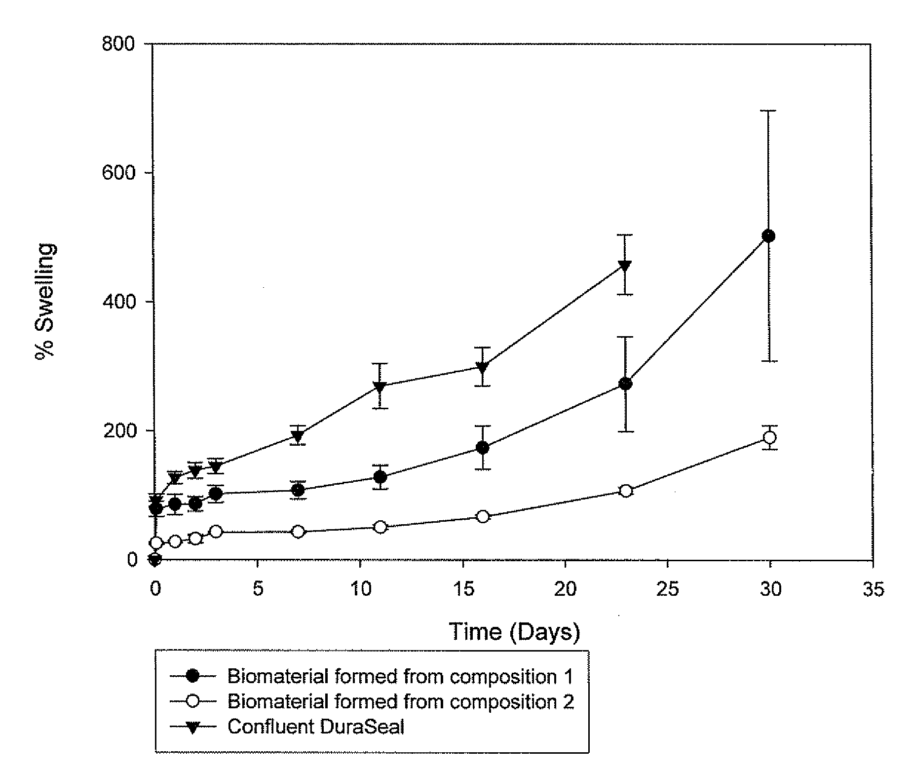
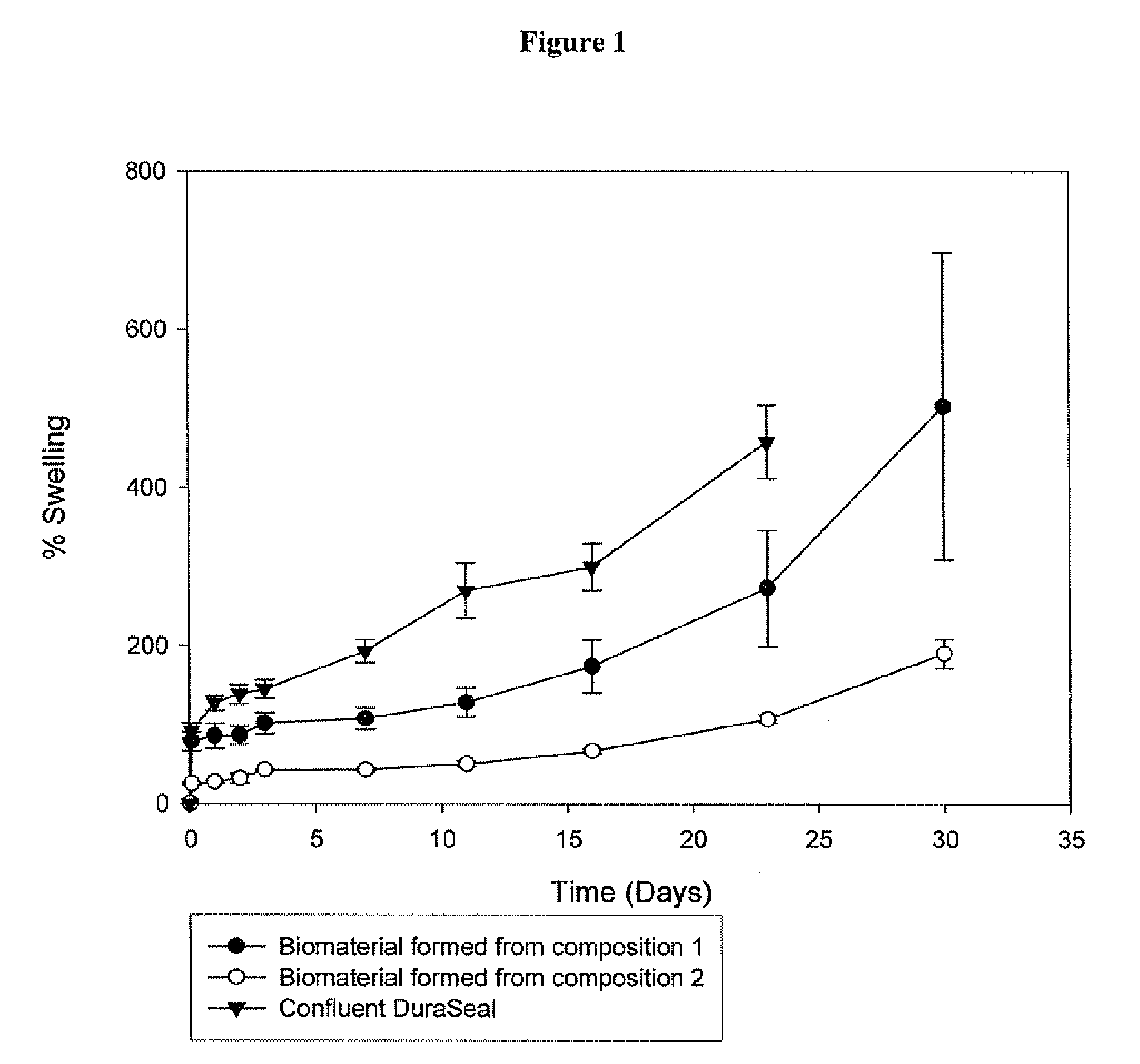
Preparation of the Biomaterial
[0191]2a. Preparation of the Biomaterial from Composition 1, 2, 3, 4 and 6
[0192]Before application of the pharmaceutical composition at the desired site, the first and second precursor molecule solutions were filled into two distinct syringes, which were connected with a coupler. The first and second precursor molecule solutions were mixed by transferring the material contained in one syringe to the other syringe (Typically, the solutions were pushed back and forward 10 times). Although, the mixture typically remains stable 10-20 minutes after its preparation, ideally the pharmaceutical composition should be used within 5 minutes after its preparation. The biomaterial was formed in situ at the desired site, by delivering to the defect site the mixture comprising the first and second precursor molecules and the activator using a two component device equipped either with a spreader tip or a sprayer tip. The biomaterial was formed in less than 1 minute aft...
example 3
Stability of the Mixture of the First and Second Precursor Molecules
First Precursor Molecule Solution
[0195]194 mg of poly(ethylene glycol) tetrasulfhydryl (“PEG-SH-5”) (mol. wt. 5 kD) were dissolved in 1 mL of 10 mM acetate buffer pH 4.9. The buffer was prepared by mixing a 100 mM acetic acid buffer and a 100 mM sodium acetate buffer to achieve pH 4.90 and diluting the buffer 1:10 v / v with water.
Second Precursor Molecule Solution
[0196]545 mg of tetronic-tetraacrylate (mol wt. 15 kD) were dissolved in 2 mL of a 20 mM acetate buffer pH 4.9. The buffer was prepared by mixing a 100 mM acetic acid and a 100 mM sodium acetate buffer to achieve pH 4.90 and diluting the buffer 1:5 v / v with water.
Basic Solution
0.3 mL of a 250 mM Carbonate Buffer pH 11.0
[0197]When only the first and second precursor molecules were mixed by vortexing 30 s, gelation was occurring within 30 min. Gelation time was measured by dipping a needle in and out of the solution, the time was measured until threads were fo...
PUM
| Property | Measurement | Unit |
|---|---|---|
| composition | aaaaa | aaaaa |
| pH | aaaaa | aaaaa |
| physiological temperature | aaaaa | aaaaa |
Abstract
Description
Claims
Application Information