Chimeric Avian-Based Screening System Containing Mammalian Grafts

a screening system and avian technology, applied in the field of chimeric model systems, can solve the problems of high cost, poor screening effect, poor screening effect, etc., and achieve the effect of convenient and efficient system, and convenient and efficient system

Active Publication Date: 2009-03-05
BAR ILAN UNIV +1
View PDF1 Cites 28 Cited by
  • Summary
  • Abstract
  • Description
  • Claims
  • Application Information

AI Technical Summary

Benefits of technology

[0013]The present invention provides novel chimeric systems and screening methods utilizing mammalian grafts transplanted into avian embryos. It is now disclosed that chimeric avian embryos comprising diseased mammalian cells or tissues serve as a convenient, efficient system for screening therapeutic drugs or treatments. It is further disclosed that the chimeric avian embryos comprising normal or aberrant mammalian tissues provide a convenient and efficient system for screening potential disease causing agents, to establish their safety or lack thereof. It is yet further disclosed that the chimeric avian embryos comprising genetically altered normal or aberrant tissues may be used to screen genetic susceptibility to or methods of treatment of diseases and disorders.

Problems solved by technology

Such animals, for example severe combined immune deficient (SCID) mice, are expensive, sickly and hard to maintain.
Because the study of human skin in vivo is limited for ethical and medical reasons, most current strategies for the testing of therapies for treating skin damage and disease involve the extensive use of experimental animals.
Animal testing often entails great expense, and involves many legal and ethical issues.
These models therefore lack important features of skin physiology, such as circulating blood cells that can participate in inflammation and in wound healing.
Furthermore, most in-vitro systems require that the epidermis be submerged in medium rather than exposed to air, precluding the possibility of using these systems to conduct experiments with topical treatments such as detergents, cosmetics, radiation, or topically applied pharmaceutical compositions.
Roux et al 2003A do not suggest the use of the CAM system to investigate wound healing in conjunction with topically applied factors, and indeed the lack of an epidermis-air interface could preclude such studies.
The fetal sheep or dog models are not practical for studying the biology of blood malignancies or for drug screening, because of the extremely high cost in money and space for the experiments.

Method used


Image

  • Chimeric Avian-Based Screening System Containing Mammalian Grafts
  • Chimeric Avian-Based Screening System Containing Mammalian Grafts
  • Chimeric Avian-Based Screening System Containing Mammalian Grafts

Examples


example 1

Culturing of Human Skin Explants on the Chorioallantoic Membrane (CAM) of a Fertilized Avian Egg

[0300]Materials and Methods:

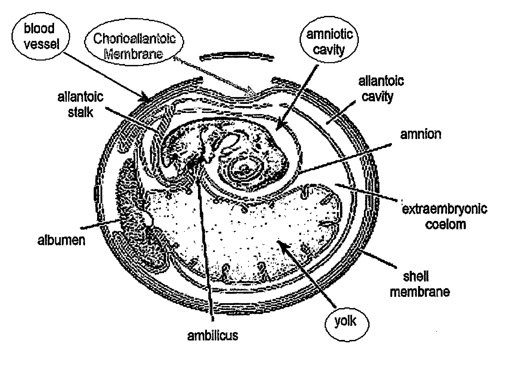
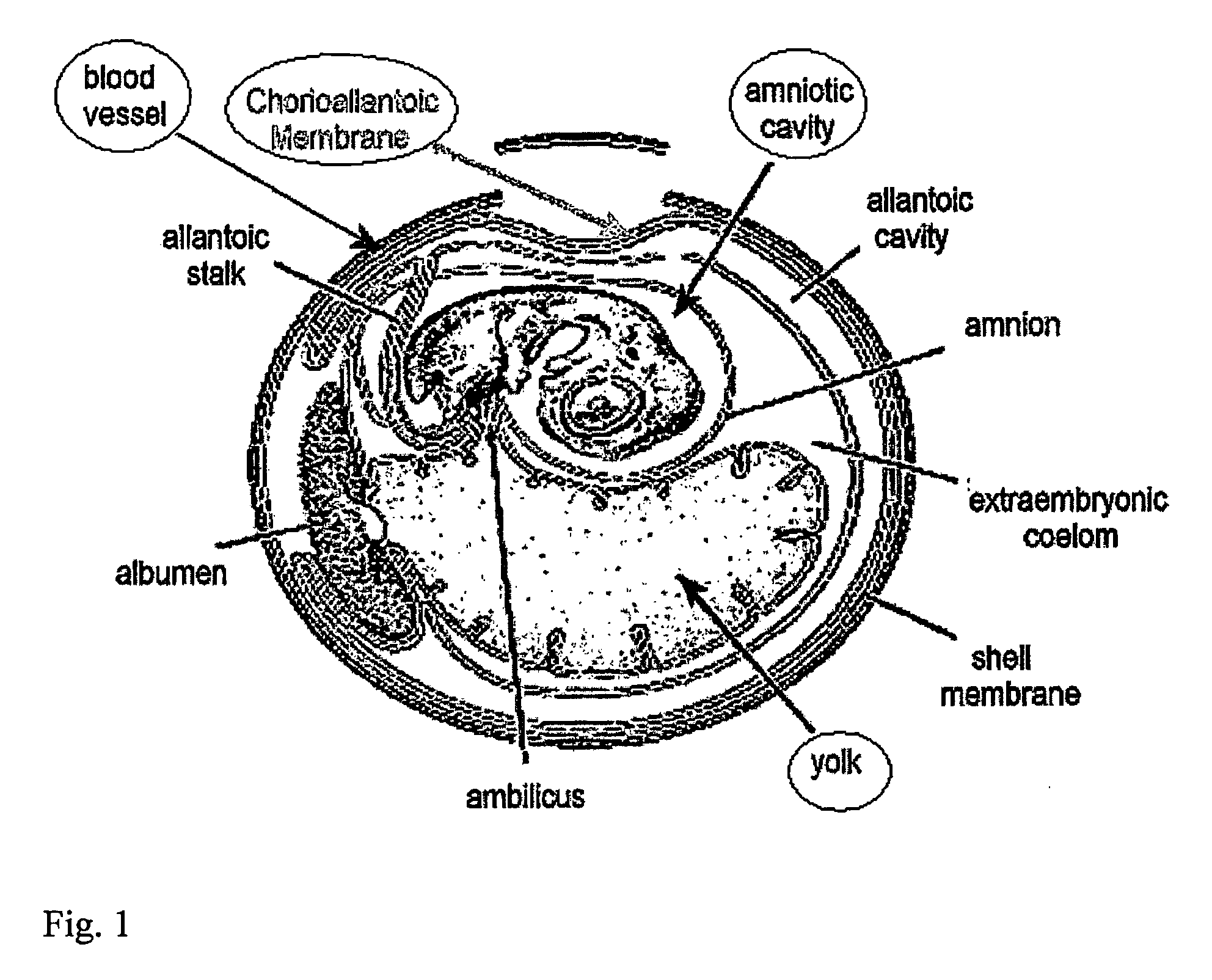
[0301]Eggs: Freshly-laid fertile chicken eggs were obtained from a local farm. Eggs were stored at 15° C. until required, warmed for one hour to room temperature, and then incubated vertically with the point down in a humid atmosphere at 37° C. for 5-10 days before use. On the third day of incubation, the eggs were turned upside down, and a small hole was made in the sharp side of the egg after cleaning with a tissue impregnated with 70% ethanol. This created an artificial air sac so that the CAM can be accessed later on without causing bleeding.

[0302]Human Skin: Human skin that is normally discarded from mastectomies and abdominal surgery was obtained from the Shaarei Zedek hospital in Jerusalem and Tel Aviv Mediccal Center in Tel Aviv. Permission was obtained from the hospitals' Medial ethic committee according to the Helsinki accords. The skin was pinned out...

example 2

Human Skin Irritation can be Quantified Using the Skin-Cam System

[0312]Punch biopsies of human skin were grafted onto the CAM of a fertilized egg as described in Example 1. The human skin samples were allowed to incubate for 2 days after grafting in order to allow for the skin to incorporate, and then the adhesive tape sealing of the samples was reopened. The detergent sodium dodecylsulfate (SDS) was applied to different skin samples at different concentrations topically using a small plastic ring cut from a pipette tip keep the irritant in place. The skin samples were then resealed and returned to the incubator for an addition three days, whereupon they underwent routine histological and immunochemical analysis using Abs for the proliferation marker PCNA. PCNA+ cells were counted in several sections, and the length of the epidermis in the section measured. Results are expressed as PCNA+ cells / mm.

[0313]As can be seen in FIG. 3A, SDS, a known irritant, significantly increases epiderm...

example 3

Wound Healing of Both Mouse and Human Skin on the CAM

[0315]Wound Healing of Mouse Skin

[0316]Mouse skin samples were cut into twenty-five millimeter square pieces, and puncture wounds were produced with a 2.5 mm dermatological biopsy punch. At this point, skin samples were grafted onto the CAM of a fertilized egg as described in Example 1. Up to three samples were grafted into each egg. The skin was then cultivated for an additional 5 days before harvesting for histological and immunocytochemical analysis.

[0317]Wound Healing of Human Skin

[0318]The above procedure was repeated for human skin.

[0319]Results

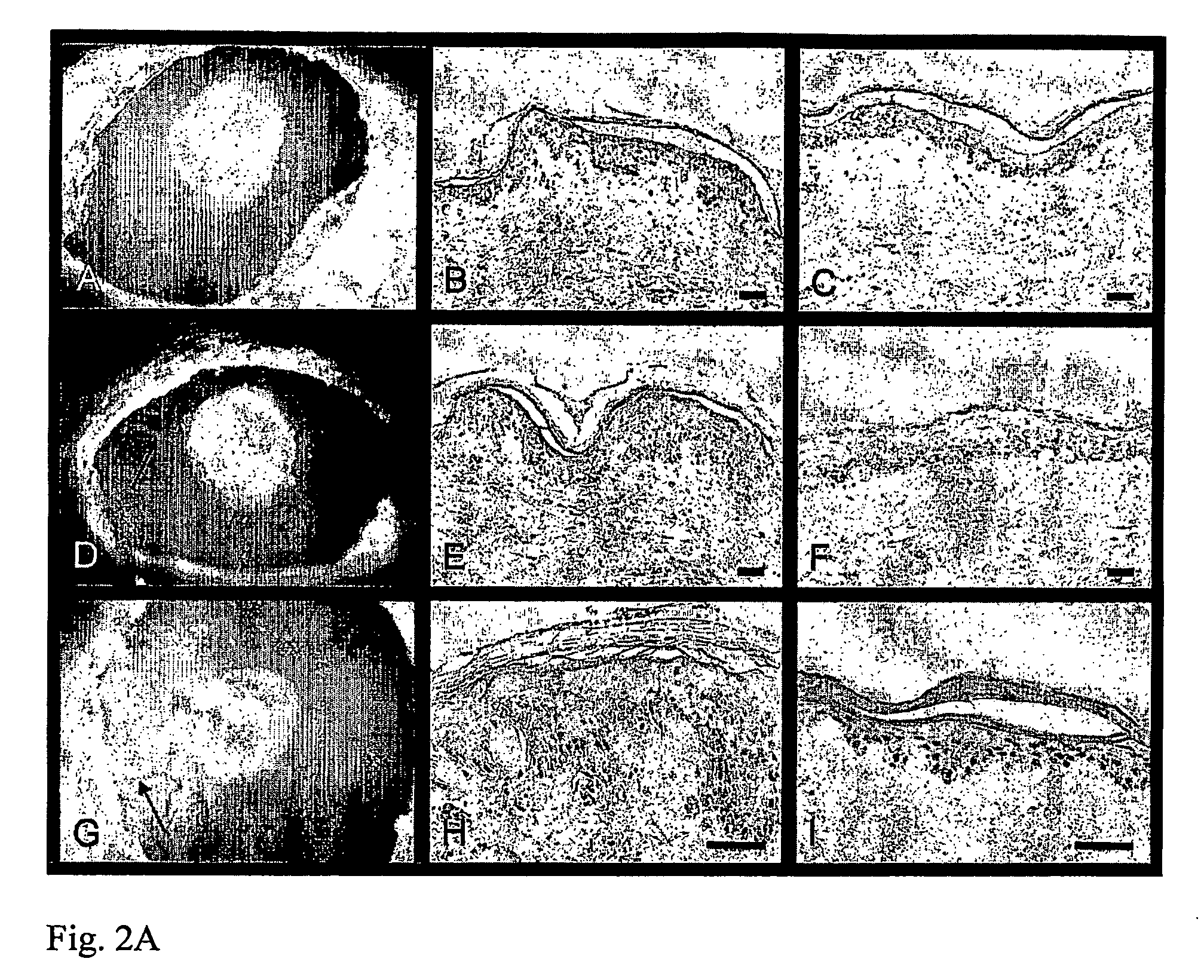
[0320]FIG. 4 illustrates wound healing in mouse and human skin grafted to the chick CAM. Panels A-D show hematoxylin and eosin-stained sections of mouse skin that had been cut with a 0.3 mm punch. In the low magnification images (panels A and B), the opening remaining after healing for 5 days is illustrated by the dashed lines. The boxed areas are enlarged in panels C and D, and the o...


PUM

PropertyMeasurementUnit
frequencyaaaaaaaaaa
timeaaaaaaaaaa
surface areaaaaaaaaaaa

Abstract

The present invention relates to animal model systems comprising a chimera between an avian embryo and a mammalian organism. Specifically, chimeric model systems comprising normal, diseased or genetically transformed mammalian cells and tissues transplanted into avian embryos, and uses thereof for in vivo testing of drugs and therapeutic modalities are disclosed.

Description

FIELD OF THE INVENTION[0001]The present invention relates to chimeric model systems comprising mammalian grafts transplanted into avian embryos, useful for in vivo testing of drugs and novel therapeutic modalities of human diseases and disorders.BACKGROUND OF THE INVENTION[0002]The embryonic chick is a widely used model for studying various biological processes. The low cost and easy availability of this system provide major advantages compared to in vivo systems utilizing sentient animals. In addition, many in vivo systems, particularly model systems for studying human tissues and organs, require transplanting an examined graft in an immune compromised animal, in order to avoid graft rejection. Such animals, for example severe combined immune deficient (SCID) mice, are expensive, sickly and hard to maintain. The development of simple, cost-effective systems for mammalian explants is therefore desirable.[0003]The skin is a complex three-dimensional organ that covers the exterior of ...

Claims


Application Information

Patent Timeline
Patent Type & Authority Applications(United States)
IPC IPC(8): A01K67/00C12Q1/02
CPCA01K67/0271A01K2227/105A01K2227/106A01K2227/30C12Q1/025C12N2799/022G01N33/5088G01N2500/00G01N2800/52A01K2267/03
Inventor GOLDSTEIN, RONTENNENBAUM, TAMARDEUTSCH, VARDA
Owner BAR ILAN UNIV
Why Eureka
Try Eureka