

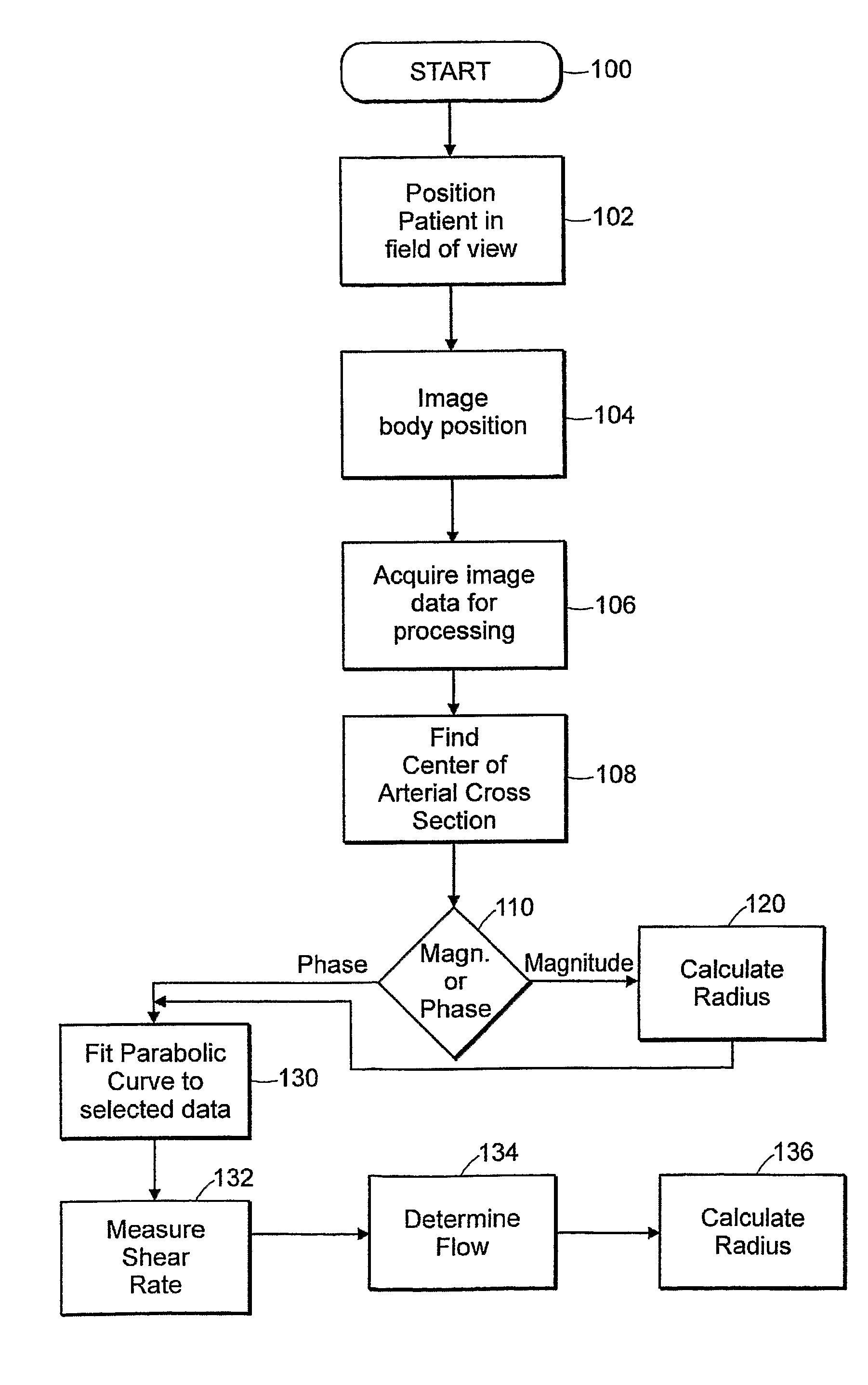
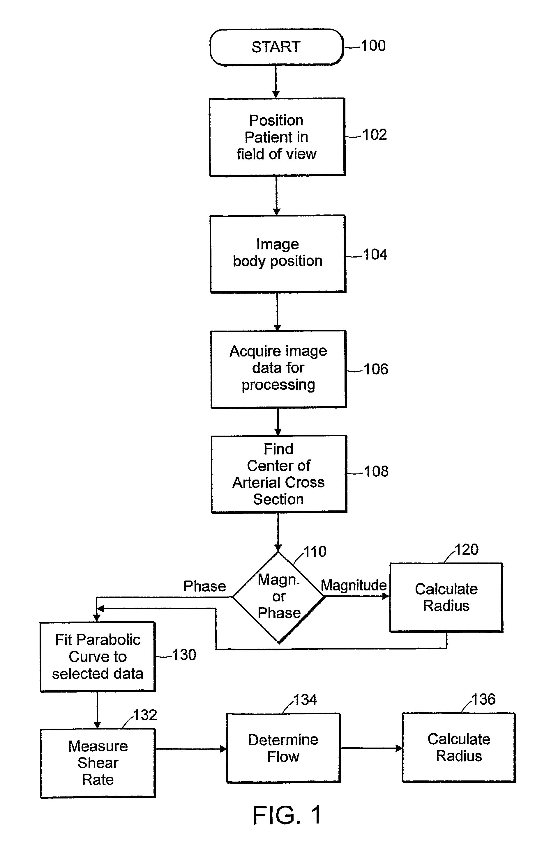
Method of Assessing Vascular Reactivity Using Magnetic Resonance Imaging, Applications Program and Media Embodying Same
a magnetic resonance imaging and vascular reactivity technology, applied in the field of noninvasive assessment of vascular reactivity, can solve the problems of large overlap in arterial dilatory response, inability to measure vascular endothelial function using ultrasound, and associated endothelial dysfunction with coronary events, etc., to achieve the effect of measuring low-flow mediated constriction, direct measurement of shear rate, and computational efficiency and precision
- Summary
- Abstract
- Description
- Claims
- Application Information
AI Technical Summary
Benefits of technology
Problems solved by technology
Method used
Image

Examples
example 1
Subjects
[0129]Thirty-three healthy subjects (16 men and 17 women), ages 20-41, with no cardiovascular risk factors including hypertension, diabetes, hyperlipidemia, smoking, obesity or cardiac disease in a first-degree relative were studied. No subject was acutely ill or was on any vasoactive medication. The study protocol was approved by the Institutional Review Board at the Johns Hopkins School of Medicine. All subjects gave written informed consent.
Study Protocol
[0130]Subjects abstained from eating or drinking except water for at least 6 hours before the study. Baseline blood pressure was recorded. PCMRI was performed using a 1.5T scanner (CV / i, General Electric Medical Systems, Milwaukee, Wis.) equipped with cardiac gradient coils (40 mT / m, 120 T / m / s). Electrocardiographic leads were placed on the thorax. A dual cardiac phased array receiving coil was placed anterior and posterior to the upper thigh. A sphygmomanometer cuff was placed on the lower thigh. Phase-contrast images we...
example 2
Methods
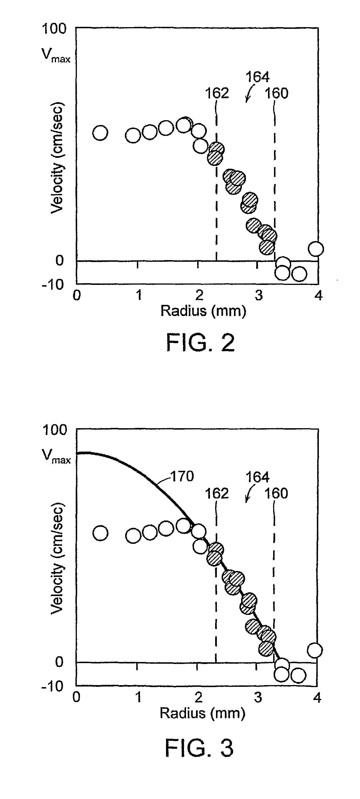
[0148]The superficial femoral artery was studied in 20 men—10 men with no risk factors aged 20-33 years, and 10 men with atherosclerotic risk factors including type II diabetics and older age, 52-69 years. A receiver coil was placed on the upper thigh, and an occluding cuff was placed distal to the receiver coil. A fixed cross-section of the artery was imaged before and during a distal occlusion. An ECG-gated PCMRI sequence was used, with 10 square cm field-of-view, matrix size 256×128, 3 mm slice thickness, VENC 60 cm / sec in the S / I direction, TR 11.4 msec, 8 views per segment, and temporal resolution 182 msec. A user-independent algorithm was employed to locate the precise center of the arterial cross-section in the magnitude image by optimizing the correlation of datapoints in a radial plot. A smoothed average of the optimized radial plot of the data was calculated. The radius of the artery was defined using the full-width, half-maximum approach (Hoogeveen R M, Bakker C J,...
example 3
[0151]In this study, we sought to determine shear-related aspects of femoral arterial reactivity that distinguish healthy men from men with vascular risk factors known to be associated with impaired endothelial function, including older age and type 2 diabetes.
Study Participants.
[0152]Twenty male subjects were studied. Ten were controls, aged 20-33, with no cardiovascular risk factors including hypertension, diabetes, hyperlipidemia, smoking, obesity or cardiac disease in a first-degree relative. Ten had risk factors for atherosclerosis including older age, 52-69 years, and type 2 diabetes. Risk factors excluded from the at-risk group were smoking, known vascular disease, and a body mass index greater than 33. None of the at-risk group had symptoms of claudication. No subject was acutely ill. The study protocol was approved by the Institutional Review Board at the Johns Hopkins School of Medicine. All subjects gave written informed consent.
Study Protocol
[0153]Subjects abstained from...
PUM
 Abstract
Description
Claims
Application Information