Use of ZnO Nanocrystals For Imaging and Therapy
a nanocrystal and nanotechnology, applied in the field of living tissue imaging, can solve the problems of limited application range in biological applications, use of fluorescence contrast agents, weak photostability,
- Summary
- Abstract
- Description
- Claims
- Application Information
AI Technical Summary
Problems solved by technology
Method used
Image


Examples
example 1
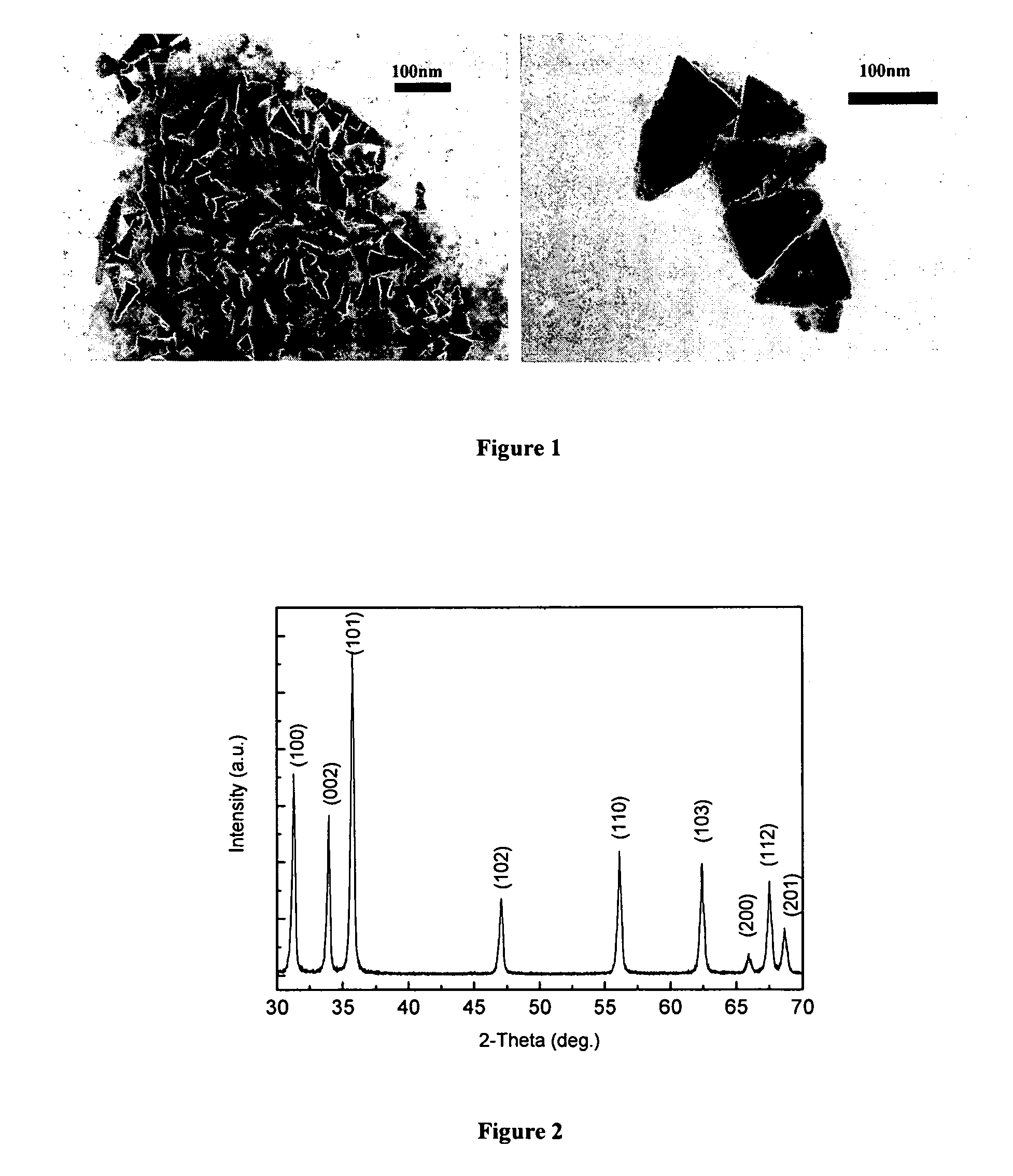
Preparation and Characterization of ZnO Nanocrystals
[0053]ZnO nanocrystals were synthesized using a non-hydrolytic sol-gel process based on the ester-elimination reaction between zinc acetate and 1,2-dodecanediol. The benzyl ester was selected as the solvent reagent because its high boiling point increased the synthesis temperature to 280 degrees Celsius, which provided high-quality samples with excellent size control, narrow size distribution, and uniform crystalline structure and dispersion properties. High (10 mmol) molar concentration of Zn acetate dehydrates results in supersaturation of the reaction solution and increases the yield of reaction.
Preparation of Aqueous Dispersions of ZnO Nanocrystals
[0054]The ZnO nanocrystals were stably dispersed in distilled water using phospholipid micelles. This aqueous dispersion was achieved by mixing a chloroform solution of ZnO nanocrystals (15 mg / mL, 100 μL) and DSPE-PEG(2000) methoxy (Avanti Polar Lipids) (20 mg / mL, 500 μL), followed by...
example 2
Imaging Using ZnO Nanocrystals
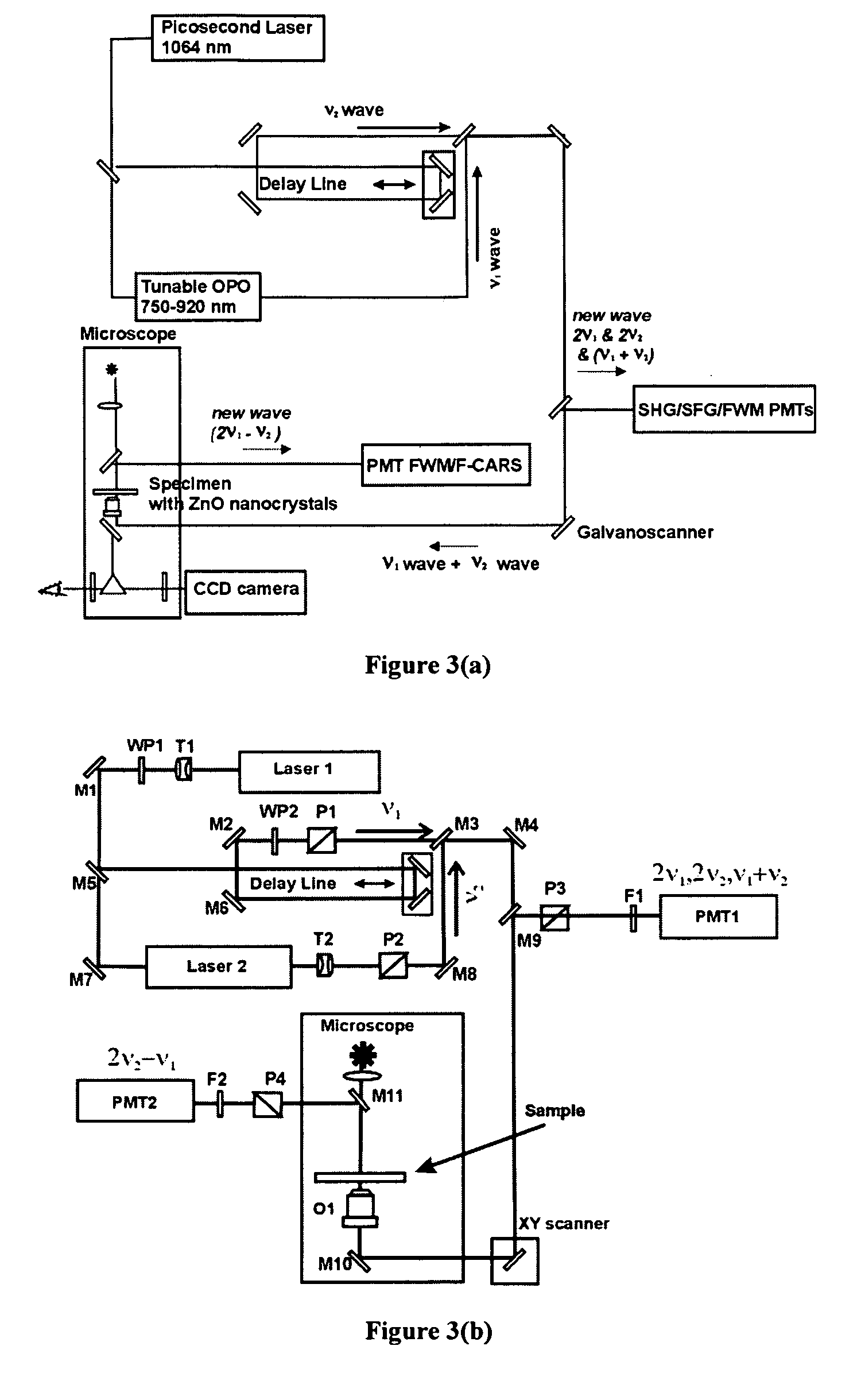
[0056]The optical setup shown in FIG. 3(b) was used to image biological samples using ZnO nanocrystals. Two picosecond lasers generated initial (input) frequencies ν1, and ν2. The ν2 wave had a fixed wavelength of 1064 nm, and ν1 wave was tunable in the 750-920 nm spectral range. Picosecond pulses with the frequencies ν1 and ν2 were time and space conjugated, and by means of dichroic mirrors, directed to the XY galvano-scanner and microscope. Both waves were coincidentally focused by a high numerical aperture objective on the specimen that had been contacted with a ZnO water dispersed sample. The nonlinear output waves generated in the sample at (2ν1−ν2), and at (ν1+ν2), 2 ν1, and 2 ν2 were collected in the backward propagation direction. They were directed to the corresponding PMT detectors by means of appropriated dichroic mirrors. Digital detection system and XY scanner were controlled by computer with appropriate software. Finally, software generate...
example 3
Preparation of Aqueous Dispersion of ZnO Nanocrystals for Targeted Imaging
[0058]ZnO nanocrystals were stably dispersed in distilled water using phospholipid micelles as the stabilizer. The phospholipid micelles are comprised of DSPE-PEG(2000) methoxy and DSPE-PEG(2000)-FA (Avanti Polar Lipids). ZnO nanocrystals in chloroform (17 mg / mL, 100 μL) was mixed with (a) 500 μL of DSPE-PEG(2000) methoxy (20 mg / mL in chloroform), or (b) a mixture of 480 μl of DSPE-PEG (20 mg / mL in chloroform) and 200 μL of DSPE-PEG(2000)-FA (2 mg / mL chloroform). Then chloroform was removed using a rotary vacuum evaporator and the dry film dispersed in distilled water (3 mL) by vortex mixing. The aqueous dispersion of ZnO nanocrystals was then sterile filtered prior to treatment of a biological specimen.
PUM
 Abstract
Description
Claims
Application Information
PatSnap Eureka turns technology decisions into work you can execute. Powered by our Innovation Knowledge Graph, it runs expert workflows across engineering, life sciences, materials and intellectual property. Get your review-ready output in minutes.


