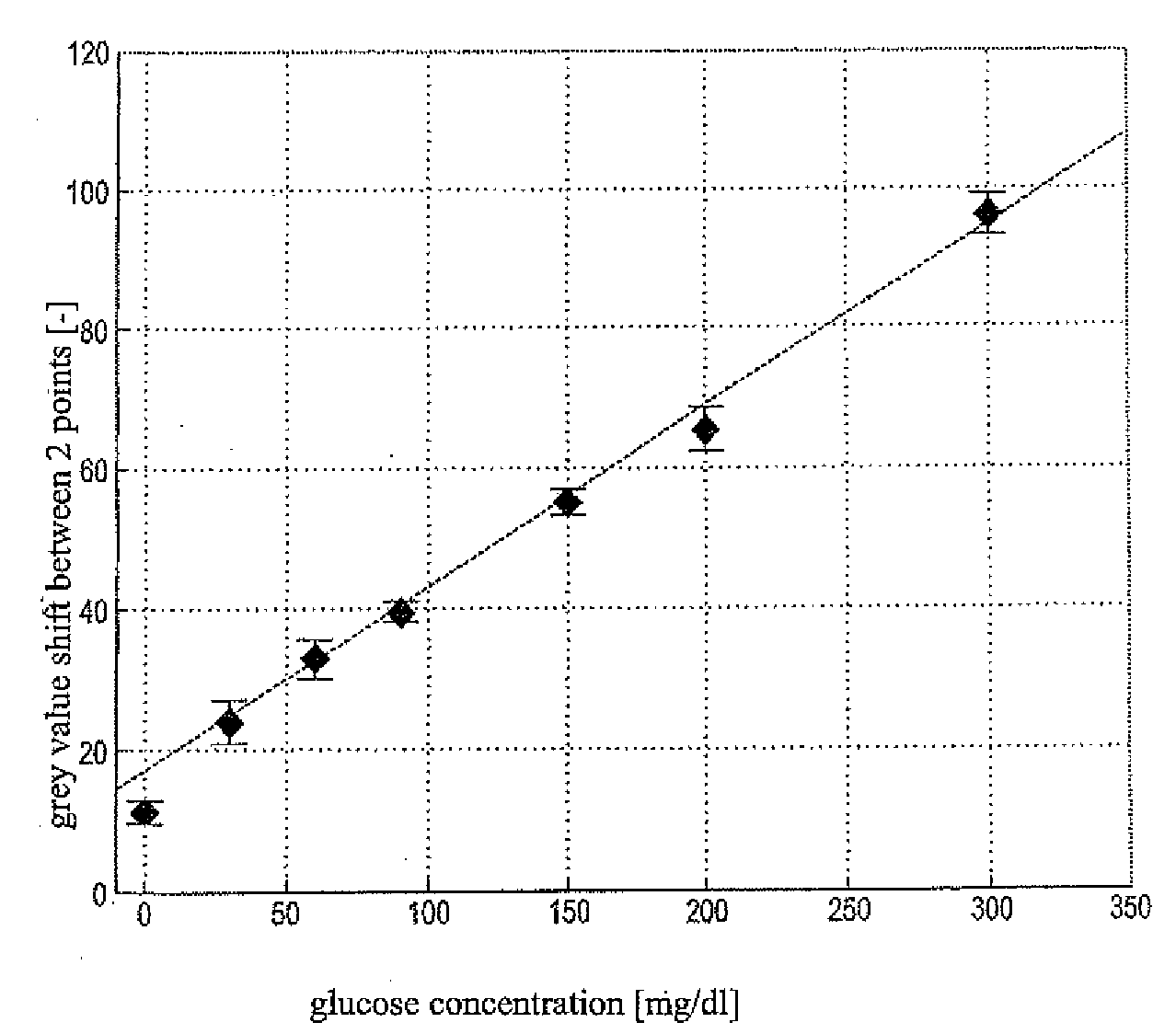
[0012]A
histogram can be used to illustrate the principle of evaluating frequencies. The light intensities (e.g., in the form of grey values) are determined and ranked into intensity intervals. The frequency of the respective
light intensity in an intensity interval is plotted against the grey value. A detection unit or an illumination unit is required for this which detects or irradiates the detection area in a spatially-resolved manner. A plurality of subareas on the detection area is examined wherein the spatial information does not have to be used for the further analysis. These subareas are not real sub-divisions of the detection area but are rather the result of the optical spatially-resolved measurement of the detection area. The number of these subareas thus depends on the number of irradiated or detected areas. The more subareas are examined, the more accurately can the differentiation of the intensity differences of various regions be determined. The intensities of the wetted subareas correlate with the concentration of the
analyte in the sample. In one embodiment, 256 intensities are distinguished. This number of intensity steps is sufficient to achieve an adequate precision / resolution to determine the concentration of the analyte. This also allows the amount of data to be kept to such a small size that it can be processed by
small data carriers which are either in an evaluation unit in the
detector or in an evaluation unit separate from the
detector. In contrast to systems of the prior art which subsequently process all intensity values to analyse the spatially-resolved measurements, in the
system according to the present disclosure preferably only certain frequencies and their associated intensities are used to calculate the concentration of the analyte. Especially in the case of time-resolved measurements in which a high
cycle rate of image taking is necessary, the analysis according to the present disclosure without storing the complete image data considerably reduces the
current consumption and memory requirements. This allows an instrument which has a low memory requirement to be produced with cheap components. Consequently the device can be manufactured and operated more cost-effectively than conventional devices.
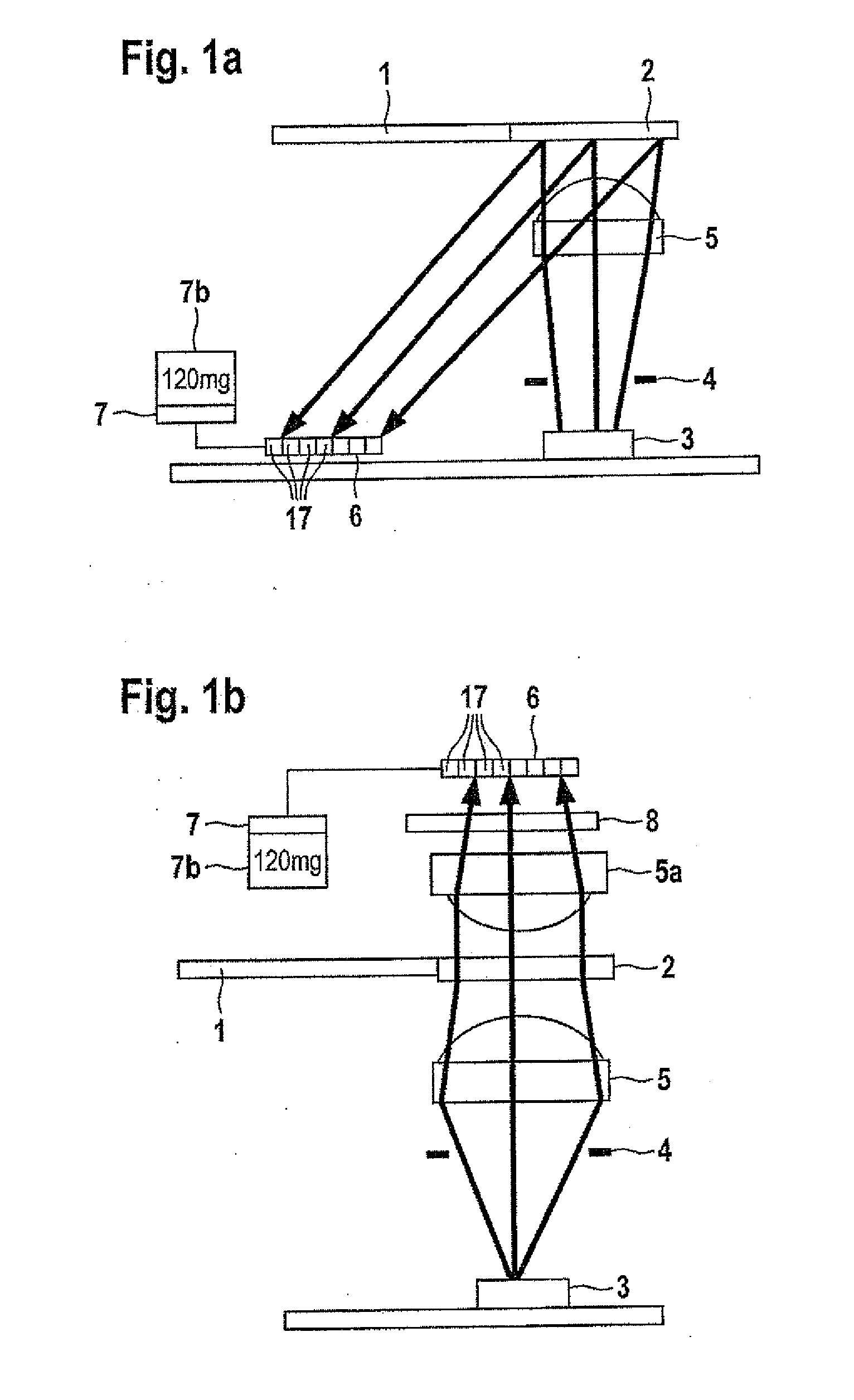
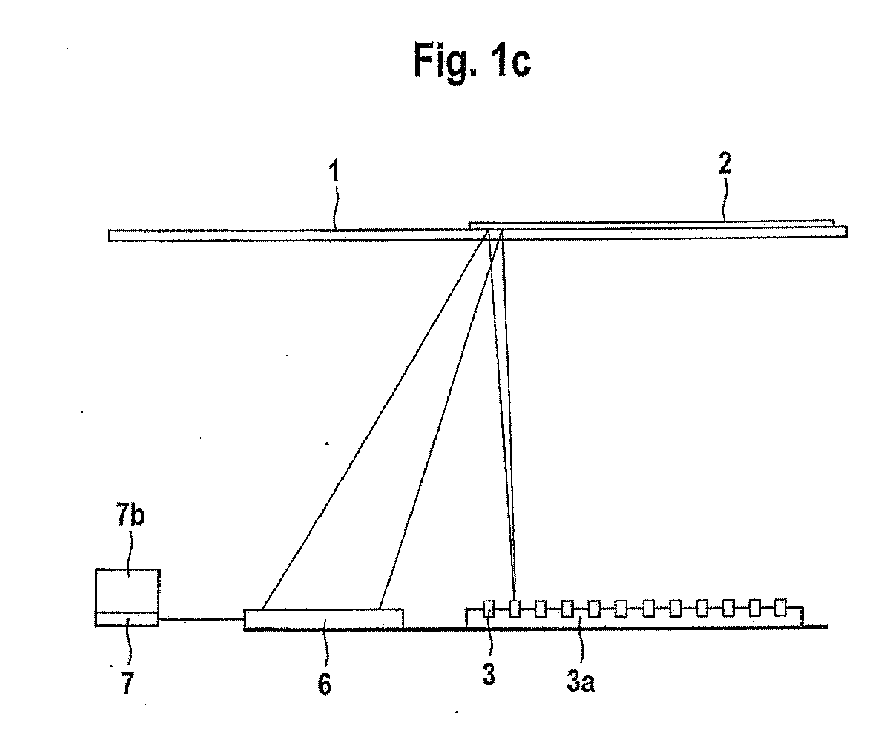
[0022]Furthermore, an instrument is described which comprises a detection unit for detecting light intensities which are radiated from subareas of a detection area of a test element and an evaluation unit which determines the concentration of the analyte on the basis of frequencies of light intensities of the light radiated from the subareas wherein the detection unit can contain a
CMOS detector the pixels of which are connected to at least one A / D converter. In addition the evaluation unit can be connected to a display unit or the display unit can be integrated into the evaluation unit. In one embodiment the detection unit and evaluation unit are integrated on a
chip, a configuration that may reduce
space requirements. Since the memory requirement is very small due to the reduced amount of data for the analysis, the
current consumption of such an integrated element is considerably lower than with conventional instruments.
[0023]Test elements such as those known from the documents EP-A 0 885 591, EP-B 0 535 480 and EP-B 0 477 322 can be used in conventional devices for determining a blood parameter. The test element contains a detection area. This detection area preferably contains all reagents and optionally auxiliary substances required for the detection reaction of the target analyte in the sample. The detection element can also contain only some of or even none of the reagents or auxiliary substances. Such reagents and auxiliary agents such as those described in the documents EP-A 0 885 591, EP-B 0 535 480 and EP-B 0 477 322 are well-known to a person familiar with the technology of analytical test elements or
diagnostic test carriers. In the case of analytes which are to be detected enzymatically, enzymes,
enzyme substrates, indicators, buffer salts,
inert fillers and suchlike can be present in the detection element. The detection element can be composed of one or more
layers and optionally contain an
inert carrier preferably on the side of the test element which is not brought into contact with the sample. In the case that the detection reaction results in an
observable change in colour (which can also be outside the
visible range) which in this connection is to be understood either as a change in colour, formation of a colour or disappearance of colour, it must be ensured through suitable measures that the carrier allows a visual or
optical observation of the detection reaction. For this purpose the
carrier material of the detection element may itself be transparent and for example have a transparent plastic foil such as for example a
polycarbonate foil or a transparent cutout on the detection side. In the case of the preferred reflection measurement, test elements such as those described in the
patent application WO 99 / 29429 can be used. These test elements contain a
pigment layer (preferably TiO2) in the detection layer. This diffusely scattering TiO2 layer increases the reflection of light which leads to a greater interaction of the incident radiated light with the reagents. This can amplify the measured effect such as the absorption of light. In a particularly preferred embodiment the dye which is formed preferably absorbs light at a
wavelength of 660 nm.
[0033]The A / D converter converts the analogue electrical
signal into a digital value. This is adequately described in the prior art. In one embodiment an 8 bit A / D converter known in the prior art is used. This A / D converter converts the electrical signals into 256 different intensity levels. The intensity levels are each of
equal size. In this manner the detected measured values can be processed further with considerably less memory capacity. In addition or alternatively it is possible to integrate an
amplifier on each pixel. This additionally results in an amplification of the signals and thus the possibility of also detecting smaller
signal changes. This
data conversion and / or amplification can considerably reduce the amount of data that is passed onto the evaluation unit. This results in the following advantages:

