Biological implantation material and method for preparing same

a technology of biological implantation and material, applied in the field of biological implantation material and method for preparing the same, can solve the problems of short durability of material and difficulty in removing cells

Inactive Publication Date: 2009-08-20
SK BIOLAND CO LTD
View PDF7 Cites 22 Cited by
  • Summary
  • Abstract
  • Description
  • Claims
  • Application Information

AI Technical Summary

Problems solved by technology

However, synthetic macromolecular materials merely substitute the body tissue with foreign substance due to lack of biological functions thereof.
However, this material has a short durability by in vivo calcification.
However, the process induces a modification of protein such as collagen due to a treatment of alkali solution having an excessive concentration (pH 12) and has a difficulty in removing cells included in a complex structure such as a substance layer due to enzyme untreatment for removal of cellular matrix.

Method used


Image

  • Biological implantation material and method for preparing same
  • Biological implantation material and method for preparing same
  • Biological implantation material and method for preparing same

Examples


example 1

Preparation of Amnion Implantation Material

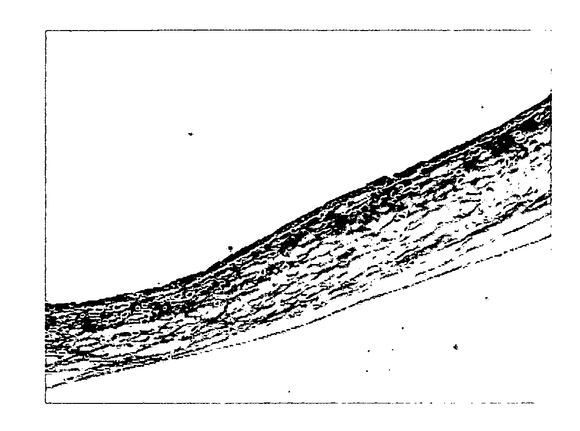
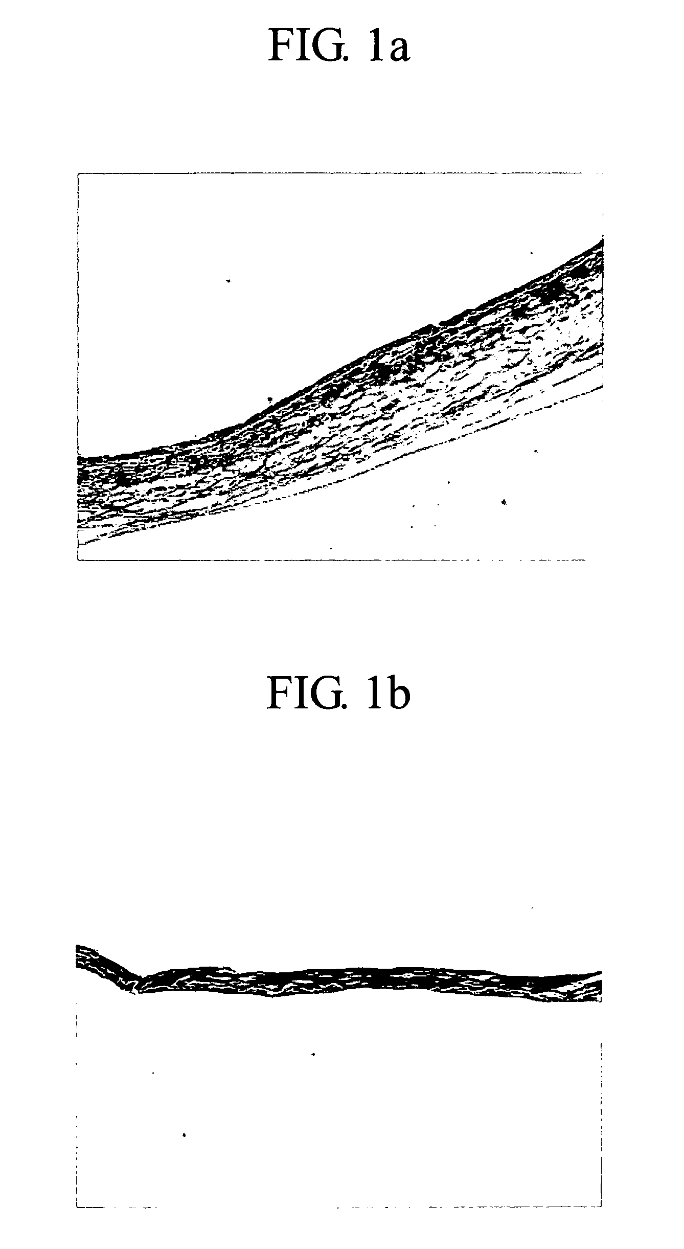
[0048]Bovine amnion samples collected from a bovine placenta were storaged in sterile saline under a cold condition and transported to the laboratory. 500 cm2 of the sample collected was treated with 1 L of 95% of ethanol and kept overnight in a cold storage to remove lipids from the bovine amnion sample. The sample was washed three times with 1 L of purified water for 10 mins and removed a substrate layer from the sample using a scrapper. The said sample was storaged in 1 L of 70% of ethanol under a cold condition to inactivate viruses and added 1 L of EDTA / sodium chloride solution (pH 11) comprising 0.2% of ethylenediamine tetraacetic acid (EDTA) and 0.9% of sodium chloride and stirred for 1 hour at 150 rpm to remove soluble alkaline impurities (step (i)). Thereafter, trypsin / EDTA / sodium chloride solution (pH 7.4) comprising 0.05% of trypsin, 0.02% of EDTA, and 0.9% of sodium chloride was treated thereto and subjected to an enzyme reactio...

example 2

Content of Lipids and Modified Collagens

[0051]The efficacy of the method according to Example 1, the method accoding to U.S. Patent Publication No. 2006 / 0024380 to Ginger A. Abraham et al. (Condition A) and U.S. Pat. No. 5,876,451 to Tooru Yui et al. (Condition B) was determined. The method according to the condition A and B is described in more detail below.

[0052]In condition A, the substrates of amnion derived from bovine placenta were removed. The sample was added to 1 L of 0.1 M EDTA / 10 mM NaOH solution per 100 cm2, stirred for 18 hours at 200 rpm and added to 1L of 1 M HCl / 10 mM NaOH solution, stirred for 8 hours at 200 rpm. The resultant sample was treated with 1 L of 1M NaCl / 10 mM phosphate buffered saline (PBS), and thereafter stirred for 18 hours, added 1 L of 10 mM PBS thereto and then stirred for 2 hours and further stirred in sterile purified water for 1 hour at 200 rpm.

[0053]In condition B, the substrates of amnion derived from bovine placenta were removed. The sample w...

example 3

Biocompatibility Test by Hypodermic Implantation to Guinea Pig

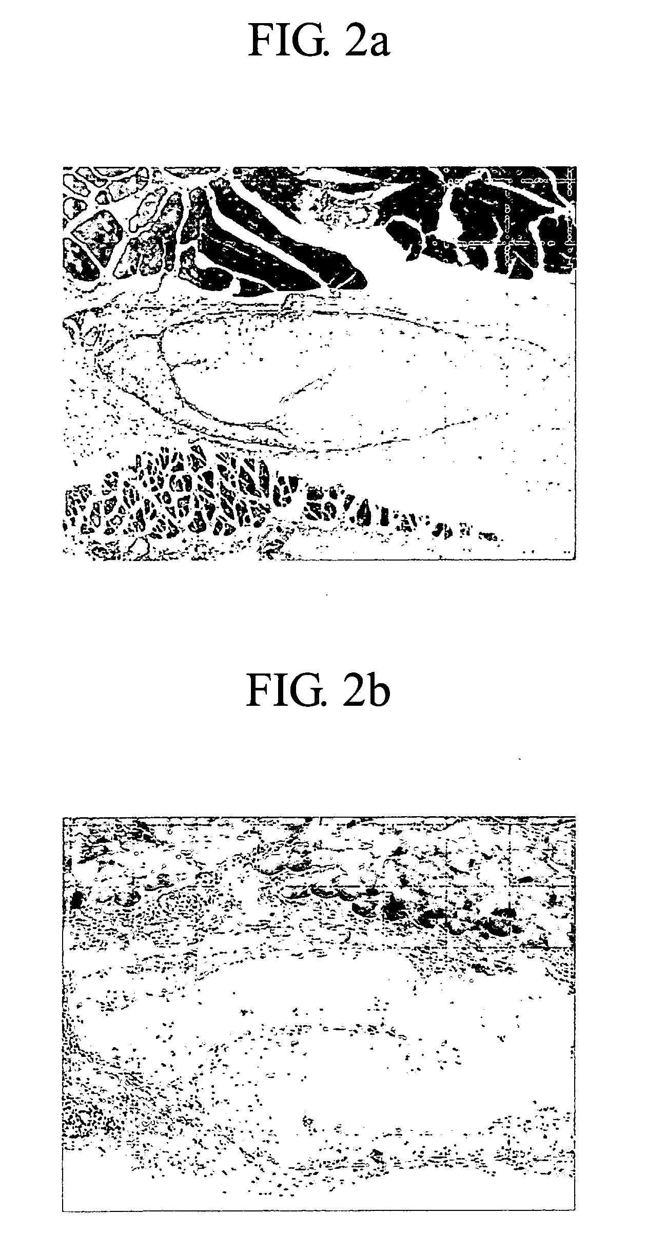
[0060]The degree of inflammatory cells and in vivo calcification produced was determined by a hypodermic implantation to guinea pig. The procedure was conducted by comparing a guinea pig tissue which was applied with the amnion implantation material prepared in Example 1 and a guinea pig which was applied with Surgisis™ (Cook Inc. USA) by the hypodermic implantation. 2 weeks and 4 weeks later, the applied tissue was procured from the each guinea pig to fix with formalin, washed and embedded with parapins. The tissue obtained in above cut into 5 μm of thickness, hematotoxyline & eosin (H&E) staining was performed and the stained tissue was then exhibited using optical microscope. After 2 weeks and 4 weeks, H&E stain photomicrograph of the tissue applied with an amnion implantation material prepared in Example 1 was shown in FIGS. 2a and 2b, respectively. Also after 2 weeks and 4 weeks, a H&E stain photomicrograph of the ti...


PUM


Abstract

The present invention relates to a biological implantation material and method of preparing the same, which comprises the steps of:
(i) treating a tissue derived from animal or human with alcohol;
(ii) contacting the said tissue with an enzyme selected from the group consisting of dispase, DNAse, RNAse and pepsin in a solvent;
(iii) treating the tissue obtained in step (ii) with alkaline solution; and
(iv) treating the tissue obtained in step (iii) with acid solution.

Description

FIELD OF THE INVENTION [0001]The present invention relates to a biological implantation material and method for preparing the same.BACKGROUND OF THE INVENTION [0002]Biological implantation material which is implantable medical prothesis and an artificial tissue to the defective tissue or organs by treating the tissue derived from animal and human with chemicals comprises substitute of heart valve, blood, ligament, and cerebral meninges and a wound dressing for treating sun burn, which are.[0003]Skin is the principal organ in the body, which prevents an outflow of body fluid, protects the body from exterior noxious substances such as bacterium and performs thermoregulation. Provided the skin is damaged by sun bum, a body fluid outflows, an infection occurs by dermis exposed to exterior noxious substances therefore the defective skin must be protected from exterior circumstances as soon as possible. Accordingly, the wound dressing used for protecting the defective tissue must have fun...

Claims


Application Information

Patent Timeline
Patent Type & Authority Applications(United States)
IPC IPC(8): A61K9/70A61P43/00
CPCA61L15/40A61L27/3604A61L2430/40A61L27/3687A61L27/365A61P43/00
Inventor KIM, IN SEOPSON, DAE GUJANG, YOUNG CHULYANG, EUN KYUNGKIM, SUNG POHONG, JONG MYOUNGJOO, JI HOONKIM, JONG SANGJUNG, SAM HYUNLEE, JONG WONKWON, MI YOUNG
Owner SK BIOLAND CO LTD
Why Eureka
Try Eureka