
Immortal unipotent porcine PICM-19H and PICM-19B stem cell lines
a stem cell line and unipotent technology, applied in the field of immortalized derivative porcine stem cell lines, can solve the problem of not being able to find effective treatment options at the momen
Patent Information
- Authority / Receiving Office
- US · United States
- Current Assignee / Owner
- Publication Date
- 2009-11-26
- Estimated Expiration
- Not applicable · inactive patent
Smart Images
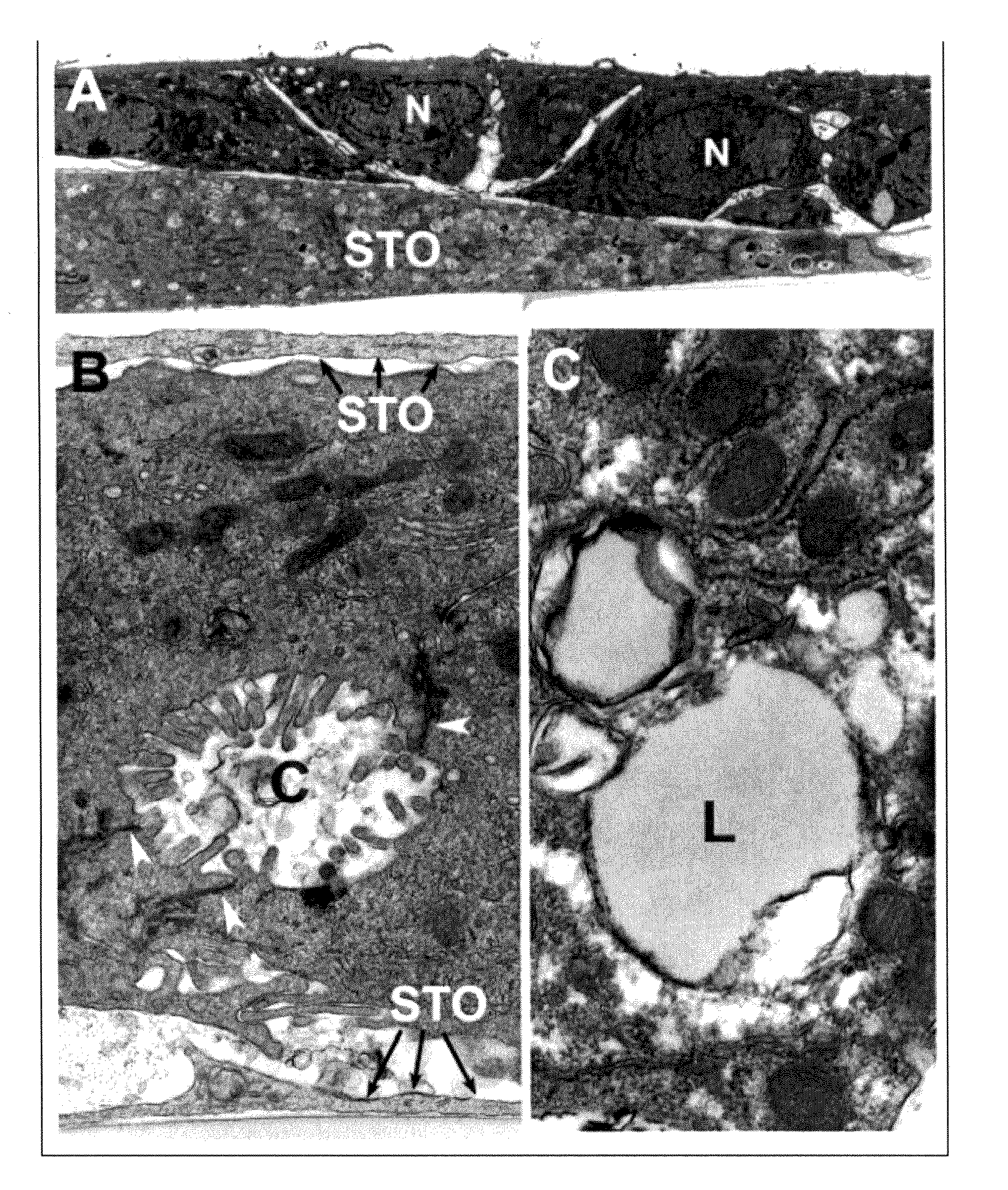
Figure 1 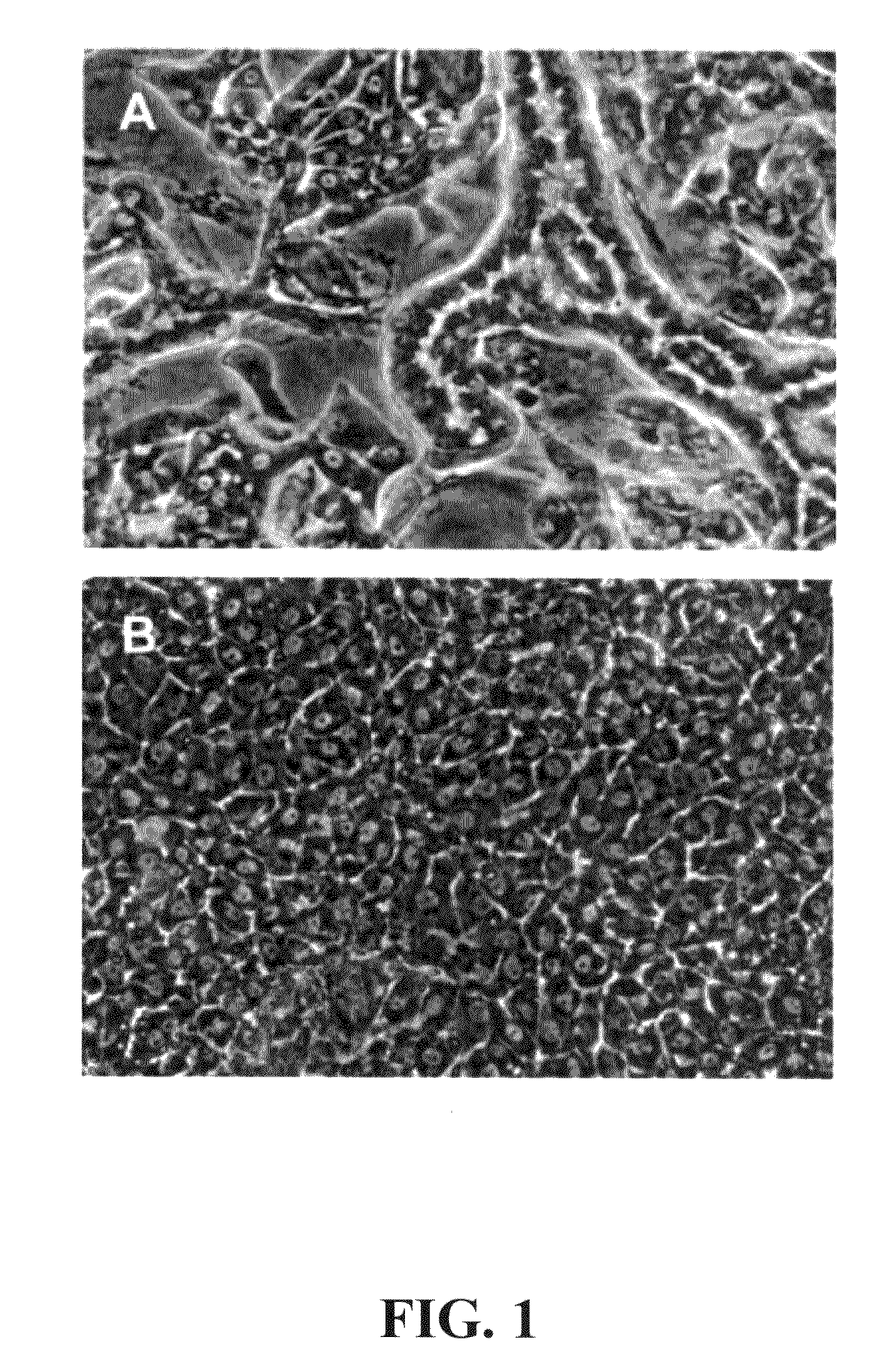
Figure 2 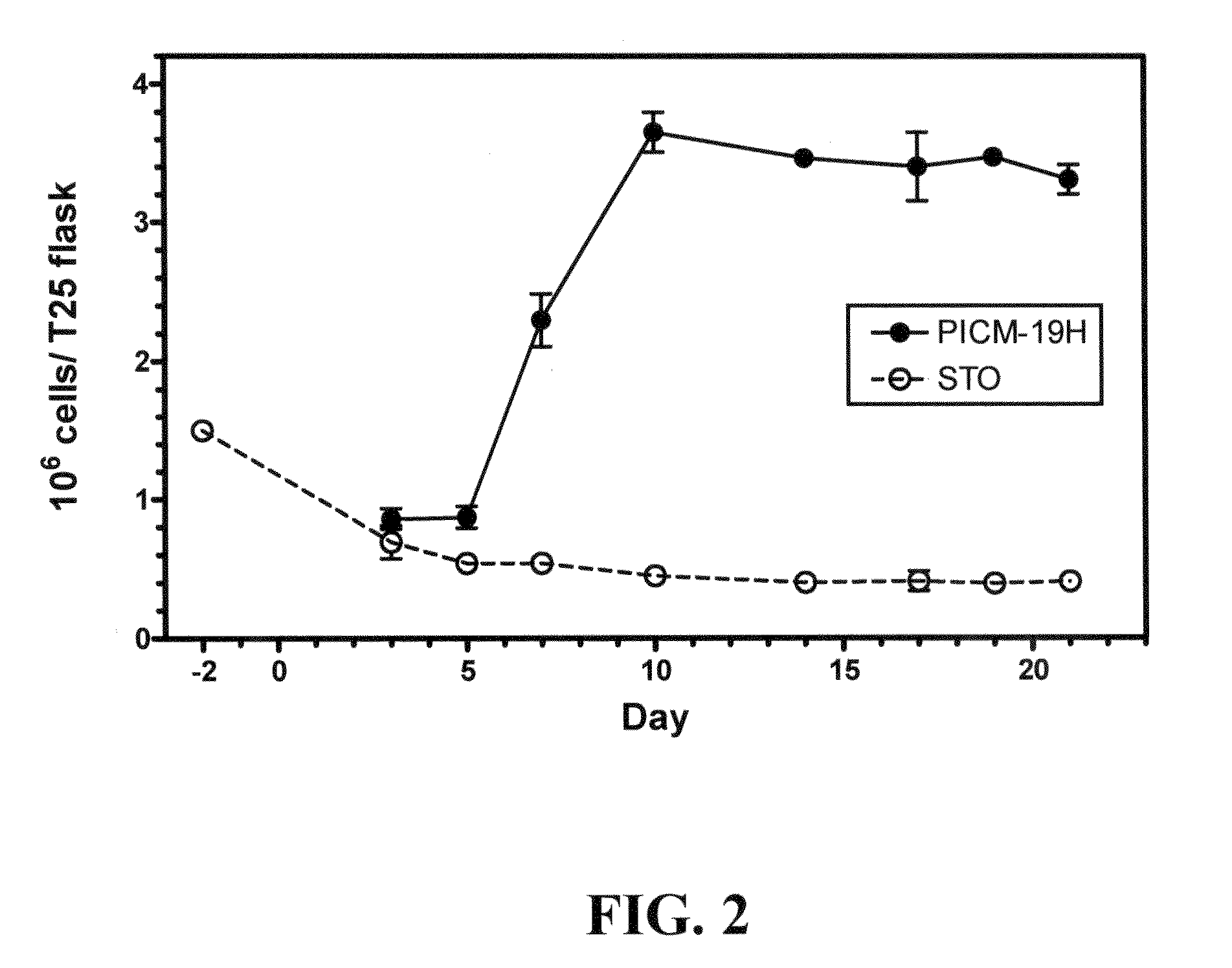
Figure 3
Abstract
Description
BACKGROUND OF THE INVENTION
[0001] 1. Field of the Invention
[0002] This invention relates to an immortalized derivative porcine stem cell line, PICM-19H, capable of differentiating exclusively into hepatocyte cells expressing hepatocyte function, for example, inducible enzyme activity, such as cytochrome P450 (CYP450) activity; an immortalized derivative porcine stem cell line, PICM-19B, capable of differentiating exclusively into bile duct cells expressing bile duct cell function and forming a complete (confluent) cell monolayer of basolaterally polarized cells; a bioartificial liver device comprising either the PICM-19H cells or the PICM-19B cells or both, a method of using the PICM-19H and / or PICM-19B stem cell lines in a bioartificial liver device or support to alleviate liver dysfunction, a method of using the PICM-19H and / or PICM-19B stem cell lines in a screening assay to detect a compound or new chemical entity which inhibits or promotes an enzyme activity involved in the metab...
Examples
example 1
Reagents Utilized for Stem Cell Culture and for Screening Assays
[0074]All cells were grown on 25 cm2 tissue culture flasks (T25; Greiner, Frickenhausen, Germany). Fetal bovine serum (FBS) and iron-supplemented calf serum were purchased from Hyclone, Logan Utah. Cell culture reagents including Dulbecco's phosphate buffered saline (PBS) without Ca++ and Mg++, media, trypsin-EDTA (0.025% trypsin, 0.43 mM EDTA), antibiotics, non-essential amino acids, and L-glutamine were purchased from InVitrogen, Gaithersburg, Md. PICM-19H cells were grown on irradiated STO mouse fibroblast (CRL 1503, American Type Culture Collection, Rockville, Md.) feeder cell layers. Feeder-layers were prepared by exposing a suspension of STO cells to 8 krad of gamma radiation and plating the cells at 6×10⁴ cells / cm2. STO feeder-layers were maintained by refeeding with 10% DMEM every 6-7 d. The growth and differentiation medium for PICM-19 cultures was a 50:50 mixture of DMEM low glucose and Medium 199 supplemented...
example 2
Establishment of PICM-19H Stem Cell Line
[0078]The PICM-19H cell line was established from a T25 mass culture of parental ARS-PICM-19 cells, i.e., approximately one million cells, that were subjected to hypothermic selection (33-34° C.) for approximately 3 wk at passage 37. The PICM-19H cell line was derived from approximately 50-100 cells that survived the temperature selection.
[0079]After expansion to mass cultures, by culturing at 37-38° C., it was observed that the PICM-19H cell line did not self-organize into multi-cellular bile ductules under standard or elevated pH culture conditions as is characteristic of the parental ARS-PICM-19 population (FIGS. 1A and B; Talbot et al. 2002, supra). Phase-contrast microscopy showed the PICM-19H cells to be generally cuboidal cells with distinct, centrally located nuclei (FIG. 1B). Like the parental ARS-PICM-19 cells, the fetal hepatocyte-like PICM-19H cells grew as a patch work of small monolayer colonies nestled in amongst the STO (mouse ...
example 3
Establishment of PICM-19B Stem Cell Line
[0081]The PICM-19B cell line was developed from a spontaneously arising morphological variant of the parental PICM-19 cells which occurred in the culture at low frequency at approximately passage 35 (FIG. 3; Talbot et al. 1994a, supra). The PICM-19B cell line was established by micropipette-mediated colony-cloning of a single colony of this variant cell type. Independent, spontaneously forming, “PICM-19B-like” outgrowths occurred at various passage levels of the parental PICM-19 cells when initiated from frozen stocks of the cells. Observation of the occurrence of the PICM-19B cells, and of many similar independent occurrences, indicated that this morphological variant appeared to arise from PICM-19 cells that were differentiating and forming into bile ductules and not from PICM-19 cells forming monolayer patches of hepatocyte-like cells (Talbot et al. 1994a, supra). To illustrate this point more clearly, FIGS. 3B, C, and D illustrates the gen...