
Zirconium-radiolabeled, cysteine engineered antibody conjugates

a technology of zirconium radiolabeled and cysteine, which is applied in the field of antibodies, can solve the problems of affecting the resolution of small animal micropets, affecting the resolution of small animals, and the half-life of readily available sup>64/sup>cu (12.7 h) is too short to provide images with good contrast in this time fram

Inactive Publication Date: 2010-05-06
F HOFFMANN LA ROCHE & CO AG
View PDF16 Cites 207 Cited by
  • Summary
  • Abstract
  • Description
  • Claims
  • Application Information

AI Technical Summary

Benefits of technology

[0013]Cysteine engineered antibodies of the invention may be site-specifically and efficiently coupled with a thiol-reactive reagent. The thiol-reactive reagent may be a radioisotope reagent, multifunctional linker reagent, a capture label reagent, a fluorophore reagent, or a drug-linker intermediate.

Problems solved by technology

Unfortunately, the half-life of readily available 64Cu (12.7 h) is too short to provide images with good contrast in this time frame.
Despite the relatively simple radioiodination techniques available for coupling 124I onto mAbs, important limitations slow a widespread pre-clinical use of this radionuclide.
1.5 and 2.1 MeV) which negatively affects the resolution of small animal microPET.
Analytical and preparative methods are inadequate to separate and characterize the antibody-drug conjugate species molecules within the heterogeneous mixture resulting from a conjugation reaction.
Furthermore, the multistep conjugation process may be nonreproducible due to difficulties in controlling the reaction conditions and characterizing reactants and intermediates.
This approach may result in loss of antibody tertiary structure and antigen binding specificity.
However, designing in cysteine thiol groups by the mutation of various amino acid residues of a protein to cysteine amino acids is potentially problematic, particularly in the case of unpaired (free Cys) residues or those which are relatively accessible for reaction or oxidation.
Furthermore, if the protein oxidatively forms an intramolecular disulfide bond between the newly engineered Cys and an existing Cys residue, both Cys groups are unavailable for active site participation and interactions.

Method used


Image

  • Zirconium-radiolabeled, cysteine engineered antibody conjugates
  • Zirconium-radiolabeled, cysteine engineered antibody conjugates
  • Zirconium-radiolabeled, cysteine engineered antibody conjugates

Examples


example 1

Preparation of Biotinylated Thiofab Phage

[0321]ThioFab-phage (5×10¹² phage particles) were reacted with 150 fold excess of biotin-PEO-maleimide ((+)-biotinyl-3-maleimidopropionamidyl-3,6-dioxaoctainediamine, Oda et al (2001) Nature Biotechnology 19:379-382, Pierce Biotechnology, Inc.) for 3 hours at room temperature. Excess biotin-PEO-maleimide was removed from biotin-conjugated phage by repeated PEG precipitations (3-4 times). Other commercially available biotinylation reagents with electrophilic groups which are reactive with cysteine thiol groups may be used, including Biotin-BMCC, PEO-Iodoacetyl Biotin, Iodoacetyl-LC-Biotin, and Biotin-HPDP (Pierce Biotechnology, Inc.), and Nα-(3-maleimidylpropionyl)biocytin (MPB, Molecular Probes, Eugene, Oreg.). Other commercial sources for biotinylation, bifunctional and multifunctional linker reagents include Molecular Probes, Eugene, Oreg., and Sigma, St. Louis, Mo.

example 2

PHESELECTOR Assay

[0322]Bovine serum albumin (BSA), erbB2 extracellular domain (HER2) and streptavidin (100 μl of 2 μg / ml) were separately coated on Maxisorp 96 well plates. After blocking with 0.5% Tween-20 (in PBS), biotinylated and non-biotinylated hu4D5Fabv8-ThioFab-Phage (2×10¹⁰ phage particles) were incubated for 1 hour at room temperature followed by incubation with horseradish peroxidase (HRP) labeled secondary antibody (anti-M13 phage coat protein, pVIII protein antibody). FIG. 8 illustrates the PHESELECTOR Assay by a schematic representation depicting the binding of Fab or ThioFab to HER2 (top) and biotinylated ThioFab to streptavidin (bottom).

[0323]Standard HRP reaction was carried out and the absorbance was measured at 450 nm. Thiol reactivity was measured by calculating the ratio between OD450 for streptavidin / OD450 for HER2. A thiol reactivity value of 1 indicates complete biotinylation of the cysteine thiol. In the case of Fab protein binding measurements, hu4D5Fabv8 (...

example 3a

Expression and Purification of ThioFabs

[0324]ThioFabs were expressed upon induction in 34B8, a non-suppressor E. coli strain (Baca et al (1997) Journal Biological Chemistry 272(16):10678-84). The harvested cell pellet was resuspended in PBS (phosphate buffered saline), total cell lysis was performed by passing through a microfluidizer and the ThioFabs were purified by affinity chromatography with protein G SEPHAROSE™ (Amersham).

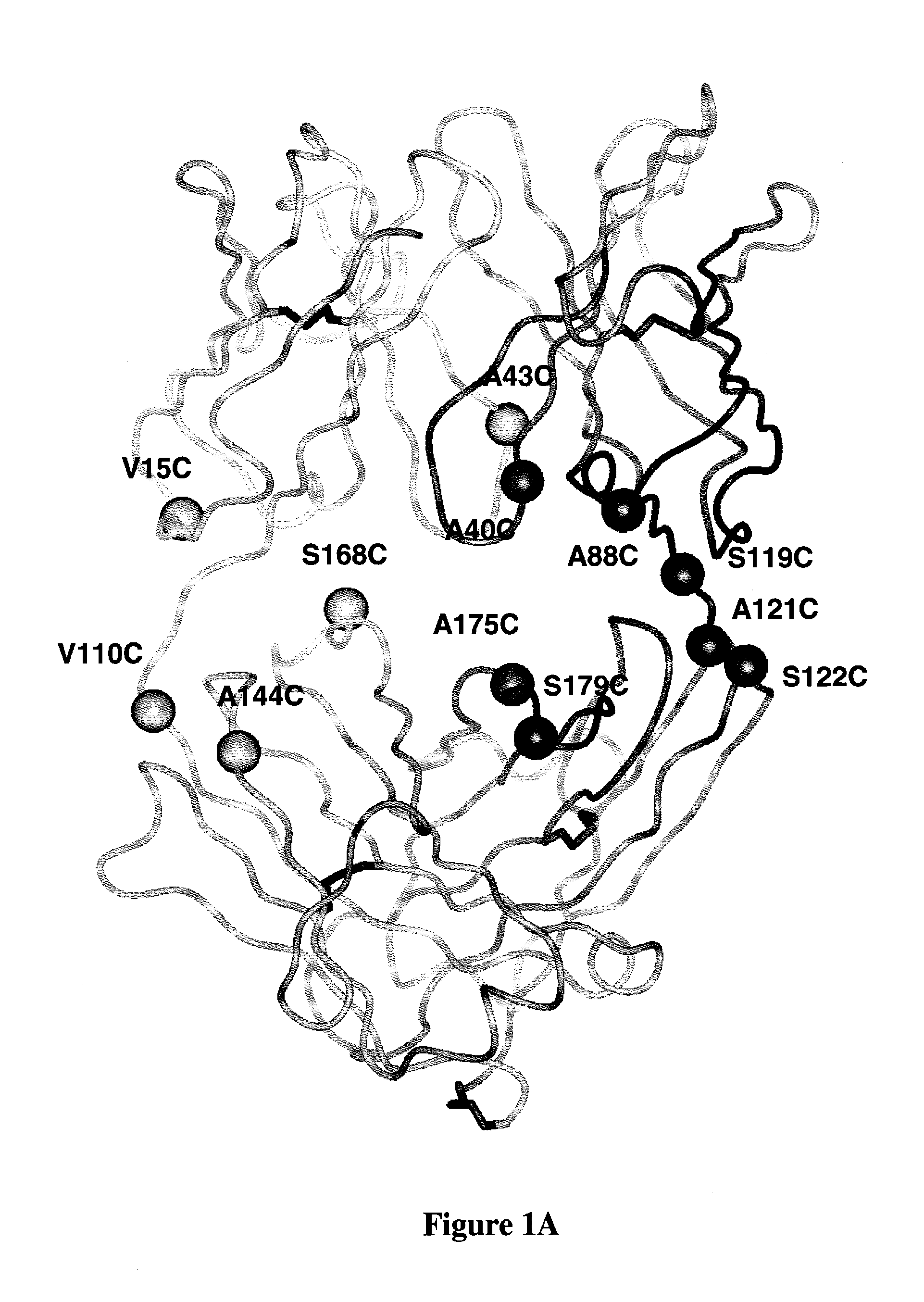
[0325]ThioFabs L-V15C, L-V110C, H-A88C, and H-A121C were expressed and purified by Protein-G SEPHAROSEcolumn chromatography. Oligomeric-Fab was present in fractions 26 to 30, and most of the monomeric form was in fractions 31-34. Fractions consisting of the monomeric form were pooled and analyzed by SDS-PAGE along with wild type hu4D5Fabv8 and analyzed on SDS-PAGE gel in reducing (with DTT or BME) and non-reducing (without DTT or BME) conditions. Gel filtration fractions of A121C-ThioFab were analyzed on non-reducing SDS-PAGE.

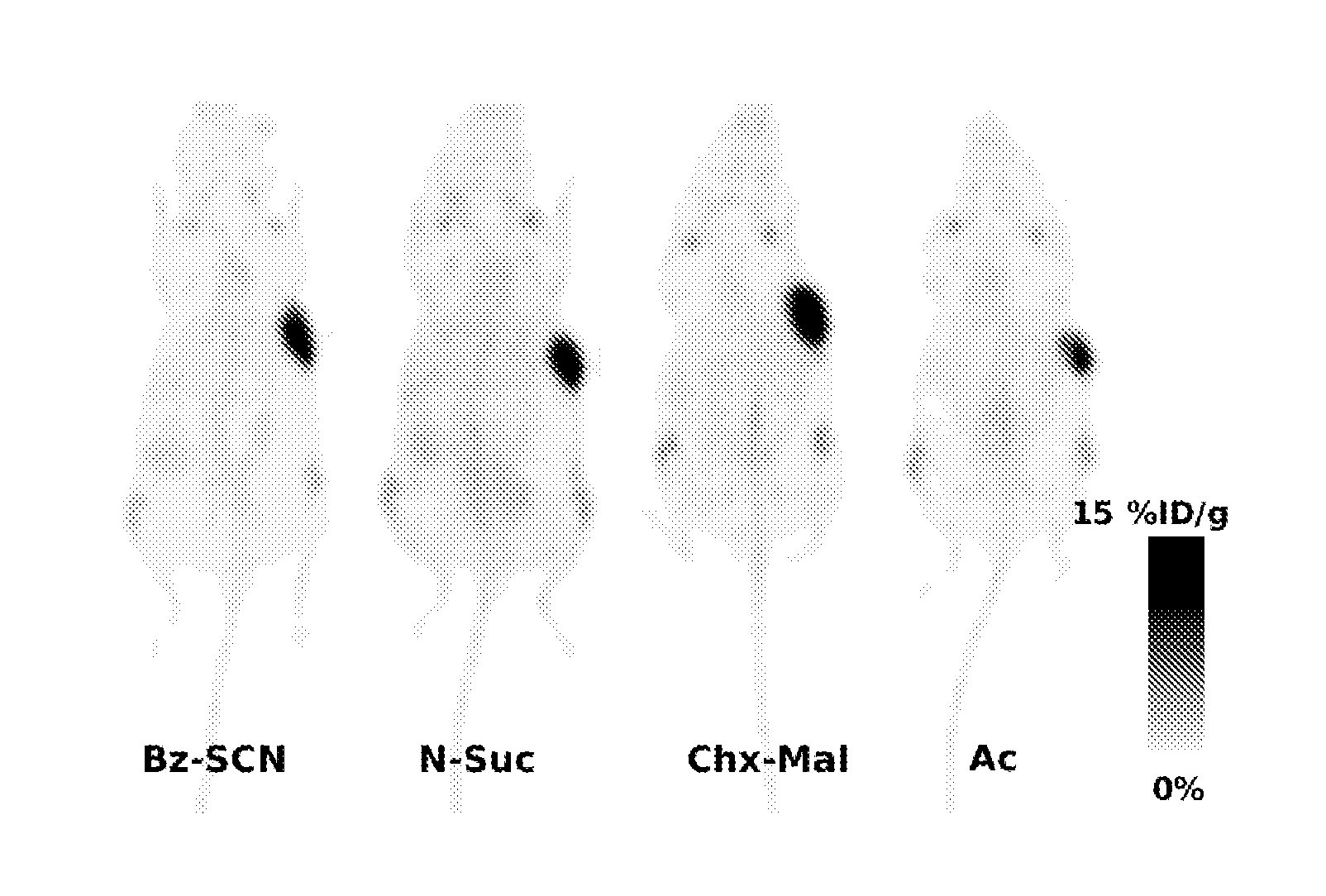
[0326]ThioFabs were conjugated ...


PUM

PropertyMeasurementUnit
pHaaaaaaaaaa
widthaaaaaaaaaa
structuresaaaaaaaaaa

Abstract

Antibodies are engineered by replacing one or more amino acids of a parent antibody with non cross-linked, highly reactive cysteine amino acids. Antibody fragments may also be engineered with one or more cysteine amino acids to form cysteine engineered antibody fragments (ThioFab). Methods of design, preparation, screening, and selection of the cysteine engineered antibodies are provided. Cysteine engineered antibodies (Ab) are conjugated with one or more zirconium complex (Z) labels through a linker (L) to form cysteine engineered zirconium-labeled antibody conjugates having Formula I:Ab-(L-Z)p  Iwhere p is 1 to 4. Imaging methods and diagnostic uses for zirconium-radiolabeled, cysteine engineered antibody conjugate compositions are disclosed.

Description

[0001]This application is a continuation-in-part of U.S. Ser. No. 12 / 399,241 filed on Mar. 6, 2009 which is a continuation of U.S. Ser. No. 11 / 233,258 filed on Sep. 22, 2005, now U.S. Pat. No. 7,521,541 issued Apr. 21, 2009, and also claims the benefit of priority under 35 USC §119(e) of U.S. Provisional Application Ser. No. 60 / 612,468 filed on Sep. 23, 2004 and U.S. Provisional Application Ser. No. 60 / 696,353 filed on Jun. 30, 2005, each of which are incorporated by reference in their entirety.FIELD OF THE INVENTION[0002]The invention relates generally to antibodies engineered with reactive cysteine residues and more specifically to antibodies with therapeutic or diagnostic applications. The cysteine engineered antibodies may be conjugated with chemotherapeutic drugs, toxins, affinity ligands such as biotin, and detection labels such as radioisotopes and fluorophores. The invention also relates to methods of using antibodies and antibody-drug conjugate compounds for in vitro, in si...

Claims


Application Information

Patent Timeline
Patent Type & Authority Applications(United States)
IPC IPC(8): A61K51/10C07K16/00C07D207/18C07C237/00C07K1/13
CPCA01K2267/0331A61K51/1027A61K51/1051A61K51/1093C07C259/06C07C323/60C07K16/00C07K16/32C07K2317/21C07K2317/51C07K2317/55C07K2317/624C07K2317/52C07C2601/14A61K38/17A61K39/395A61K47/42A61K48/00A61K49/14A61K49/16C07K14/435C07K16/18C07K16/28C07K19/00
Inventor GILL, HERMANJUNUTULA, JAGATH R.LOWMAN, HENRY B.MARIK, JANTINIANOW, JEFFWILLIAMS, SIMON
Owner F HOFFMANN LA ROCHE & CO AG
Learn More