Method for Obtaining Xeno-Free Hbs Cell line

a cell line and stable technology, applied in the field of stable hbs cell line, can solve the problems of increasing the risk of transferring non-human pathogens in any clinical application, increasing the likelihood of graft rejection, and the number of people suffering from disorders suitable for treatment by these methods,

Inactive Publication Date: 2010-05-27
CELLARTIS AB (SE)
View PDF5 Cites 3 Cited by
  • Summary
  • Abstract
  • Description
  • Claims
  • Application Information

AI Technical Summary

Problems solved by technology

Unfortunately, the number of people suffering from disorders suitable for treatment by these methods by far outstrips the number of organs available for transplantation.
However, all currently available human blastocyst-derived stem cell (hBS) lines have at some point during their derivation and / or maintenance been directly or indirectly exposed to animal material.
One potential consequence of using xeno-contaminated hBS cells in patients is an increased likelihood of graft rejection [Martin J M et al, 2005].
Moreover, xeno-exposure increases the risk of transferring non-human pathogens in any clinical application.
Thus, the predicted hazards associated with using xeno-exposed hBS cells in patients, makes these cells unsuitable for clinical applications.
However, it has so far not been possible to derive and continuously culture hBS cell lines in a completely xeno-free system.

Method used


Image

  • Method for Obtaining Xeno-Free Hbs Cell line
  • Method for Obtaining Xeno-Free Hbs Cell line
  • Method for Obtaining Xeno-Free Hbs Cell line

Examples


example 1

Obtaining and Culture Xeno-Free hBS Cells

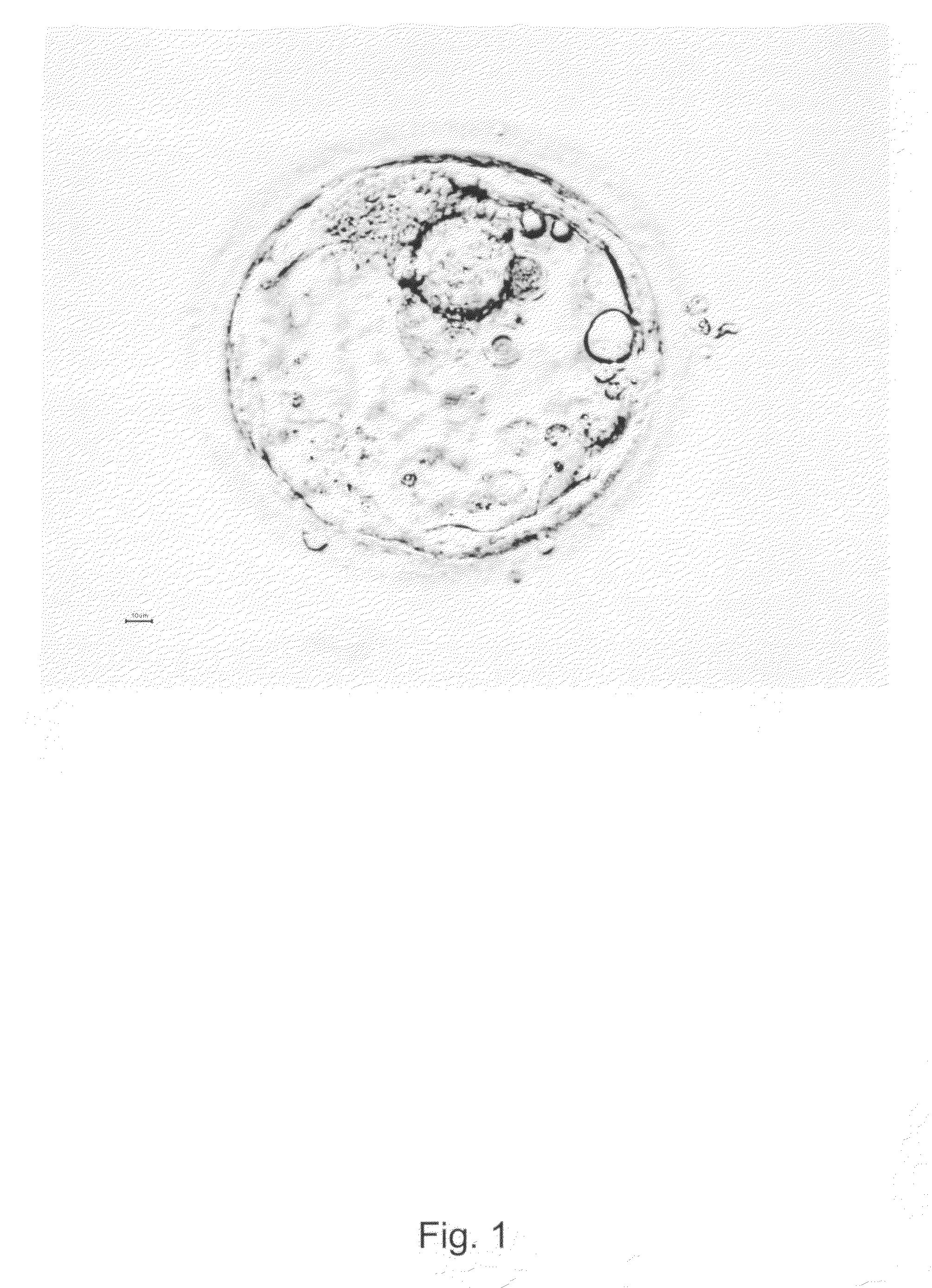
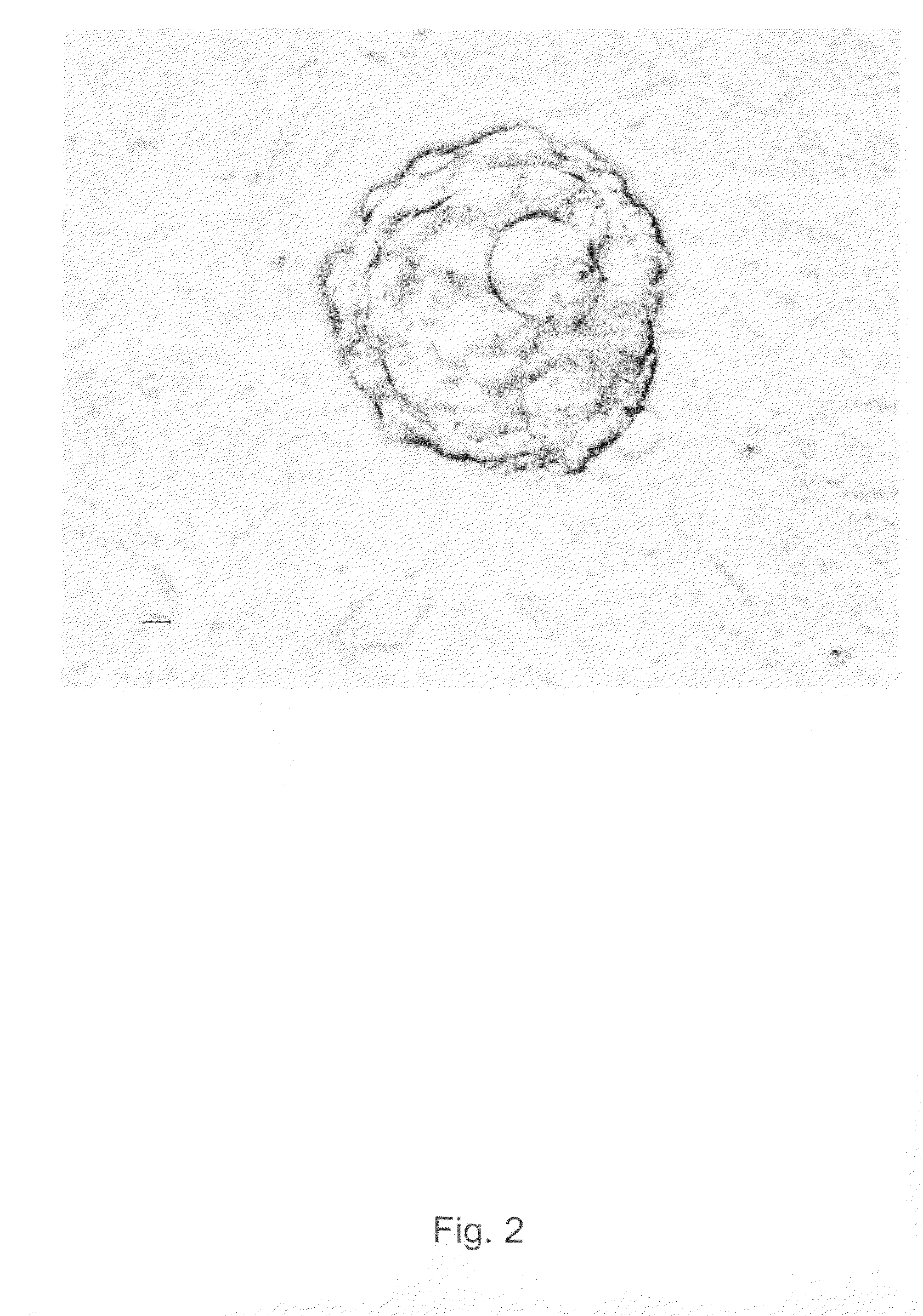
[0206]Surplus human embryo from clinical in vitro fertilization (IVF) treatment was donated after informed consent and approval from the local ethics committee at Göteborg University. The donated embryo was cultured to blastocyst until the age of 5 days in media traditionally used in IVF treatment. The blastocyst was graded according to Gardner as 4AA and according to WO2003055992 as a blastocyst of quality A (expanded blastocyst with many distinct tightly packed ICM cells and a cohesive trophectoderm with many cells). The blastocyst was treated with acid Tyrode's solution (Medicult) solution (ready-to-use concentration) for 15-30 seconds in room temperature in order to remove the zona pellucida and parts of the trophectoderm (see FIGS. 1 and 2) before placing the inner cell mass cells onto mitomycin-C inactivated xenofree human foreskin fibroblast feeders in xeno-free serum containing a DMEM with osmolarity of around 270, supplemented with 2...

example 3

Feeder Layer Preparation

[0209]Prior to plating the xenofree human fibroblast feeders, the tissue cultured wells are coated with 0.1% recombinant human gelatin (Fibrogen) for a minimum of 1 hour at room temperature. Confluent monolayers of xenofree hFF003 (fifth to eight passage) cells grown in IMDM, 10% human serum and 1% penicillin-streptomyocin were then treated with mitomycin-C (Sigma) for 2.5 hours. Mitomycin-C treated feeders were plated on IVF wells (Becton Dickinson), 200 000 cells per 2.89 cm2 in a medium which was based on DMEM (as above) supplemented with 10% (v / v) human serum, 1% penicillin-streptomyocin, 1% Glutamax, 0.5 mmol / l β-mercaptoethanol and 1% non-essential amino acids (Gibco Invitrogen Corporation). Prior to the placing blastocysts with their inner cell mass cells and cells derived therefrom or hBS cells, the medium was changed to a DMEM (as above), now instead supplemented with 20% (v / v) human serum, 10 ng / mL human recombinant bFGF, 1% penicillin-streptomyocin...


PUM

PropertyMeasurementUnit
timeaaaaaaaaaa
timeaaaaaaaaaa
timeaaaaaaaaaa

Abstract

A method for obtaining a stable xeno-free hBS cell line, xeno-free hBS cell lines obtained according to said method and use thereof. The method comprises the steps of:
    • i) removing the zona pellucida from a blastocyst to obtain trophectoderm-enclosed inner cell mass by a xeno-free procedure,
    • ii) at least partly removing the trophectoderm to obtain isolated inner cell mass cells by a xeno-free procedure,
    • iii) placing the inner cell mass cells on a layer of human feeder cells in a xeno-free medium,
    • iv) co-culturing of the inner cell mass cells with human feeder cells for a time period of from about 5 days to about 50 days in a xeno-free medium,
    • v) releasing the inner cell mass cells or cells derived thereof from trophectoderm overgrowth, if any, by a xeno-free procedure,
    • vi) selectively, transferring the inner cell mass cells or cells derived thereof to a fresh layer of human feeder cells in a xeno-free medium to obtain xeno-free hBS cells,
    • vii) propagating the xeno-free hBS cells by co-culturing with human feeder cells in a xeno-free medium to obtain a xeno-free hBS cell line.
The xeno-free hBS cell line is suitable for use in medicine and in in vitro testing.

Description

FIELD OF THE INVENTION[0001]The present invention relates to a method to obtain a stable xeno-free hBS cell line, xeno-free hBS cell lines obtained according to said method and use thereof.BACKGROUND OF THE INVENTION[0002]A stem cell is a cell type that has a unique capacity to renew itself and to give rise to specialized or differentiated cells. Although most cells of the body, such as heart cells or skin cells, are committed to conduct a specific function, a stem cell is uncommitted until it receives signals to develop into a specialized cell type. What makes the stem cells unique is their proliferative capacity, combined with their ability to become specialized. For years, researchers have focused on finding ways to use stem cells to replace cells and tissues that are lost, damaged or diseased. So far, most research has focused on two types of stem cells, embryonic and somatic stem cells. Embryonic stem cells are derived from the pre-implanted fertilized oocyte, i.e. blastocyst, ...

Claims


Application Information

Patent Timeline
Patent Type & Authority Applications(United States)
IPC IPC(8): C12N5/071C12N5/0735C12N5/077
CPCC12N5/0606C12N5/0656C12N2502/13C12N2509/00C12N2502/1323A61P1/16A61P25/16A61P25/28A61P3/10A61P43/00A61P9/00A61P9/10
Inventor SEMB, HENRIKKILMARE, EVA KARINSTREHL, RAIMUNDELLERSTROM, CATHARINAFREJ, KATARINAMOYA, KARINAHYLLNER, SVEN JOHAN
Owner CELLARTIS AB (SE)
Why Eureka
Try Eureka