Tissue Cavitation Device and Method

a tissue cavitation and tissue technology, applied in the field of surgical devices and methods, can solve the problems of weakened bone prophylactic fixation in this manner, fractures under normal physiologic loading conditions, skeletal fractures, etc., and achieve the effect of minimal invasiv

Inactive Publication Date: 2010-09-23
MIDDLETON LANCE +1
View PDF101 Cites 46 Cited by
  • Summary
  • Abstract
  • Description
  • Claims
  • Application Information

AI Technical Summary

Benefits of technology

[0014]During minimally invasive surgery, the flexible cutting element of the cavitation device can be adapted to assume a first shape for insertion of the device into tissue through a tube placed percutaneously, thereby creating only a relatively small access opening in the tissue. Depending on the application and size, the insertion tube can be a trochar, a cannula, or a needle. As the device is inserted beyond the distal end of the insertion tube, the flexible cutting element is adapted to assume a second shape for forming a cavity in tissue upon rotation of the shaft. When it assumes the second shape, the flexible cutting element extends or projects away from the longitudinal axis of the shaft. Thus, the diameter of the cavity is greater than the diameter of the initial access opening or pilot hole. In addition to cutting, a flexible cutting element is capable of displacing and impacting tissue away from the axis of the shaft.
[0015]According to one method of the present invention, the periphery of the target tissue, such as bone, can be accessed with an insertion tube placed percutaneously, and a pilot hole can be formed in the bone with a standard surgical drill and drill bit. Next, the cavitation device of the present invention is inserted to the depth of the pilot hole and rotated. As the flexible cutting element of the device moves from its first shape to its second shape, portions of the cutting element forcefully extend away from the longitudinal axis of the shaft, thereby forming a tissue cavity. Emulsified bone can be removed through known irrigation and suction methods. In the case of bone harvesting, the abated bone is used at another surgical site to promote healing of a bony deficit or to promote joint fusion. The cavity can then be filled with a suitable bone substitute that is injectable and hardens in situ. In the case of removing and replacing osteoporotic bone, the cavity is filled with structural synthetic bone or bone cement. Since the device and methods of the present invention are minimally invasive, they can be used for the prevention of osteoporosis related fractures in individuals at high risk. Skeletal structures where osteoporosis related fractures are common include the radius, femur, and vertebral bodies.
[0017]The objects and advantages of the present invention include simplicity, wherein a flexible cutting element eliminates the need for complex assemblies with numerous moving parts. The shape-changing behavior of the flexible cutting element enables the device to be adapted to a shape suitable for minimally invasive placement in tissue. The inherent outward forces associated with the shape change of the flexible cutting element assist in the cutting and displacement of tissue during the process of forming a cavity.

Problems solved by technology

The disease leads to skeletal fractures under light to moderate trauma and, in its advanced state, can lead to fractures under normal physiologic loading conditions.
Without the availability of minimally invasive surgical procedures, however, the prophylactic fixation of osteoporosis-weakened bone in this manner would not be practical because of the increased morbidity, blood loss and risk of complications associated with conventional procedures.
Existing devices for forming a cavity within soft or hard tissue are relatively complex assemblies consisting of multiple components.
The complexity of the device leads to increased manufacturing costs and may also raise concerns regarding the potential for malfunction.
Such high rotational speeds can only be produced by a powered surgical drill and certainly cannot be produced by manual rotation.
Thus, the Mirza device does not permit the surgeon to exercise the precise control that can be attained through manual rotation.
Moreover, there may be a concern for structural failure or loosening of the relatively small hinge assembly at such a high rotational speed when operated in bone.
The high rotational speed of the Mirza device may also generate excessive heat that could damage healthy tissue surrounding the cavity.
The Reiley et al. device, however, is not intended to cut tissue, and at least a small cavity must therefore be cut or otherwise formed in the tissue in order to initially insert the Reiley et al. device.

Method used


Image

  • Tissue Cavitation Device and Method
  • Tissue Cavitation Device and Method
  • Tissue Cavitation Device and Method

Examples


second embodiment

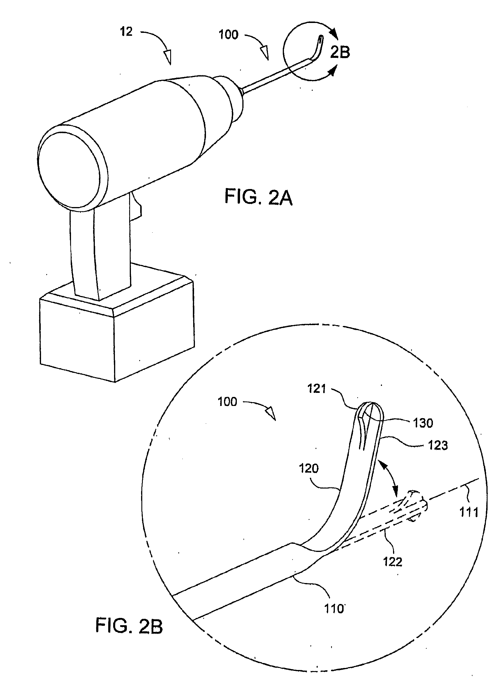
[0039]FIGS. 4A to 4C show the present invention, cavitation device 200, comprising rotatable shaft 210 and a flexible cutting element 220 having a free end 221 and a cutting tip 230. Flexible cutting element 220 is formed from a material, such as Nitinol, which is capable of shape change arising from thermal shape-memory behavior. Rotatable shaft 210 has a longitudinal axis 211. FIG. 4A shows cavitation device 200 at rest, with flexible cutting element 220 deformed below the transformation temperature to a first shape 222 in which flexible cutting element 220 is substantially colinear with longitudinal axis 211. When flexible cutting element 220 is in first shape 222, cavitation device 200 can be easily passed telescopically through the interior of an insertion tube 14, as shown in FIG. 4B. Referring now to FIG. 4C, as flexible cutting element 220 extends past distal end 15 of insertion tube 14, applied heat 24 activates the thermal shape-memory properties of flexible cutting elemen...

fifth embodiment

[0042]Another flexing means for biasing a flexible cutting element to move from a first shape toward a second shape is centrifugal force arising from rotational velocity of the shaft. Centrifugal force is the force that tends to impel a thing or parts of a thing outward from a center of rotation. FIG. 8A shows the invention, cavitation device 600, comprising rotatable shaft 610 with longitudinal axis 611 and flexible cutting element 620 having a cutting tip 630 and cutting flutes 632. Flexible cutting element 620 has a generally circular cross-section. FIG. 8B shows the cross-section of flexible cutting element 620 at the distal end of shaft 610 and illustrates that flexible cutting element 620 is a standard cable structure with a uniform helical arrangement of wires 622 concentrically stranded together. This type of cable structure has high strength and high flexibility. In additional, the cable structure has a naturally abrasive quality to aid in tissue cutting. Continuing to refe...

sixth embodiment

[0043]the present invention, cavitation device 700, is shown in FIGS. 9A and 9B. Referring to FIG. 9A, a plurality of flexible cutting elements 720 are generally colinear with the rotatable shaft 710 to form a first shape suitable for minimally invasive placement of the device within tissue. The proximal ends of flexible cutting elements 720 are rigidly attached to rotatable shaft 710, and the distal ends of the flexible cutting elements 720 are attached to a spindle 730. Referring now to FIG. 9B, when cavitation device 700 is rotated at a sufficient rotational velocity, flexible cutting elements 720 have a tendency to bow outward under the influence of centrifugal force. In this embodiment, the operator can also advance rotatable shaft 710 toward spindle 730 to assist in moving the flexible cutting elements 720 from the first shape toward a second shape, in which the flexible cutting elements extend outwardly from the axis of rotation.


PUM


Abstract

A percutaneous surgical device and method for creating a cavity within tissue during a minimally invasive procedure. A cavitation device includes a shaft interconnected to a flexible cutting element. A flexible cutting element has a first shape suitable for minimally invasive passage into tissue. The flexible cutting element has a means to move toward a second shape suitable for forming a cavity in tissue. When used in bone, the resulting cavity is usually filled with bone cement or suitable bone replacement material that is injectable and hardens in situ. The disclosed cavitation device and methods can be used for the following applications: (1) treatment or prevention of bone fracture, (2) joint fusion, (3) implant fixation, (4) tissue harvesting (especially bone), (5) removal of diseased tissue (hard or soft tissue), and (6) general tissue removal (hard or soft tissue).

Description

PRIORITY[0001]The application claims priority to and is a continuation of U.S. Non-Provisional patent application Ser. No. 10 / 818,452, entitled “Tissue Cavitation Device and Method,” filed Apr. 5, 2004, which is a continuation of U.S. Non-Provisional patent application Ser. No. 09 / 872,042, entitled “Tissue Cavitation Device and Method”, filed Jun. 1, 2001, now U.S. Pat. No. 6,746,451, the disclosures of which are herein incorporated by reference in their entirety.FIELD OF THE INVENTION[0002]The present invention relates generally to surgical devices and methods and, more particularly, to minimally invasive surgical devices and methods for creating a cavity within hard or soft tissue.BACKGROUND OF THE INVENTION[0003]Surgeons are using minimally invasive surgical techniques on an increasing basis for the treatment of a wide variety of medical conditions. Such techniques typically involve the insertion of a surgical device through a natural body orifice or through a relatively small in...

Claims


Application Information

Patent Timeline
Patent Type & Authority Applications(United States)
IPC IPC(8): A61B17/00A61B17/58A61B17/16A61B17/88
CPCA61B17/1617A61B17/1635A61B17/1668A61B17/1664A61B17/8802A61B2017/00261A61B17/1671
Inventor MIDDLETON, LANCEMIDDLETON, LAURA
Owner MIDDLETON LANCE
Why Eureka
Try Eureka