
Method for Searching and Constructing 3D Image Database

a three-dimensional image and database technology, applied in the field of three-dimensional (3d) image database, can solve the problems of micro-meso-scale biological research that remains understudied, biologists often cannot obtain images (information) of an organism's internal structure without damaging the organism itself, physical limitations of laboratory equipment, etc., to facilitate multi-angle observation

Active Publication Date: 2010-11-11
NATIONAL TSING HUA UNIVERSITY +1
View PDF12 Cites 27 Cited by
  • Summary
  • Abstract
  • Description
  • Claims
  • Application Information

AI Technical Summary

Benefits of technology

[0009]The 3D image database, a proper computer-aided graphic platform, will not only facilitate the management of the huge amount of categorized data but also rationally excavate the hidden information cloaked within.
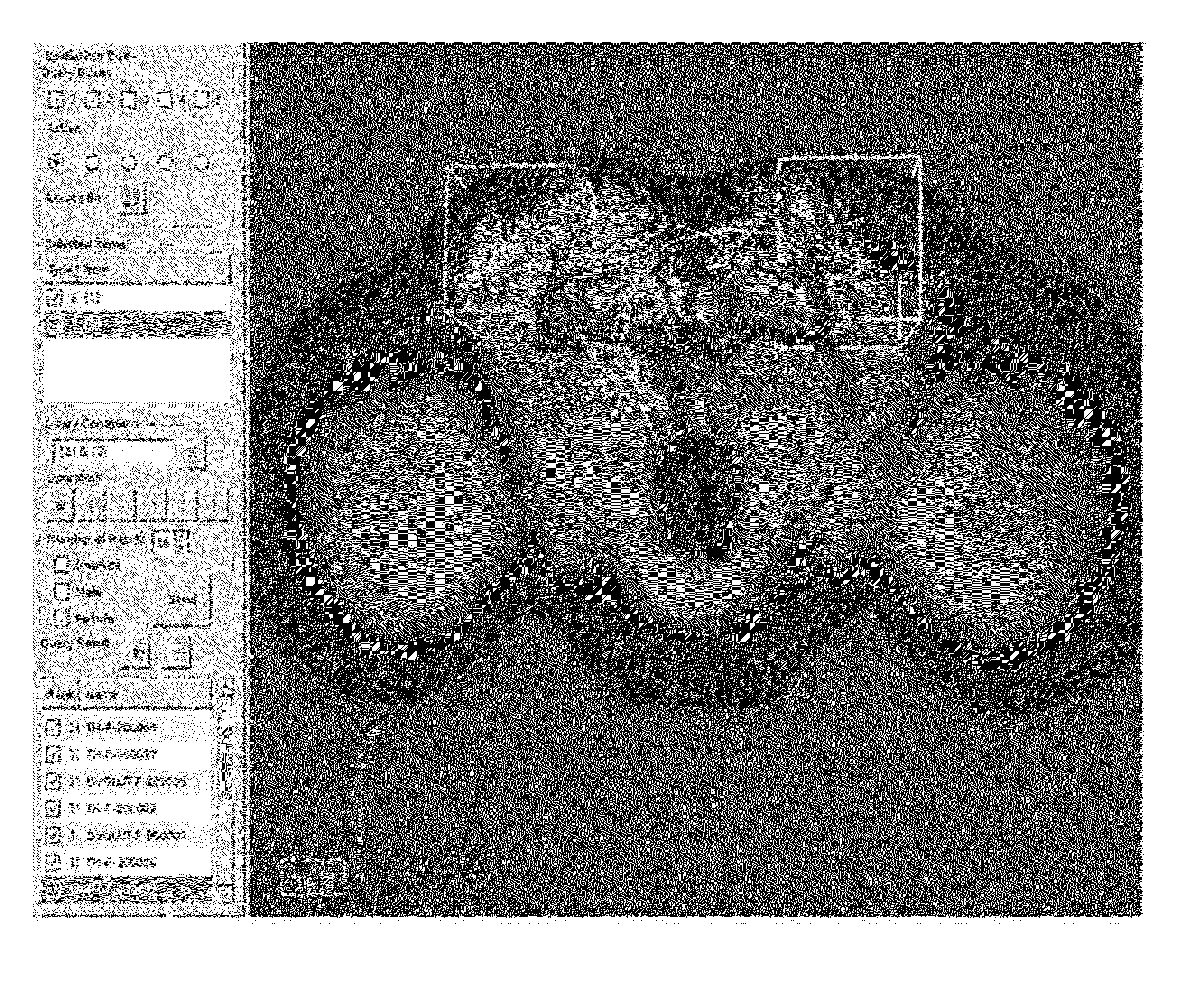
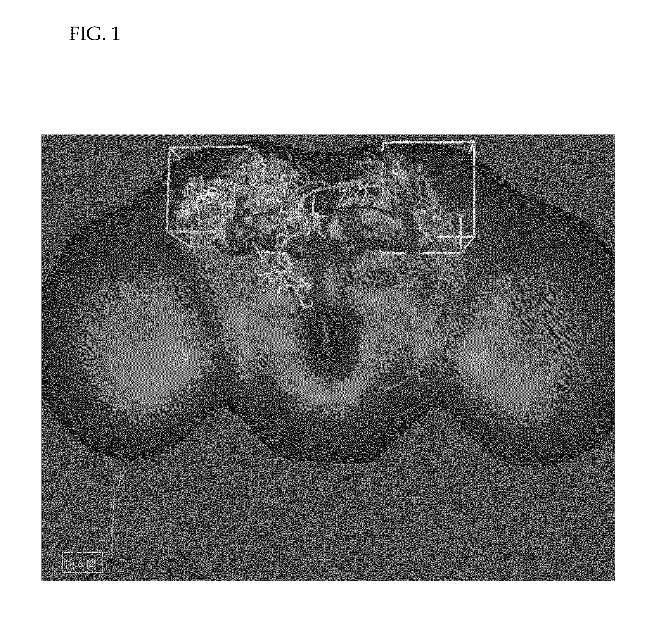
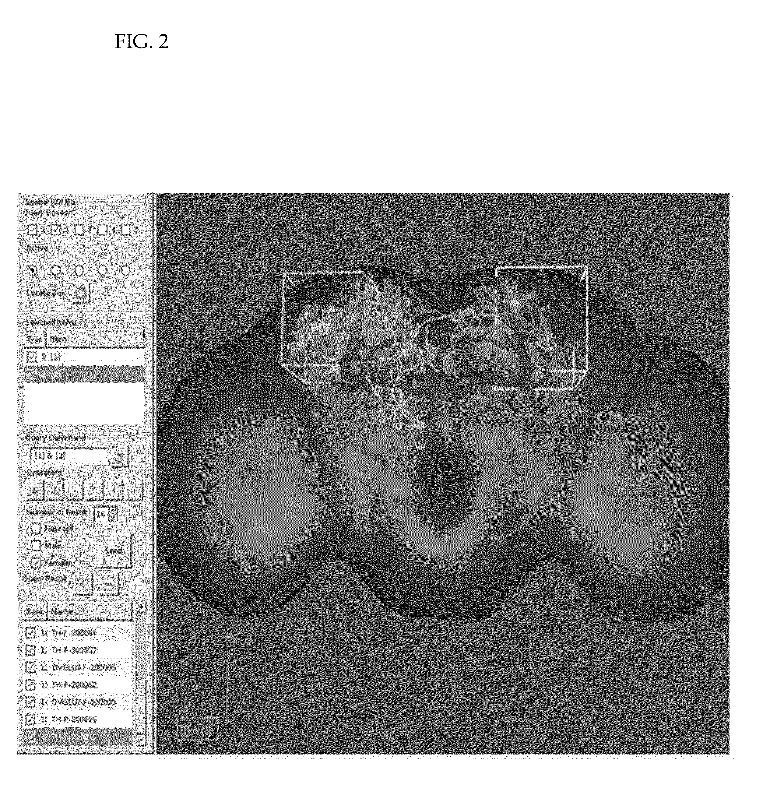
[0022]The visualization interface displays the neurons / neuropils in 3D and allows operations such as rotation and zooming from different angles to facilitate multi-angle observation. The visualization interface can also display the neural images using semi-transparent effect to illustrate numerous network motif images simultaneously on the visualization interface.
[0049]The 3D query interface provided users the capability to select the target neuron / neuropils directly from 3D scene instead of finding the desire target from a very long name list. Please refer to FIG. 4, here presents an innervation-query interface, which enables users to build paths of neuron innervations step-by-step. Users may start from one neuron / neuropil and find out the connections of said neuron / neuropil with other neurons / neuropils. The figure illustrates two neurons (DVGLUT-F-000029, lemon yellow and DVGLUT-F-0000189, orange) innervating the mushroom body (MB_r, maroon). DVGLUT-F-000029 neuron is found to connect to a selected box, which occupies the space belonging to another neuropil-optic tubercle (OpTu—1, green). A third neuron (DVGLUT-F-000134, red) is found with its terminals innervating OpTu_l from the right side. A third neuropil (Lob_l, blue) is found to connect with DVGLUT-F-000134 neuron through its nerve terminals. A column on the left of the visualization interface shows part of neurons also innervating Lob—1.

Problems solved by technology

Although scientists have a clear understanding of macro-scale biology such as animal behavior and micro-scale biology such as gene expression and perspectives of biology, micro-meso-scale biological research remains under-studied owing to technical limitations which including the difficulty to acquire the 3D structure of nerve cells and cerebral neural networks.
Biologists often can not obtain images (information) of an organism's internal structure without damaging the organism itself.
Furthermore, when acquiring biological images, physical limitations of laboratory equipment could only generate a serial of two-dimensional (2D) images instead of three dimensional images; as a result, the spatial information between organs is not immediately made available.
Although the information processing and transmission of the human brain fascinates scientists the most, the fact that the human brain has 100 billion neurons, plus human's relatively longer life span and genes that cannot be manipulated at will, has limited neuroscience research on human brain structures at the cellular level.

Method used


Image

  • Method for Searching and Constructing 3D Image Database
  • Method for Searching and Constructing 3D Image Database
  • Method for Searching and Constructing 3D Image Database

Examples


example 1

Generating 3D Images

[0030]The 3D image was generated by inputting Drosophila neuronal image obtained from micro-imaging device. Said image was acquired from a fluorescent-labeled specimen scanned by a laser scanning microscope. During the scanning process, at least part of the sample was scanned by laser. The cross-section of different depths of the sample was scanned in accordance with a predetermined order; the resulting scanned images were numerous plane images at different depths. Images from different slices of the same stack were combined to form a complete image; and then the resulting 3D image consisting of different cross-sections was generated by computer software such as AVIZO (Visualizaiton Science Group, Merignac Cedex, France).

example 2

Constructing 3D Image Database

1. Aligning 3D Images to a Standard Coordinate

[0031]The 3D images generated from image processor programs, such as AVIZO, were aligned to a standard coordinate. The alignment correction on 3D images made different image sets to have common space coordinates.

[0032]In a preferred embodiment, the standardized coordinate was generated by demarcating a standard Drosophila brain space according to Cartesian axis x, y and z. (Wu, C. C. et al. 2008 Algorithm for the creation of the standard Drosophila brain model and its coordinate system. 5th International Conference on Visual Information Engineering VIE, Xi' an, China, pp. 478-483).

[0033]After aligning to the standardized coordinate, each raw 3D image was corrected to fit the standardized coordinate. As a result, each voxel of the 3D image would designate a point location (X,Y,Z). The spatial and intensity information of the voxel (with gray level intensity value above threshold) of 3D images was then stored ...

example 3

Searching 3D Image Database

[0045]The present invention provided a method for query of neuronal image database. A preferred embodiment was an interactive method for searching a 3D brain neuronal image database of Drosophila.

[0046]The database in the present invention stored the information of connectivity relationships between the brain neuronal networks of Drosophila. Users can query the database through a 3D interactive interface. In this database, users could query the neuronal transmission paths of neuronal signal stimulated by the binding of olfactory receptors with certain molecules. The results showed the information regarding neural transmission paths to olfactory glomeruli, and users could further query which site of mushroom body was the recipient of the stimulated signal.

[0047]FIG. 5 illustrated system architecture for interactive search of the 3D image database. The said interactive search device 10 was linked via the internet 60 for data transmission, and automatically s...


PUM


Abstract

The present invention relates to methods for searching and constructing a 3D motif image database, wherein said 3D motif image database can be used to understand the connection relationship of a 3D network, e.g. a neural network comprising biological neural networks or artificial neural networks. The searching and constructing methods are applied on the 3D motif image database, a proper computer-aided graphic platform. The database not only facilitates the management of the huge amount of categorized data but also rationally excavates the hidden information cloaked within.

Description

FIELD OF THE INVENTION[0001]The present invention relates to a method for query and construction of a three-dimensional (3D) image database.BACKGROUND OF THE INVENTION[0002]Research in brain function may be roughly divided into various levels (from microscopic to macroscopic), e.g. gene expressions, protein biochemical reactions, neuronal functions, brain neural network organizations and animal behavior. Molecular biology, flourished since the 1960s, allows genetic manipulation to reflect biological functions at different scales. In such ways, researchers can now make use of technology to identify Drosophila memory genes of olfaction and memory and alter these genes to influence its behavior. Although scientists have a clear understanding of macro-scale biology such as animal behavior and micro-scale biology such as gene expression and perspectives of biology, micro-meso-scale biological research remains under-studied owing to technical limitations which including the difficulty to ...

Claims


Application Information

Patent Timeline
Patent Type & Authority Applications(United States)
IPC IPC(8): G06K9/62G06F7/00G06N3/02G06T17/00
CPCG06T2200/24G06T19/00
Inventor WANG, SHENG-CHUANCHIANG, ANN-SHYNLEE, PING-CHANGZHUANG, CHAO-JUNCHANG, HSIU-MINGCHEN, YUNG-CHANGTSAI, KUEN-LONGHSIEH, CHANG-HUAINLIN, CHING-YAOCHING, YU-TAI
Owner NATIONAL TSING HUA UNIVERSITY
Learn More