Biphasic implant device providing gradient

a prosthetic device and gradient technology, applied in the direction of powder delivery, medical preparations, pharmaceutical delivery mechanisms, etc., can solve the problems of joint inability to move, stress and friction, and inability to effectively repair the articular cartilage, so as to prevent the formation of tissue, prevent the formation of incorrect tissue types, and prevent bone voids

Active Publication Date: 2010-12-30
DSM IP ASSETS BV
View PDF62 Cites 10 Cited by
  • Summary
  • Abstract
  • Description
  • Claims
  • Application Information

AI Technical Summary

Benefits of technology

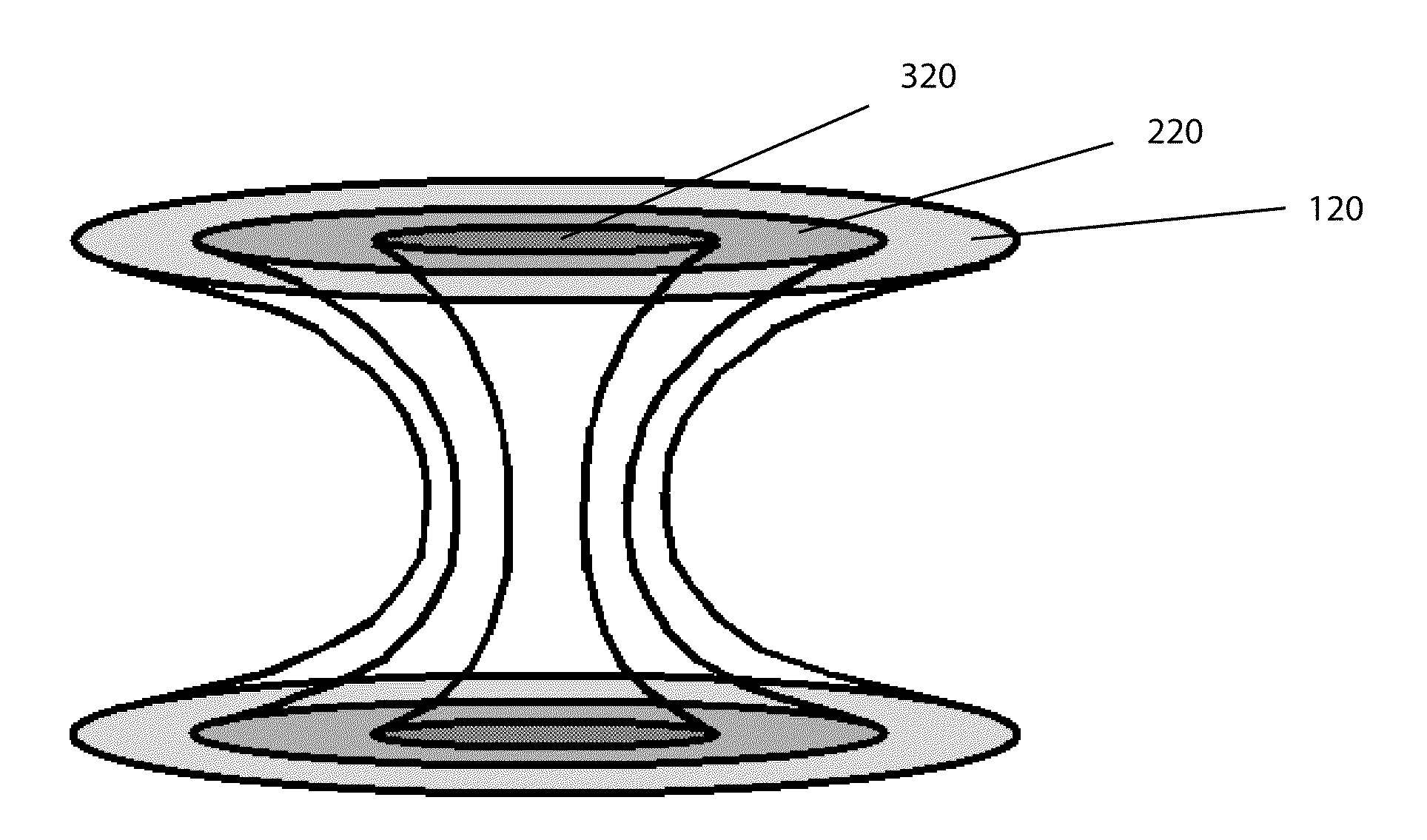
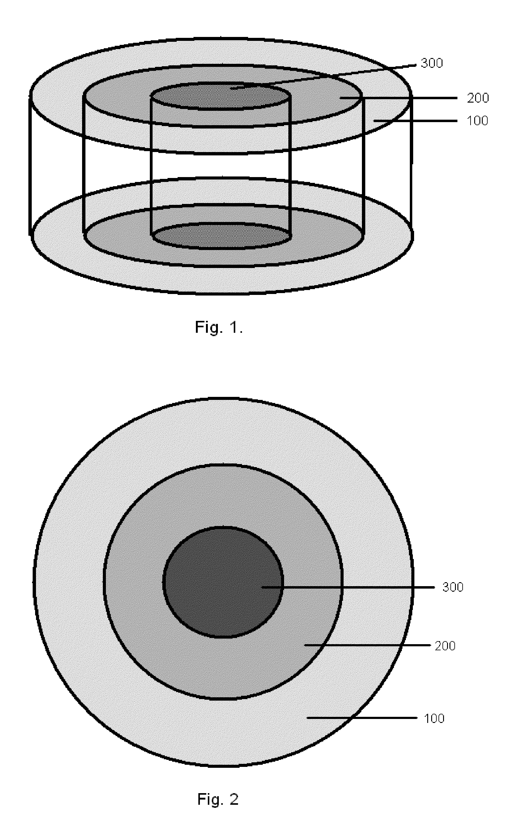
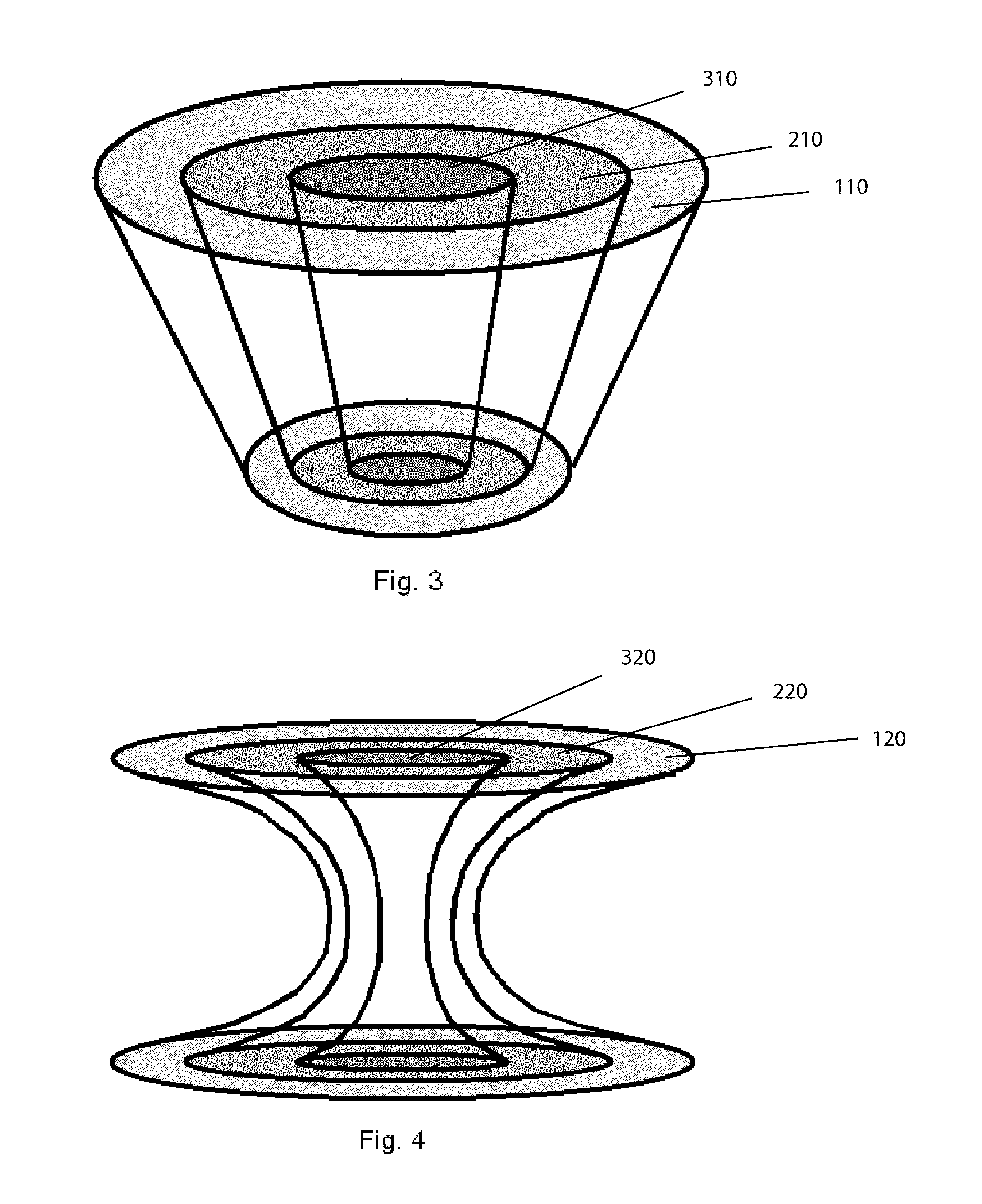
[0034]In another aspect of the invention, the implant provides for the systematic tissue conduction and growth from the surrounding cartilage tissue, and retards the formation of tissue in the interior of the implant. In this manner, it is believed that the growth of the incorrect type of tissue can be avoided, and better ensure that only the desirable hyaline cartilage is formed. In an embodiment, the device may comprise a gradient, where the gradient is arranged to retard the tissue formation most at or near the center of the implant (when viewed top down), and transitions to little or no retardation of tissue formation towards the perimeter of the implant, adjacent to normal cartilage tissue. The gradient may be in the form of a circular gradient, and may be uniform throughout the device from upper surface to lower surface, or alternatively may vary from top to bottom. The gradient may be a smooth transition or gradual gradient, or alternatively a stepwise gradient having well defined regions within the gradient. The gradient may be a concentration gradient, such as biologically active agents, additives, or combinations thereof. The gradient may be a physical gradient, such as porosity, density, expansion, swelling, elasticity, hardness, compressibility, and combinations thereof. The gradient may be a material gradient, or chemical gradient, such as molecular weight, cross-linking, hydrophobicity, polarity, crystalinity, and combinations thereof. The gradient may be part of the first phase of a multiphasic device, and corresponds to the cartilage region, and may be attached to a rigid base corresponding to the bone region.
[0035]In another aspect of the invention, the multiphasic implant provides for the transmission or conduction of pressure forces through the device, down to the underlying bone tissue below the device; in this manner, bone tissue loss below the device, such as may occur due to stress-shielding, may be minimized or avoided. One embodiment of an implant device capable of transmitting such forces would present a bone region presenting a porous material and a rigid penetrating force conductive material capable of transmitting the forces received from a malleable cartilage region to the underlying tissue. The forces to be transmitted may be hydrostatic and directed through channels running through the bone region material, or alternatively force transmission may be in the form of kinetic pressure pulses through the rigid conductive material arranged in the bone phase. The rigid conductive material may be in the form of columns arranged perpendicular to the top and bottom surfaces of the implant, and may flare out to a wider dimension at the junction with underlying bone. The rigid conductive material may be in the form of a rigid multi-facetted web structure oriented perpendicular to the top and bottom surfaces of the implant. In another embodiment, the rigid conductive material is a wedge or cone that transmits the forces through the implant to the underlying bone, but may also transmit some forces laterally as an outward force to the porous bone region material.
[0036]In yet another embodiment the multiphasic device capable of transmitting pressure forces presents at least a first material in the form of at least two porous rigid scaffolds, where the first material is separated by at least a second material in the form of a malleable elastic hydrogel, and where the hydrogel is capable of transferring hydrostatic pressure pulses through the bone region of the device in order to prevent bone voids from forming in external underlying bone tissue.

Problems solved by technology

Bone is very sensitive and the sharp pain of arthritis often comes from irritation of bone nerve endings and since human tissue has a very limited capacity to heal without a blood supply, articular cartilage cannot repair itself effectively.
Without articular cartilage, stress and friction would occur to the extent that the joint would not permit motion.
As stated above, articular cartilage has only a very limited capacity to regenerate.
If this tissue is damaged or lost by traumatic events, or by chronic and progressive degeneration, it usually leads to painful arthrosis and decreased range of joint motion.
Articular cartilage repair following injury or degeneration represents a major clinical problem, with treatment modalities being limited and joint replacement being regarded as appropriate only for the older patient.
(Extensive use of cortisone not only has a wide variety of harmful effects, but is also believed to harm cartilage.)
These were safer than Aspirin and cortisone but had potent side effects, especially causing bleeding within the stomach and intestinal ulcers.
While much safer and seemingly more effective, Vioxx was found to have significant cardiac side effects and is no longer available.
However, these anti-inflammatory medications only treat the symptoms of cartilage damage and arthritis and do not promote repair.
Once again, this procedure only treats the symptoms of cartilage damage and arthritis and does not promote repair.
But this is not a final solution as the degenerative process continues to wear away at the articular cartilage.
Although successful, the window of opportunity for this procedure is often missed, as the few clinical symptoms showing the need for this treatment are not evident until the defect deepens to involve the underlying bone, thus the damage encountered upon detection is frequently too extensive for repair through ACI.
Studies have shown that microfracture techniques do not fill in the chondral defect fully and the repair material formed is fibrocartilage.
The fibrocartilage tissue can temporarily return function for activities such as running and a sport play, but ultimately fails, as fibrocartilage is unable to mechanically share and disipate loading forces as effectively as the original hyaline cartilage.
Fibrocartilage is much denser and isn't able to withstand the demands of everyday activities as well as hyaline cartilage and is therefore at higher risk of breaking down.
Several plugs can fill up rather larger defects and will grow to re-supply a new joint surface.
Unfortunately, this procedure leaves defects of equal or worse proportions elsewhere and often the harvested tissue is not viable due to the traumatic harvesting procedure.
Due to the problems associated with current state of the art treatments, much work has been done to produce a synthetic off-the-shelf scaffold to be used in place of the harvested osteochondral plug.
However, these single-phase scaffold implants proved unsuccessful in healing of the complex multiphasic articular cartilage along with the underlying bone.
While these showed an improvement over single phase devices, it is evident that these devices do not take into consideration how cells will be migrating into the scaffolds as well as how their presence influences the surrounding, uninvolved tissue.
Additionally, prior art scaffolds did not take into consideration the joint fluid and how it impacts maturation and maintenance of healthy hyaline cartilage.
Although prior art synthetic scaffolds, whether single phase, multi-phase, or of gradient construction have proven suitable for growth and maturation of cells within a bioreactor, these prior art devices are unsuitable for direct implantation, for at least the reasons that follow.
Although some success in establishing hyaline cartilage can be seen in small defects of 5 millimeters or less, larger defects show tell tale signs of collapse or dimpling in the center of a repair plug, as the less desirable fibrocartilage, which has grown within the prior art devices, succumbs to the forces within the joint.
Additionally, prior art devices show a halo or ring of collapsed tissue around the periphery of the device do to lack of intimate contact with the uninvolved tissue that has retracted away from the defect site.
Another discovery of applicants is that prior art devices do not address the instantaneous articular cartilage tissue contraction that occurs when the surface of hyaline cartilage is cut or torn.
Anomalous mechanical loading of these tissues often leads to pathology.
For example, the lack of mechanical stimulation of a joint leads to suppression of proteoglycan synthesis and release of mediators responsible for degradation of cartilage matrix components.
This is believed to be the cause of collapse or dimpling of the newly formed cartilage seen with prior art devices.
Furthermore, there is a dearth of knowledge about the modes of mechanical signal transduction in chondrocytes.
Thus, this places a limit of success for prior art devices having a matrix equal to, or less stiff than the surrounding host tissue to 5 millimeters in diameter.
However, any device having a cartilage scaffold matrix greater in stiffness than the surrounding host tissue will not be properly influenced by mechanical signal transduction and will either form calcified tissues or disorganized fibrocartilage that collapses as the matrix degrades and the tissue experiences stress loading.
Within the bone layer, known prior art devices failed to recognize the impact a rigid scaffold has on the surrounding uninvolved tissue.
As this occurs, the bones lose minerals, heaviness (mass), and structure, making them weaker and increasing their risk of collapse and or breaking.
The theory behind this pathology formation is that stress shielding, caused by the presence of porous tissue scaffolds, results in bone density loss.
Two functional problems identified with rigid porous scaffolds of prior art devices are as follows.
First these rigid devices do not contain elongated channels and thus they tend to dissipate and dampen the hydrostatic pressure pulses that would normally flow through viscous fluids.
Secondly these devices are too rigid through the cartilage region thus not allowing for initial compression to establish a pressure wave through the bone marrow.
Concerning the synovial fluid, prior art devices fail to recognize the role this substance plays in maintaining healthy articular cartilage.

Method used


Image

  • Biphasic implant device providing gradient
  • Biphasic implant device providing gradient
  • Biphasic implant device providing gradient

Examples


Embodiment Construction

[0052]A device and methods are disclosed for treating tissue deficiencies, defects, voids and conformational discontinuities produced by congenital deformities, tissue pathology, traumatic injuries and surgical procedures, particularly those located in mammalian bone and cartilage. In one embodiment, the device is to provide the means by which hyaline cartilage tissue can be conducted across a tissue specific first scaffold region by controlled migration of chondrocytes and / or cartilage precursor cells. Additionally, in an embodiment, the scaffold region can be designed to affect the concentration, location and activity of fluids, factors, molecules or other biologically active agents received from, or delivered to, the extracellular fluids, especially synovial fluid. Thus, the device provides means to regenerate a first specific form of tissue.

[0053]A tissue specific second scaffold region may be attached to the first region for controlled migration of osteoblasts and / or bone precu...


PUM


Abstract

Tissue implants prepared for the repair of tissues, especially avascular tissues such as cartilage. One embodiment presents an electric potential capable of receiving and accumulating desirable factors or molecules from surrounding fluid when exposed to dynamic loading. In another embodiment the implant promotes tissue conduction by retarding, restricting and controlling cellular invasion through use of gradients until competent tissue forms. Further embodiments of the tissue implants may be formed into a multi-phasic device that provides deep tissue mechanical stimulus by conduction of mechanical and fluid forces experienced at the surface of the implant.

Description

BACKGROUND OF THE INVENTION[0001]What is disclosed is a device for repairing and replacing lost or damaged tissue. Particularly, one embodiment is directed to a multi-phasic prosthetic device for repairing or replacing cartilage or cartilage-like tissues. Said prosthetic devices are useful as articular cartilage substitution material and as a scaffold for regeneration of articular cartilaginous tissues.[0002]Cartilage is found throughout the body, such as in the supporting structure of the nose, ears, ribs (elastic cartilage), within the meniscus (fibrous cartilage), and on the surfaces of joints (hyaline cartilage or articular cartilage). A joint is a bending point where two bones meet. The knee, hip, and shoulder are the three largest joints.[0003]The specialized covering on the ends of bones that meet to form an articulating joint is called hyaline or articular cartilage. It is the cartilage that is damaged and wears as one ages, or sustains an injury. Articular cartilage is uniq...

Claims


Application Information

Patent Timeline
Patent Type & Authority Applications(United States)
IPC IPC(8): A61K9/14
CPCA61K9/0024
Inventor CASTIGLIONE-DODD, EMME M.BRADIOA, GINOEBRAHIML, ALIRINGEISEN, TIMOTHY A.
Owner DSM IP ASSETS BV
Why Eureka
Try Eureka