
N-Linked Glycosylation Alteration in E0 and E2 Glycoprotein of Classical Swine Fever Virus and Novel Classical Swine Fever Virus Vaccine
a technology of vaccines, which is applied in the field of n-linked glycosylation alteration in e0 and e2 glycoproteins of novel classical swine fever virus vaccines, can solve problems such as inefficient glycosylation, and achieve the effect of delaying the onset or severity of cs
- Summary
- Abstract
- Description
- Claims
- Application Information
AI Technical Summary
Benefits of technology
Problems solved by technology
Method used
Image

Examples
example 1
Viruses and Cell Cultures
[0090]Swine kidney cells (SK6) (Terpstra et al., supra) free of Bovine Viral Diarrhea Virus (BVDV) were cultured in Dulbecco's Minimal Essential Medium (DMEM, GIBCO, Grand Island, N.Y.) with 10% fetal calf serum (FCS, Atlas Biologicals, Fort Collins, Colo.). CSFV Brescia strain (obtained from the Animal and Plant Health Inspection Service, Plum Island Animal Disease Center) was propagated in SK6 cells and used for the construction of an infectious cDNA clone (Risatti et al. 2005a, supra). Growth kinetics were assessed on primary swine macrophage cell cultures prepared as described by Zsak et al. (1996. J. Virol. 70: 8865-8871). Titration of CSFV from clinical samples was performed using SK6 cells in 96-well plates (Costar, Cambridge, Mass.). Viral infectivity was detected, after 4 days in culture, by an immunoperoxidase assay using the CSFV monoclonal antibodies (mAbs) WH303 (Edwards et al. 1991. Vet. Microbial. 29:101-108) and the Vectastain ABC kit (Vector...
example 2
Construction of CSFV Glycosylation Mutants
[0091]A full-length infectious clone of the virulent Brescia isolate (pBIC) (Risatti et al. 2005a, supra) was used as a template in which putative O- and N-linked glycosylation sites in the E2 glycoprotein were mutated. Glycosylation sites were predicted using analysis tools from the Center for Biological Sequence Analysis (http: / / www.cbs.dtu.dk / services / ). Mutations were introduced by site-directed mutagenesis using the QuickChange XL Site-Directed Mutagenesis kit (Stratagene, Cedar Creek, Tex.) performed per manufacturer's instructions and using the following primers (only forward primer sequences are shown); O1v: CATCATTACATAAGG ACGCTTTAGCCACTTCCGTGACATTCGAGC (SEQ ID NO:4); N1v: CCCTGTAGT CAAGGGAAAGTACGCCACAACCTTGTTGAATGGTAG (SEQ ID NO:5); N2v: AAA GTACAACACAACCTTGTTGGCTGGTAGTGCATTCTACCTAGT (SEQ ID NO:6); N3v: ATTCTACTGTAAATGGGGGGGCGCTTGGACATGTGTGAAAGGTGA (SEQ ID NO:7); N4v: ATAGGTAAGTGCATTTTGGCAGCTGAGACAGGTTACAGAATAGTG (SEQ ID NO:8); N5v...
example 3
In Vitro Rescue of CSFV Brescia and Glycosylation Mutants
[0092]Full-length genomic clones were linearized with Srfl and in vitro transcribed using the 17 Megascript system (Ambion, Austin, Tex.). RNA was precipitated with LiCl and transfected into SK6 cells by electroporation at 500 volts, 720 ohms, 100 watts with a BTX 630 electroporator (BTX, San Diego, Calif.). Cells were seeded in 12-well plates and incubated for 4 days at 37EC and 5% CO2. Virus was detected by immuno-peroxidase staining as described above, and stocks of rescued viruses were stored at −70EC.
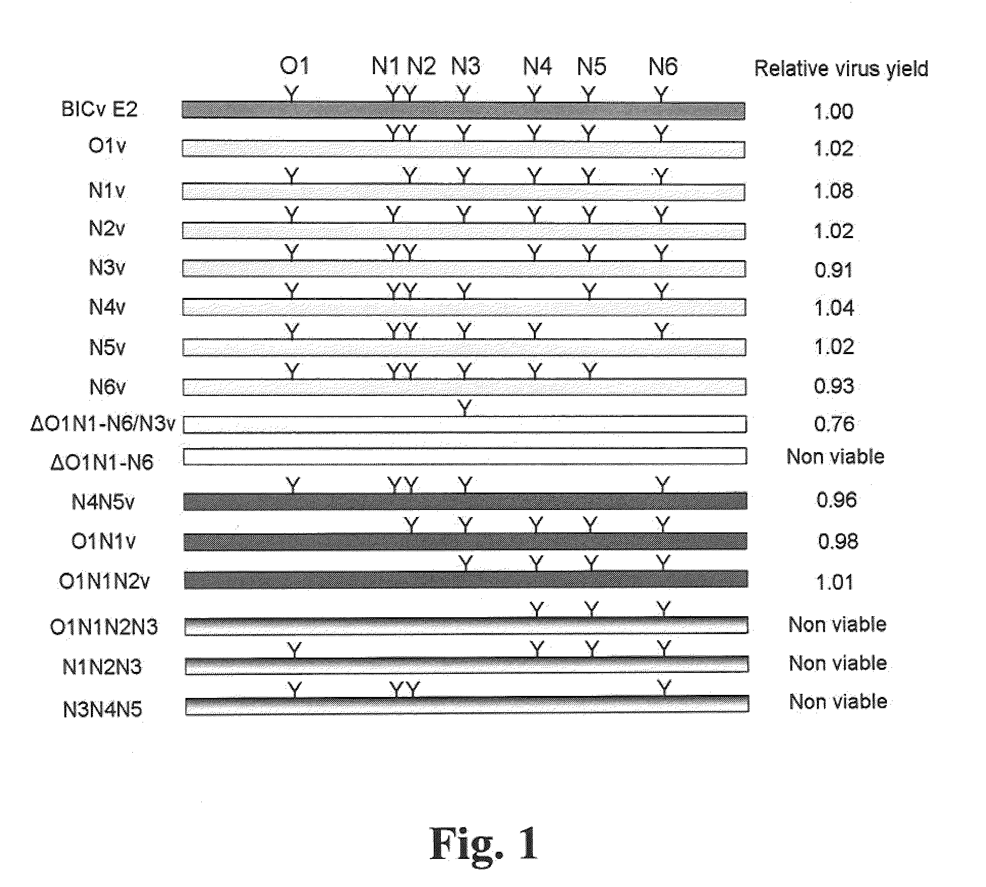
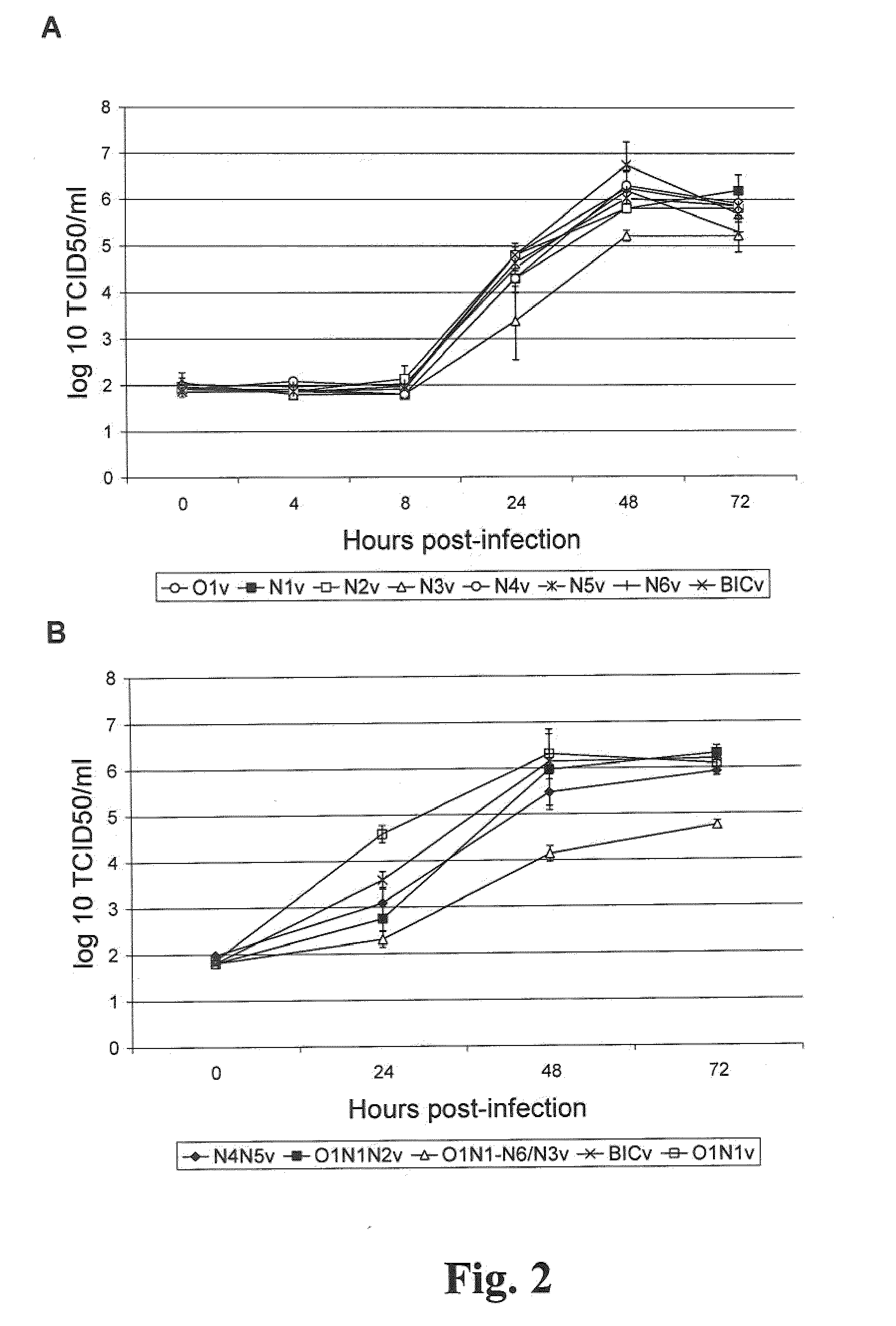
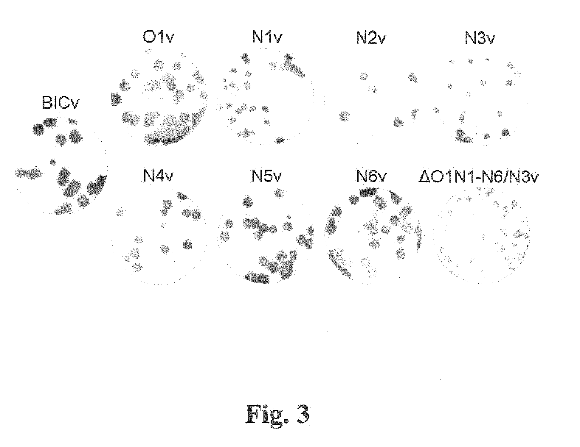
[0093]Infectious RNA was in vitro transcribed from full-length infectious clones of the CSFV Brescia strain or a set of glycosylation mutants (Table 1, FIG. 1) and used to transfect SK6 cells. Mutants referred to as O1, N1, N2, N3, N4, N5, and N6 represent each of seven putative glycosylation sites starting from the N terminus of E2 (Table 1), whereas multiple mutants are represented by combinations of indicated sites (FIG. 1...
PUM
| Property | Measurement | Unit |
|---|---|---|
| temperature | aaaaa | aaaaa |
| Tm | aaaaa | aaaaa |
| temperatures | aaaaa | aaaaa |
Abstract
Description
Claims
Application Information