
Photokinetic ocular drug delivery methods and apparatus

a biologically active substance and ocular drug technology, applied in the field of photokinetic delivery of biologically active substances, can solve the problems of visual impairment, eye drops may not be an effective method for administering drugs of larger molecular weight into the eye, and the minimally invasive technique only allows for less than 5%, so as to achieve less cost.

Active Publication Date: 2011-05-26
BOARD OF RGT THE UNIV OF TEXAS SYST
View PDF9 Cites 66 Cited by
  • Summary
  • Abstract
  • Description
  • Claims
  • Application Information

AI Technical Summary

Benefits of technology

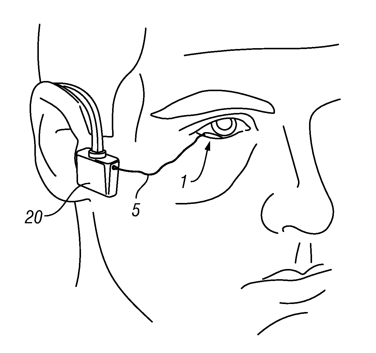
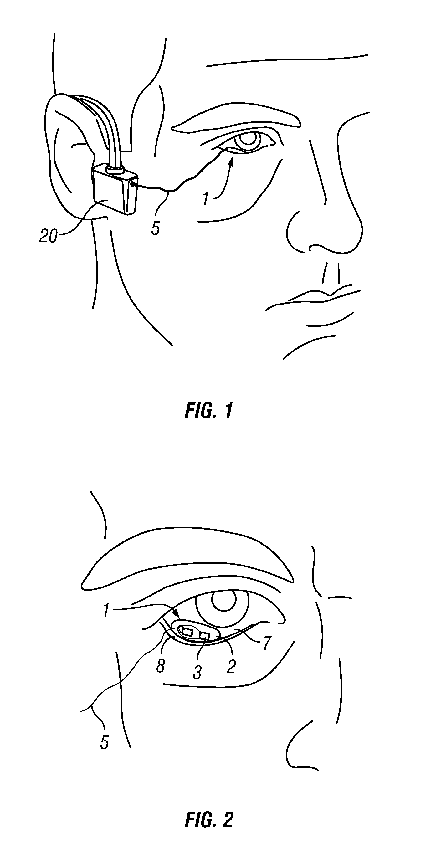
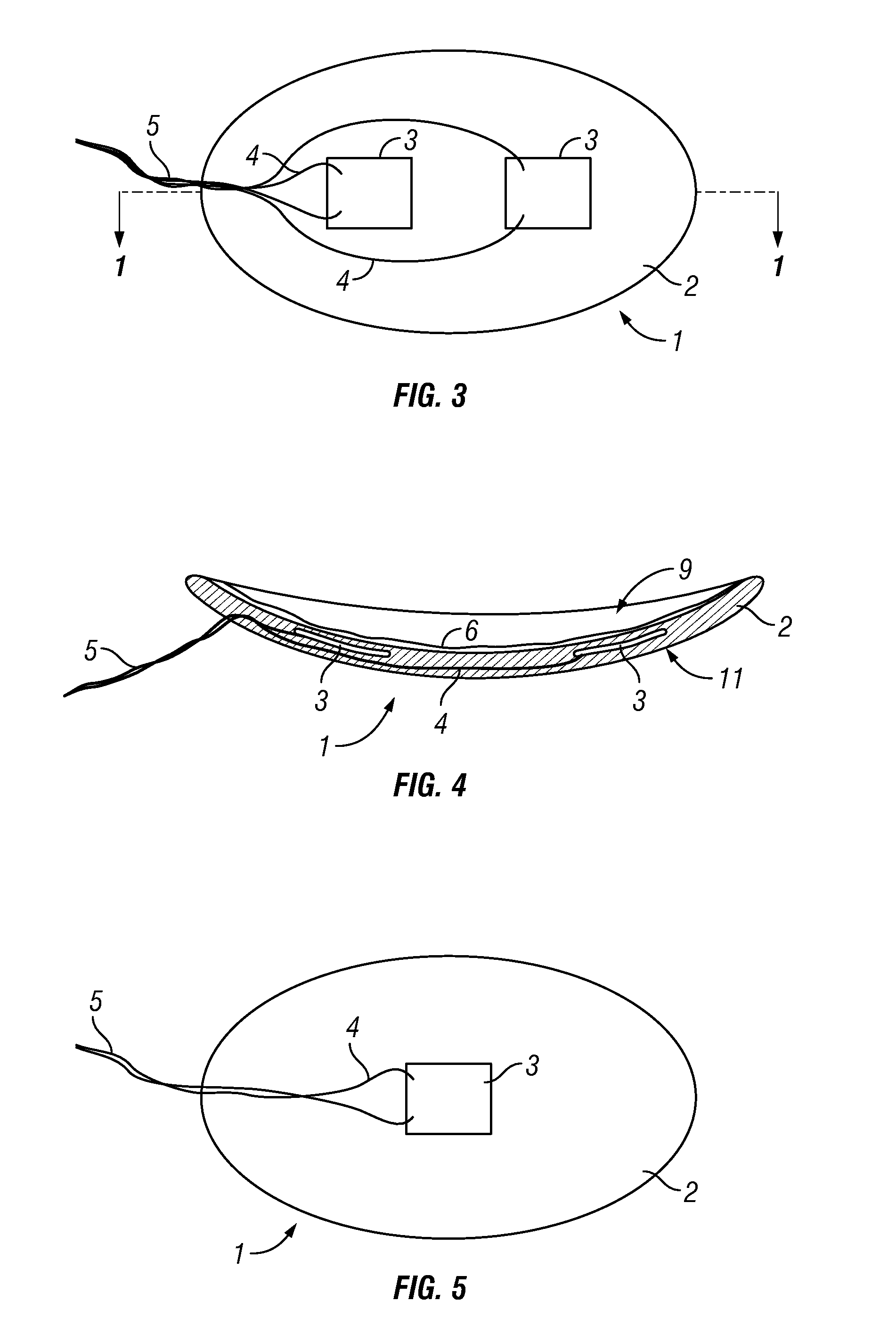
[0019]The novel technology described herein generally relates to devices and methods for transscleral / transcorneal needleless drug administration. Specifically, the technology is an ocular drug delivery method wherein a drug applied to scleral / corneal tissue is illuminated with a selected narrow wavelength light from a LED source and pulsed at a selected frequency that then causes the drug to permeate into and through the tissue. The technology comprises in vitro methods for the selection of optical properties of light emitting devices applied to specific drug formulations thus defining in vivo administration systems. The technology provides a non-invasive method using light and drug reservoir devices to introduce drugs into the eye without the use of needles. The system is safer and less costly than ocular drug administration by needle injection.

Problems solved by technology

Millions of people worldwide suffer from ocular diseases, many of which lead to visual impairment.
However, this minimally invasive technique only allows for less than about 5%, and many times less than 1%, of the administered drug to reach the drug target site before being washed away by tear formation or being absorbed systemically by the surrounding eye tissues.
Eye drops may not be an effective method for administering larger molecular weight drugs into the eye for treatment of posterior segment eye diseases such as age-related macular degeneration, diabetic retinopathy, retinitis pigmentosa, and primary ocular lymphoma.
Systemic administration of an eye targeted drug has very poor bioavailability within the eye due to blood-ocular barriers that normally protect the eye from circulating antigens, inflammatory mediators, and pathogens.
Typically, systemic administration does not yield therapeutic drug levels in the posterior vitreous, retina, or choroid, and although systemic administration can deliver drugs to the posterior eye, the large systemic doses necessary to achieve intraocular therapeutic levels are often associated with significant side effects.
Intravitreal administration of drugs by needle injection is associated with an entirely new set of potentially catastrophic side effects such as infection, intravitreal hemorrhage or retinal detachment.
Additionally, ocular injections are painful and costly.
The combination of low power and slow cyclical stimulation of the molecular optical sites would result in reversible conformational changes causing the molecule to bend and flex resulting in gross physical movement.
While not intending to be bound by any theories, it is hypothesized that if a drug molecule in a pharmacologically acceptable formulation is placed on the surface of the sclera / cornea and cyclically illuminated with a selected wavelength of light at a selected pulse rate, the resulting cyclic physical shape change of the molecule may cause gross movement and result in the migration of the molecule across the sclera membrane.
Although ionotophoresis provides a method for controlled drug delivery transdermally, irreversible skin damage can occur from galvanic and pH burns resulting from electrochemical reactions that occur at the electrode and skin interface.
Consequently, its application to ocular therapies has been limited, with limited reports of its use in delivering molecules into the eyes of patients.
However, many of these approaches suffer limitations as well, such as being suitable only for delivering therapeutic molecules of a limited size (e.g., molecular weights of less than 200 Da), or unappealing side affects or potential for added eye damage for the patient seeking treatment.

Method used


Image

  • Photokinetic ocular drug delivery methods and apparatus
  • Photokinetic ocular drug delivery methods and apparatus
  • Photokinetic ocular drug delivery methods and apparatus

Examples


example 1

Methotexate Transcleral Delivery

[0120]Materials and Methods

[0121]A traditional “up and down” vertical Franz cell skin perfusion apparatus as described above was adapted and used for this sclera tissue permeation model. The absorption spectrum of the test drug, methotrexate (MTX, a drug used in the treatment of primary ocular lymphoma) was determined. Two wavelengths of light were selected of this facilitated permeation study. Samples of the Franz recipient solution were taken at 15, 30 and 60 minutes of photokinetic exposure and passive controls and analyzed by HPLC.

[0122]Instruments and Materials

[0123]Franz cells (PermeGear, Inc, Bethlehem Pa.) having an 11.28 mm diameter permeation area, 1.0 cm2 were used in the MTX experiment. A photokinetic ocular drug delivery (PODD)-modified Franz cell testing device was configured so that it accommodated the placement of LEDs within the donor chamber. The cells were placed within an aluminum block heater 35.5° C. on a magnetic stir bar setup ...

example 2

Delivery of Insulin

[0154]Two pertinent ocular drugs were selected for testing the hypotheses. Methotrexate (MW=454 Daltons) is used for the treatment of primary ocular lymphoma. Insulin (MW=5808 Daltons, 5.808 kDa) and insulin like growth factors have been implicated as a possible preventive treatment for diabetic retinopathy. The transscleral delivery of methotrexate has been described in Example 1, above.

[0155]A traditional “up and down” vertical Franz cell (11.28 mm diameter permeation area, 1.0 cm2) perfusion apparatus (FIG. 6) was adapted and used for this sclera tissue permeation model. PODD modified Franz cell testing device was configured so that it accommodated the placement of the selected discreet wavelength LEDs within the donor chamber. Discrete LEDs at 351 nm and 370 nm were used for the MTX study and 405 nm and 450 nm for the insulin study (peak emitting wavelengths at ±10 nm at the 50% radiance output). The LEDs were driven by a square wave pulse generator set at 24 ...

example 3

Transcleral Insulin Delivery of High Molecular Weight Drugs

[0170]Using the same procedures as set out above, and the apparatus of FIG. 6, scleral tissue permeation determinations of methotrexate over a short time period, as well as the scleral tissue permeation of vancomycin (1 mg / mL), insulin, Insulin Like Growth Factor-1 (IGF-1), Avastin™ (bevacizumab; Genentech, San Francisco, Calif.), and hyaluronic acid (HA) were conducted. High molecular weight hyaluronic acid (5,000 Da+in size) was selected as a test compound because the sodium salt of hyaluronic acid (SH) is a high molecular weight biopolymer made of repeating disaccharide units of glucuronic acid and N-acetyl-β-glucosamine, and which is present in the vitreous body and the aqueous humor. Hyaluronic acid is a natural polymer which, due to its water retaining capability, binds to cell membranes and can therefore be considered to be a putative vehicle for controlled ocular delivery (Durrani, et al., Int. J. Pharm., Vol. 118 (2...


PUM

PropertyMeasurementUnit
Fractionaaaaaaaaaa
Fractionaaaaaaaaaa
Massaaaaaaaaaa

Abstract

The present invention relates generally to transscleral, transcorneal, and transocular delivery of biologically active substances through the tissues, blood vessels and cellular membranes of the eyes of patients without causing damage to the cellular surface, tissue or membrane. The invention provides compositions and methods for enhanced transscleral, transcorneal and transocular delivery of biologically active substances using pulsed incoherent light, and particularly the transcleral, transcorneal or transocular delivery of high molecular weight biologically active substances to a patient using pulsed incoherent light. The invention further provides a device for the application of the pulsed incoherent light to cellular surfaces and membranes of the eye of a subject using those compositions and methods.

Description

CROSS REFERENCE TO RELATED APPLICATIONS[0001]This provisional patent application claims priority to U.S. Provisional Patent Application Ser. No. 61 / 250,371, filed Oct. 9, 2009, and U.S. Provisional Patent Application Ser. No. 61 / 328,625, filed Apr. 27, 2010, both of which are incorporated herein by reference in their entirety.STATEMENT REGARDING FEDERALLY SPONSORED RESEARCH OR DEVELOPMENT[0002]Not applicable.REFERENCE TO APPENDIX[0003]Not applicable.BACKGROUND OF THE INVENTION[0004]1. Field of the Invention[0005]The inventions disclosed and taught herein relate generally to photokinetic delivery of biologically active substances across a mammalian ocular surface. More particularly, the invention provides methods, apparatus, and compositions for the transcleral / transcorneal, ocular delivery of biologically active substances, such as therapeutic agents, using pulsed incoherent light.[0006]2. Description of the Related Art[0007]Millions of people worldwide suffer from ocular diseases, ...

Claims


Application Information

Patent Timeline
IPC IPC(8): A61F9/00C12N13/00
CPCA61F9/0017A61N5/062A61K41/00A61F9/0079A61K38/12A61K38/14A61K45/06A61K31/167A61K31/58A61K31/7042A61K31/728A61K9/0048A61K47/16A61K47/36A61K31/519A61K31/573A61K38/28A61K38/30A61N2005/0651A61N2005/0656A61N2005/0659A61N2005/0661A61N2005/0662A61K41/0057
Inventor KRAFT, EDWARD R.KULP, GABRIELA A.GODLEY, BERNARD F.KOUTROUVELIS, ARISTIDES P.
Owner BOARD OF RGT THE UNIV OF TEXAS SYST
Learn More