Method and apparatus for monitoring processes in living cells

a living cell and process monitoring technology, applied in the field of surface plasmon resonance (spr) technique, can solve the problems of limiting the operation of visible and nir wavelengths, labeling can alter the physiological activity of interacting ligands, and the complexity of cells poses an enormous difficulty in evaluating the precise mod

Inactive Publication Date: 2011-08-04
YISSUM RES DEV CO OF THE HEBREWUNIVERSITY OF JERUSALEM LTD
View PDF21 Cites 21 Cited by
  • Summary
  • Abstract
  • Description
  • Claims
  • Application Information

AI Technical Summary

Benefits of technology

[0058]In yet another specific preferred embodiment, tomographic measurements are performed at various wavelengths, each at optimal incidence angle, such that the measurement provides SPR information on slices at different heights above the surface of the metallic film, and thus allows identifying the location of the different biomolecules and organelles within cells.
[0059]In a further preferred embodiment of the invention, the high sensitivity of the FTIR-SPR apparatus of the present invention is used for two-dimensional microscopy, by employing micron-scale gold film patches placed on the base of the SPR prism, whereas the patch size is determined upon ability to produce a detectable signal and the thickness of the gold patches matches the condition for the observation of SPR reflection minima, thereby enabling the detection and the monitoring of cells cultured on said patches.

Problems solved by technology

The complexity of cells poses an enormous difficulty in evaluating the precise mode by which molecules of life and other materials such as drugs, toxins and pathogens interact with them.
A major challenge in modern pharmacology is to develop new experimental strategies for monitoring the dynamic interactions between molecules of biomedical significance and cognate targets in living cells.
However, there is always a risk that labeling can alter the physiological activity of the interacting ligands.
These conventional applications typically utilize glass-based optics, which limits their operation to the visible and NIR wavelength range (near-infrared 0.75-1.4 μm wavelength range).
However, visible-range SPR is not suitable for studying processes occurring inside living cells since the cells' size considerably exceeds the penetration depth of visible-range surface-plasmon waves.

Method used


Image

  • Method and apparatus for monitoring processes in living cells
  • Method and apparatus for monitoring processes in living cells
  • Method and apparatus for monitoring processes in living cells

Examples


example 1

[0165]Real-time monitoring of transferrin-induced endocytic vesicle formation

Experimental Setup

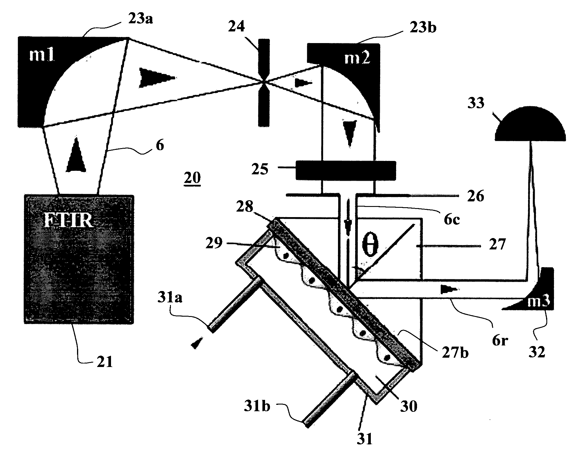
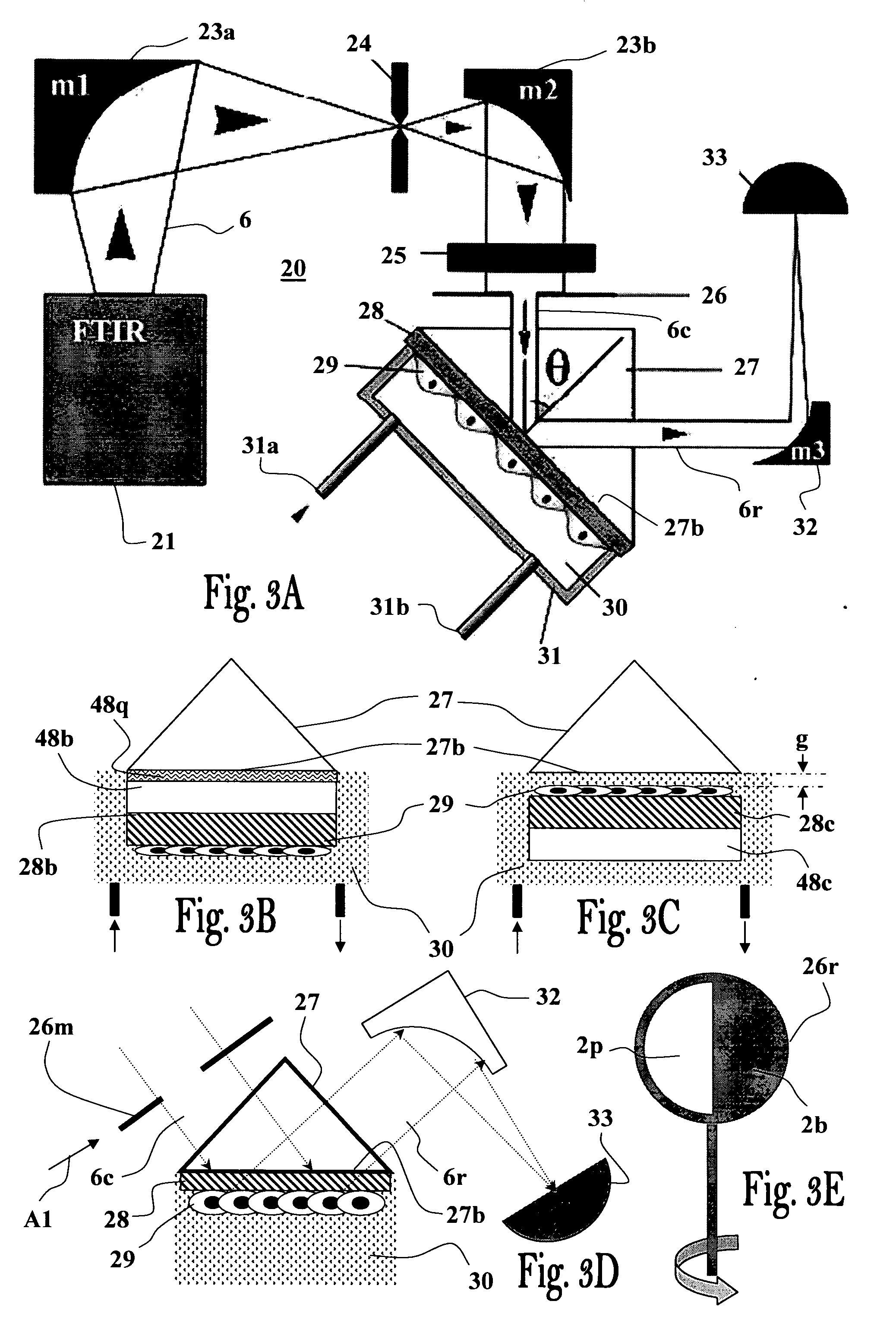
[0166]Surface plasmon (SP) was excited using Kretchmann's geometry as illustrated in FIG. 3A employing the Bruker FTIR spectrometer (Equinox 55—Bruker Optik GmbH, Ettlingen, Germany) equipped with a KBr beam splitter as mid-IR source, and a right-angle ZnS prism having a 20×40 mm base (ISP Optics, Inc., Irvington, N.Y, US) coated with an 18-nm-thick gold film (electron-beam evaporation). Cells were cultured on the gold surface, as described below. The prism and cells were attached to a flow chamber mounted on a goniometer, in such way that the cells on the gold-coated base faced the flow chamber's volume (0.5 ml). The flow chamber was filled with cell culture medium, resulting in direct contact between the medium and the gold layer, or cells cultured on that layer. The medium was passed through the chamber at a constant flow rate (5 μl / min) during the entire experiment, using a motorized b...

example 2

[0189]The following examples provide several biological applications of the FTIR-SPR technique of the invention. The experimental setup is based on the setup shown in FIG. 3A, utilizing the Bruker Equinox 55 FTIR spectrometer with a tungsten lamp equipped with the KBr beam splitter as a light source. For the 1-mm-diameter pinhole, the beam diameter is about 3-4 mm and the beam divergence is Δθdiv=0.8°. The collimated beam passes through the grid polarizer (Specac, Ltd.) and is reflected from the right-angle ZnS prism (ISP Optics, Inc.) mounted on a θ-2θ rotating table. The additional parabolic mirror focuses the reflected beam on the liquid-nitrogen-cooled MCT (HgCdTe) detector. A temperature-stabilized flow chamber (with a volume of about 0.5 ml) is in contact with the gold-coated base of the prism. The gold film thickness is chosen according to the targeted wavelength (FIG. 17). To operate this setup, an appropriate incident angle was chosen. Then the sample was mounted and the re...

example 3

Glucose Uptake by Erythrocytes

[0192]In the following section measurements of glucose uptake by erythrocyte suspension in the PBS medium are described. Briefly, fresh human red blood cells (RBCs) were washed four times in PBS by centrifugation. The supernatant and buffy coat cells were discarded and RBCs were resuspended in glucose-free PBS to yield ce=5% v / v. For complete glucose depletion, cells were incubated for 60 min at 22° C. in PBS. Then, RBCs were centrifuged and resuspended in fresh PBS containing the required concentrations of D-glucose supplemented with L-glucose, to keep the total glucose concentration (osmolarity) equal to 20 mM. The SPR measurements were performed at 22° C. to slow down cellular glycolysis.

[0193]The measurements were performed using a ZnS prism coated with a 12-nm thick Au film. It was decided to operate the FTIR-SPR setup with λ=4 μm wavelength at which the surface plasmon penetration depth, δzd=4.5 μm (FIG. 20), is comparable to the typical erythrocy...


PUM

PropertyMeasurementUnit
wavelength rangeaaaaaaaaaa
thicknessaaaaaaaaaa
thicknessaaaaaaaaaa

Abstract

The invention provides a method and apparatus for monitoring processes in living cells by measuring optical reflectivity by Surface Plasmon Resonance at the surface and/or inside living cells attached to a thin metal film (28), wherein said thin metal film is attached or optically coupled to a base of a prism (27) such that a collimated and optically polarized light beam (6c) in the near-infrared and/or mid-infrared wavelength ranges directed to a side surface of the prism is internally reflected by said prism at its base (27b) and measured by detector means (33) capable of measuring the intensity and optionally also polarization or phase of the reflected beam (6r).

Description

FIELD OF THE INVENTION[0001]The present invention relates to a Surface Plasmon Resonance (SPR) technique for real time monitoring of dynamic processes at the surface of, and inside living cells. More particularly, the present invention relates to a SPR method and system operating in the mid-infrared wave length range and capable of detecting dynamic processes occurring in living cells, and to methods and configurations for culturing the living cells.BACKGROUND OF THE INVENTION[0002]Cells, even the simplest ones, display a remarkable degree of complexity, reflected by their unique and specific abilities to interact with each other, and with various molecules of life (e.g., proteins, lipids, carbohydrates, etc). The complexity of cells poses an enormous difficulty in evaluating the precise mode by which molecules of life and other materials such as drugs, toxins and pathogens interact with them. A major challenge in modern pharmacology is to develop new experimental strategies for mon...

Claims


Application Information

Patent Timeline
Patent Type & Authority Applications(United States)
IPC IPC(8): G01N21/55
CPCG01N21/05G01N2021/3595G01N21/554G01N21/553
Inventor DAVIDOV, DANAROETI, BENJAMINGOLOSOVSKY, MICHAELLIRTSMAN, VLADISLAV
Owner YISSUM RES DEV CO OF THE HEBREWUNIVERSITY OF JERUSALEM LTD
Why Eureka
Try Eureka