Method for manufacturing a porous three-dimensional scaffold using powder from animal tissue, and porous three-dimensional scaffold manufactured by same

a three-dimensional scaffold and animal tissue technology, applied in the field of manufacturing a porous three-dimensional scaffold, can solve the problems of affecting the quality of patients' lives, unable to maintain the vitality of blood clots (including stem cells) in cartilage repair, and unstable physical properties of blood clots formed in this technique, etc., to achieve the effect of regenerating hyaline cartilage tissue, enhancing the effect of cartilage regeneration

Inactive Publication Date: 2011-08-11
AJOU UNIV IND ACADEMIC COOP FOUND
View PDF4 Cites 32 Cited by
  • Summary
  • Abstract
  • Description
  • Claims
  • Application Information

AI Technical Summary

Benefits of technology

[0045]The porous three-dimensional scaffold of the present invention manufactured by an animal tissue, for example, an animal cartilage powder can be advantageously applied in clinical fields, depending upon its purpose for treatment or use since it has various size, porosity, shape and structure. The present invention can provide the porous three-dimensional scaffold with enhanced effects capable of regenerating a hyaline cartilage tissue without any limitation of its physical property, porosity, shape, structure and size.
[0046]In addition, the porous three-dimensional scaffold of the present invention manufactured by an animal tissue, for example, an animal cartilage powder is remarkably biocompatible and clinically applicable without any immune rejection or inflammation when being implanted since the porous three-dimensional scaffold is manufactured by physically and / or chemically decellularizing an animal tissue before or after powdering it, or simultaneously with powdering it after isolating an animal tissue, for example, an animal cartilage. Furthermore, it is remarkably effective upon regenerating cartilage, as compared with scaffolds composed of collagen and other synthetic polymers.
[0047]Besides, the porous three-dimensional scaffold of the present invention manufactured by an animal tissue, for example, an animal cartilage powder can provide an environment suitable for cell migration, cell growth, and cell differentiation because it is manufactured to have a three-dimensional structure. Furthermore, the porous three-dimensional scaffold can be comprised of growth factors and proper components suitable for regenerating cartilage. The porous three-dimensional scaffold of the present invention can be usefully applied for a tissue-engineered scaffold of treating cartilage loss, due to its outstanding bio-compatibility, bio-degradable ability and three-dimensional structure.

Problems solved by technology

Once cartilage is damaged, its self-regeneration is extremely limited to ultimately cause osteoarthritis, which largely affects the quality of patients' lives.
This method is advantageous due to its simple operation procedure and short operation time, but has a critical limation being unable to maintain blood clots (including stem cells) well that are essential to cartilage repair.
Furthermore, the blood clots formed in this technique are physically unstable and not likely to regenerate efficiently a normal cartilage.
Hence, it is difficult to expect a successful healing of cartilage defect by using this bone marrow stimulation techniques.
This technique is outstanding to treat the damaged cartilages, but problematic to provoke a secondary damage during the isolation of osteochondral tissues.
However, it also has a problem that the area of cartilage damage should be covered with periosteum and tightly sutured after injecting chondrocytes.
Moreover, the periosteum may allow chondrocytes to overgrow, which may cause pain at damaged areas after the surgery.
The ACI is also disadvantageous to undergo two steps of surgery processes; isolating chondrocytes under an arthroscopic operation and culturing them for a long time in vitro, and then transplanting a cell suspension into damaged areas.
Unfortunately, a certain cellular antigen may cause an inflammation reaction or an immune rejection by being recognized by a host, when implanting a xenogenic and allogenic tissue to the host.
The physical method can destruct cell membranes and expose cellular components.
But, the physical method is usually considered as insufficient for complete decellularization.
Enzymatic digestion using trypsin, or chemical treatment using an ionic solution can destruct cell membranes and break the connection between the inner side and the outer side of cells.
The prior arts could reduce possible immune rejection since a porous three-dimensional scaffold is manufactured by decellularizing a natural tissue, however, they have a problem that there are limitations concerning the size, porosity, shape and structure of the scaffold due to using a natural tissue itself.
Accordingly, it was difficult to apply the prior arts to commercial purpose and therapeutic use.
But, the prior arts of simply decellularizing natural cartilage tissue could not meet the aforesaid needs because of obstructing diversification for treatment.
However, the above-mentioned methods have a problem that there are limitations concerning the size, porosity, shape and structure of the scaffold due to using extracellular matrix obtained by decellularizing a natural tissue itself.
Accordingly, it was difficult to apply the above-mentioned methods to commercial purpose and therapeutic use.

Method used


Image

  • Method for manufacturing a porous three-dimensional scaffold using powder from animal tissue, and porous three-dimensional scaffold manufactured by same
  • Method for manufacturing a porous three-dimensional scaffold using powder from animal tissue, and porous three-dimensional scaffold manufactured by same
  • Method for manufacturing a porous three-dimensional scaffold using powder from animal tissue, and porous three-dimensional scaffold manufactured by same

Examples


reference example 1

Isolation of Porcine Cartilage

[0063]In order to isolate porcine cartilage, pig cartilage was purchased and utilized from a facility satisfying standards referred to EN 12442 “Animal tissues and their derivatives utilized in the manufacture of medical devices, Part 1; Analysis and management of risk, Part 2; Controls on sourcing, collection and handling”.

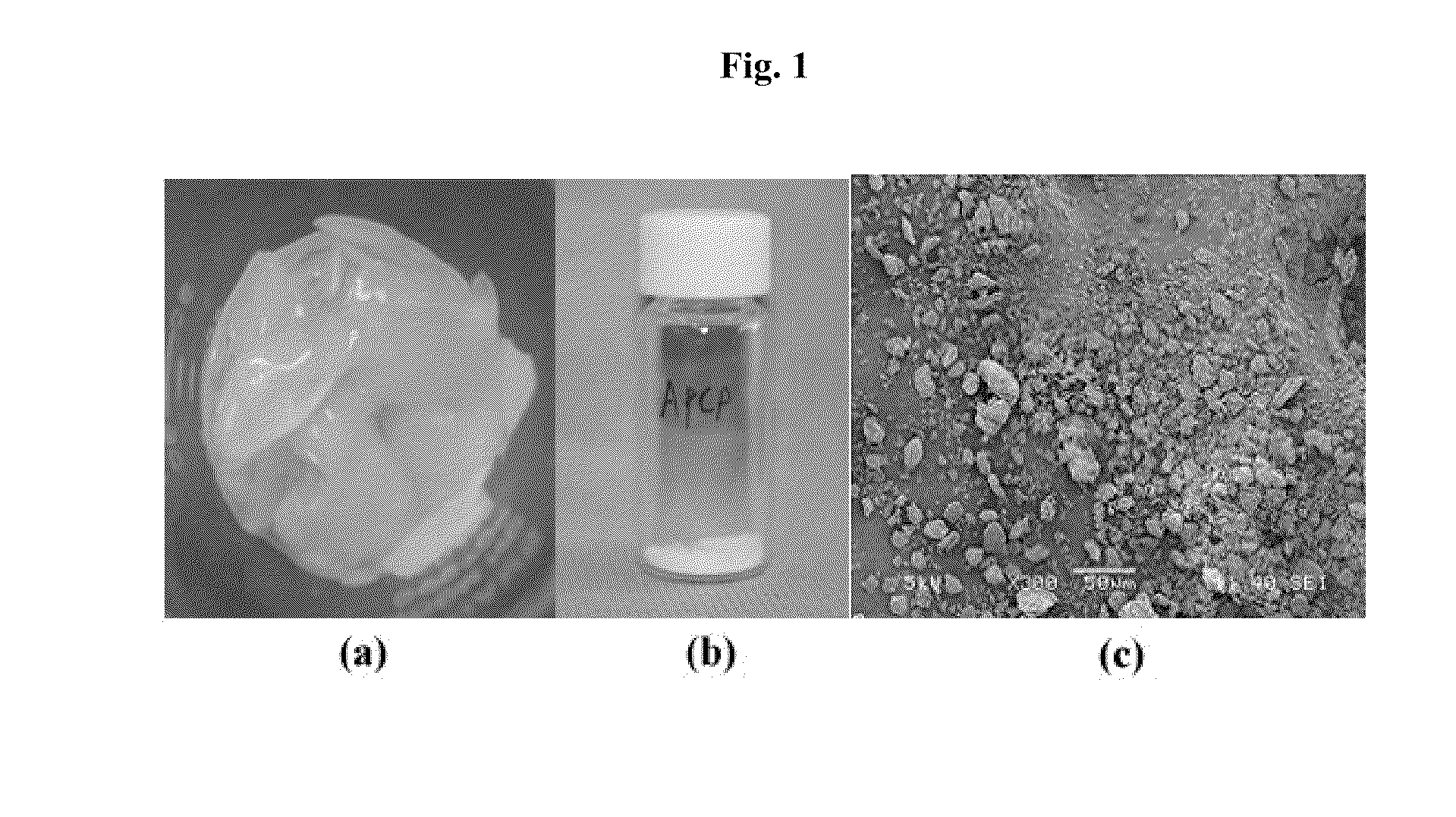
[0064]Cartilage tissue was isolated from the porcine cartilage and cut into pieces (about 20×30 mm) and then, washed 3 times for 10 minutes by a saline solution. The resulting cartilage fragment was immersed in PBS solution containing antibiotic-antimycotic agents and finally stored at −80° C. in an ultralow-temperature refrigerator (See FIG. 1(a)).

example 1

Pulverizing Porcine Cartilage

[0065]The cartilage fragment washed out was pulverized by a pulverizer that is commercially available and well-known to those skilled in this arts (Hood Mixer HMF-505, Hanil Co. Ltd. Korea) to reduce its size to about 2×2 mm. The pulverized cartilage fragment was freeze-dried and freeze-dried cartilage fragment was finally reduced to powder having its size of about 10 μm by a freezing mill (JAI, JFC-300, Japan).

[0066]1-1. Morphological Analysis of Porcine Cartilage Powder

[0067]The resulting porcine cartilage powder was morphologically analyzed under a scanning electron microscope. The porcine cartilage powder prepared in Example 1 was fixed by 2.5% glutaraldehyde for about 1 hour, and washed by phosphate buffer solution. The sample was dehydrated, dried and observed under a microscope (JEOL, JSM-6380, Japan; 20 KV) to measure the size and shape of the powder. The powder was observed in the size of about 10 μm (FIG. 1(c)).

example 2

Decellularization and Characterization of Porcine Cartilage Power

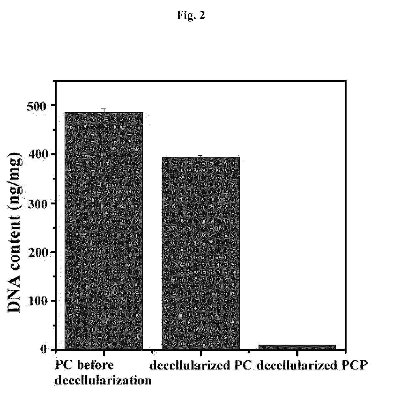
[0068]2-1. Decellularization of Porcine Cartilage Power

[0069]In order to remove chondrocytes and genetic components and obtain pure extracellular matrix, decellularization process was performed as below.

[0070]The porcine cartilage powder prepared in Example 1 was added to 1 l of 0.1% SDS (sodium dodecyl sulfate, Bio-Rad, USA) (per 10 g of the porcine cartilage powder) and stirred at 100 rpm for 24 hours. After treating SDS, the resultant was washed 5 times by tertiary distilled water at 100 rpm for 30 minutes.

[0071]In order to precipitate the cartilage powder for exchanging washing solution, the cartilage powder was centrifuged at 10,000 rpm for 1 hour with an ultracentrifuge (US-21SMT, Vision, Korea).

[0072]200 ml of 200 U / ml DNase (Sigma, USA) was added to the cartilage powder and stirred at 100 rpm at 37° C. for 24 hours. The resultant was washed 5 times by tertiary distilled water at 100 rpm for 30 minutes. The wash...


PUM

PropertyMeasurementUnit
temperatureaaaaaaaaaa
pHaaaaaaaaaa
sizeaaaaaaaaaa

Abstract

The present invention provides a method for manufacturing a porous three-dimensional scaffold using animal tissue powder, comprising powdering an animal-derived tissue, decellularizing the animal-derived tissue before or after powdering it, or simultaneously with powdering it, and forming the decellularized animal-derived tissue powder into a porous three-dimensional scaffold by a particle leaching method.

Description

TECHNICAL FIELD[0001]The present invention relates to a method for manufacturing a porous three-dimensional scaffold using powder from animal-derived tissue and a porous three-dimensional scaffold manufactured by the same, more particularly to a method for manufacturing a porous three-dimensional scaffold and a porous three-dimensional scaffold manufactured by the same, in which powder from animal-derived tissue is manufactured into a porous scaffold in a three-dimensional structure using a particle leaching method so as to construct a porous three-dimensional scaffold that may have various size, porosity, shape and structure, depending upon its purpose for treatment or use.BACKGROUND OF THE INVENTION[0002]Articular chondrocytes are specialized mesoderm-derived cells found exclusively in cartilage. Cartilage is an avascular tissue composed of extracellular matrix produced by chondrocytes. It neither causes an inflammation reaction, nor induces a self-regeneration, when being damaged...

Claims


Application Information

Patent Timeline
Patent Type & Authority Applications(United States)
IPC IPC(8): A61F2/00A61K35/12
CPCA61L27/3604A61L27/3612A61L27/3654A61L27/3683A61L27/3817A61L2430/40A61L27/56A61L2400/08A61L2400/18A61L2430/06A61L27/3852A61L27/00A61L27/36A61L27/38A61F2/00
Inventor MIN, BYOUNG-HYUNJANG, JI WOOK
Owner AJOU UNIV IND ACADEMIC COOP FOUND
Why Eureka
Try Eureka