
Antibody binding to envelope protein 2 of hepatitis c virus and method for identifying genotype of hepatitis c virus using the same

Inactive Publication Date: 2011-08-18
TORAY IND INC +1
View PDF0 Cites 2 Cited by
  • Summary
  • Abstract
  • Description
  • Claims
  • Application Information

AI Technical Summary

Benefits of technology

[0022]According to the present invention, simple and very accurate identification of HCV genotype 1a, HCV genotype 1b, and HCV genotype 2a is made possible and hepatitis C patients for whom interferon therapy is appropriate can be efficiently selected. In particular, adverse reactions can be alleviated and the chance of selecting a new therapeutic method can be provided for hepatitis C patients infected with HCV of genotype 1a or 1b, for which no therapeutic effects can be expected from interferon therapy.

Problems solved by technology

Pathological conditions exhibited by half or more of chronic hepatitis C patients will almost certainly go from bad to worse and are known to progress to cirrhosis or cancer of the liver.
However, HCV antibody tests are problematic in that when a subject has experienced HCV infection in the past, the subject would unavoidably test positive for hepatitis C, even after being completely cured.
HCV antibody tests are also problematic, since an anti-HCV antibody in blood is detected only when 1 to 3 months have passed after infection.
If a test is conducted before such time, HCV cannot be detected and the subject would test negative for hepatitis C.
Furthermore, even when a subject tests positive for HCV in an HCV antibody test and an HCV core antigen test, it is currently impossible to identify the HCV genotype.
Particularly on HCV genotype 1a and HCV genotype 1b, effective antiviral action cannot be exerted, and patients rather suffer from adverse effects of interferon.
Meanwhile, a nucleic acid amplification test is problematic in relation to insufficient preservative quality and stability for test samples, since the test uses serum RNA of a subject as a target molecule.
The nucleic acid amplification test also presents various problems, and precautions are necessary in regards to the use of an RT-PCR method.
For example, PCR may be carried out after transcription of RNA as a target molecule to DNA, resulting in a false negative result due to RNA degradation or inactivation and / or inhibition of a reverse transcriptase or a false positive result due to cross contamination of a reaction system.
Hence, the nucleic acid amplification test is thought to be inferior to an HCV antibody test or an HCV core antigen test using a protein as a target molecule in terms of accuracy.

Method used


Image

  • Antibody binding to envelope protein 2 of hepatitis c virus and method for identifying genotype of hepatitis c virus using the same
  • Antibody binding to envelope protein 2 of hepatitis c virus and method for identifying genotype of hepatitis c virus using the same
  • Antibody binding to envelope protein 2 of hepatitis c virus and method for identifying genotype of hepatitis c virus using the same

Examples


example 1

Preparation of Vector for Expression of Fusion Protein of Antigen E2 Protein of HCV Strain and Labeling Protein

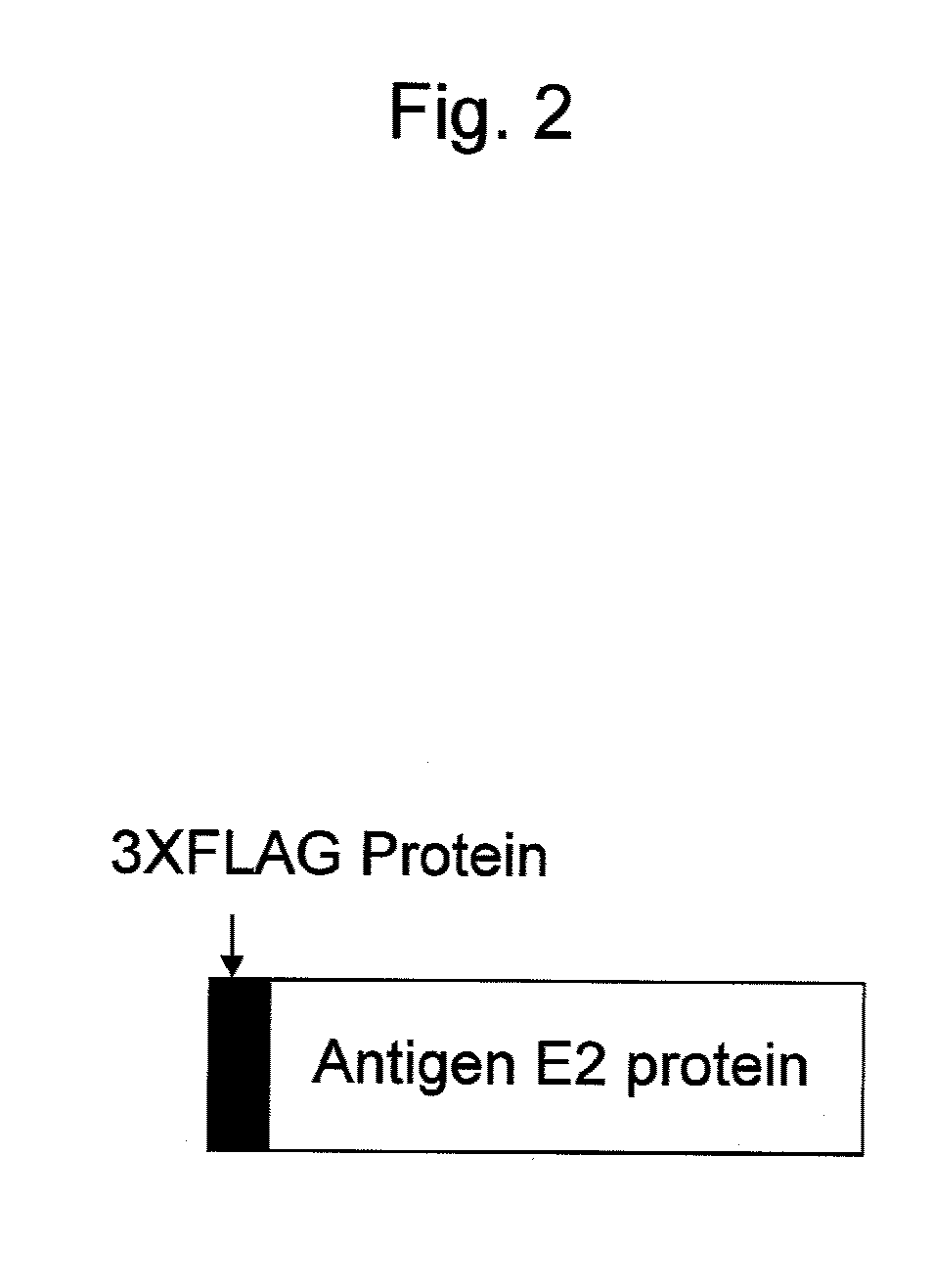
[0096](1) Construction of Vector for Expression of Fusion Protein of Antigen E2 Protein Derived from J6CF Strain of HCV2a and 3xFlag Tag

[0097]The antigen E2 protein derived from the J6CF strain of HCV2a; that is, a protein consisting of the region without transmembrane region of the E2 protein of the J6CF strain of HCV2a, was prepared as described below.

[0098]First, a gene encoding a protein consisting of a region corresponding to amino acid positions 384 to 720 of the precursor protein (SEQ ID NO: 5) of the J6CF strain, when the initiation methionine at the N-terminus was determined to be the 1st amino acid, was amplified by a PCR method using the cDNA of the genomic RNA of the J6CF strain of HCV2a (GenBank Accession No. AF177036) as a template, an Advantage GC2 PCR kit (Takara Bio Inc.), and J6E2dTM-s (SEQ ID NO: 6: CACAAGCTTCGCACCCATACTGTTGGGG) and J6E2dTM-as (SEQ ID NO:...

example 2

Expression of Fusion Protein of Antigen E2 Protein and Labeling protein

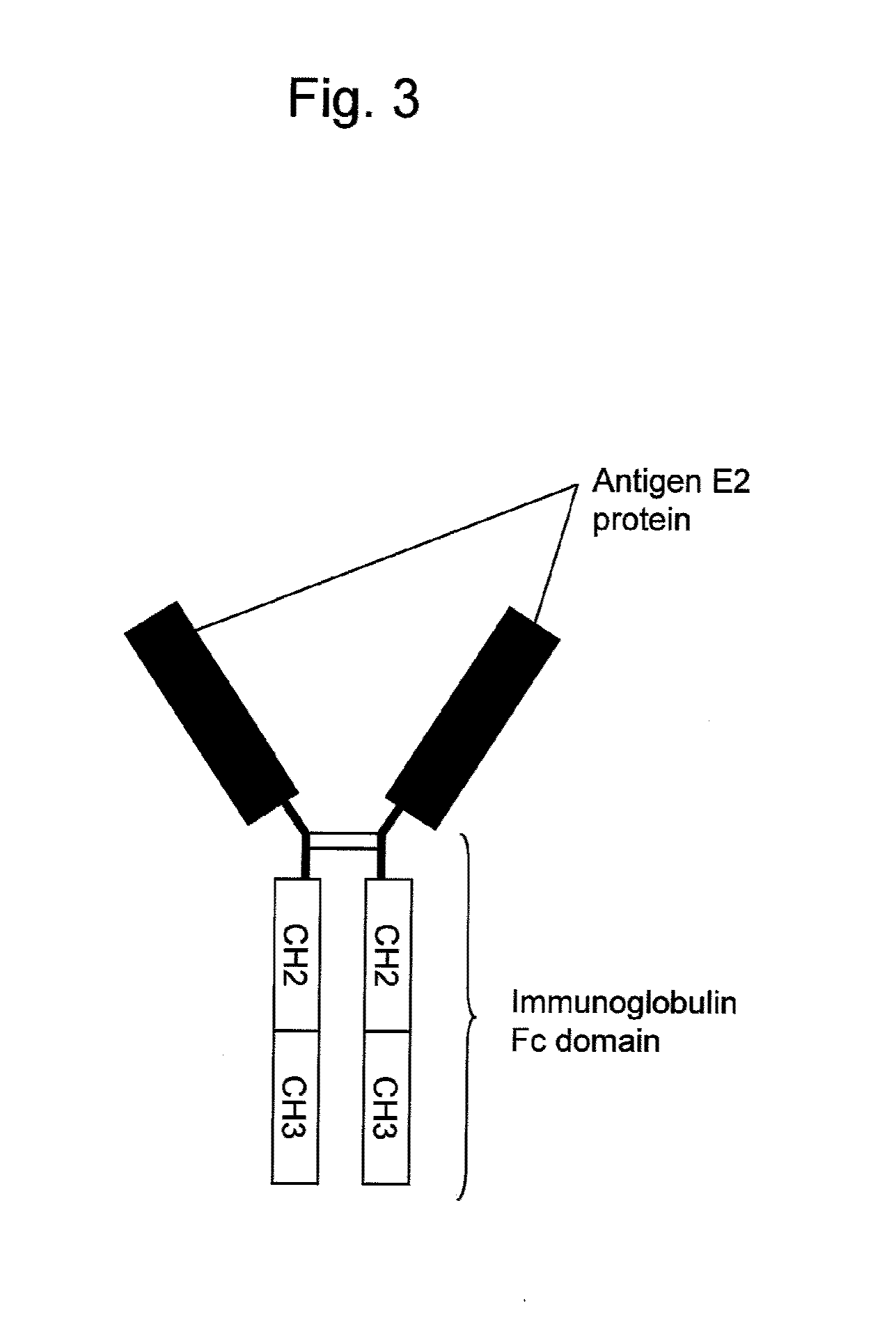
[0124]CMV-3xFLAGJ6E2dTM, CDM-J6E2Fc, CDM-JFH1E2Fc, CDM-THE2Fc, CDM-Con1E2Fc, SRα-J1E2Fc, and SRα-H77E2Fc constructed in Example 1 were introduced into monkey kidney-derived COS1 cells and then each fusion protein was expressed as described below.

[0125]First, COS1 cells were subcultured in RPMI1640 medium (Invitrogen Corporation) containing 10% fetal calf serum (Invitrogen Corporation), 100 U / ml penicillin, and 100 μg / ml streptomycin. On the day before the gene transfer, COS1 cells were seeded in 150 cm2 culture flasks (Corning Coaster Corporation) at a split ratio of 1:2 and then cultured overnight at 37° C. in a 5% CO2 incubator.

[0126]Subsequently, DEAE dextran (GE Healthcare) and chloroquine (Sigma) were added to RPMI1640 medium at final concentrations of 400 μg / ml and 100 μM, respectively. 50 μg of the above expression vector (CMV-3xFLAGJ6E2dTM, CDM-J6E2Fc, CDM-JFH1E2Fc, CDM-THE2Fc, CDM-Con1E2Fc, SRα-J1E2Fc, o...

example 3

Purification of Fusion Protein of Antigen E2 Protein and Labeling Protein

[0129]The culture supernatant of cells into which CMV-3xFLAG-J6E2dTM had been introduced was subjected to purification using anti-FLAG M2 agarose (Sigma) as described below.

[0130]First, 1 ml of anti-FLAG M2 agarose was added to 500 ml of the culture supernatant and then allowed to react at 4° C. for 2 hours during stirring in a spinner bottle. After 2 hours, a mixture of the supernatant and anti-FLAG M2 agarose was transferred to Econocolumn (Bio-Rad Laboratories Inc.), the Void fraction was removed, and then anti-FLAG M2 agarose was collected.

[0131]Next, anti-FLAG M2 agarose was washed twice with 10 ml of TBS (50 mM Tris-HCl, 150 mM NaCl, pH 7.4). Six fractions (anti-FLAG antibody column elution fractions 1-6) were eluted with 0.1 M Glycine-HCl (pH 3.5) to 1 ml / fraction. Immediately after elution, 1M Tris-HCl (pH 9.5) was added to return the pH to neutral. 20 μl of each fraction was fractionated under reductiv...


PUM


Abstract

The present invention provides an antibody that specifically binds to envelope protein 2 of HCV of genotype 2a but does not immunologically react with envelope protein 2 of HCV of genotype 1a.

Description

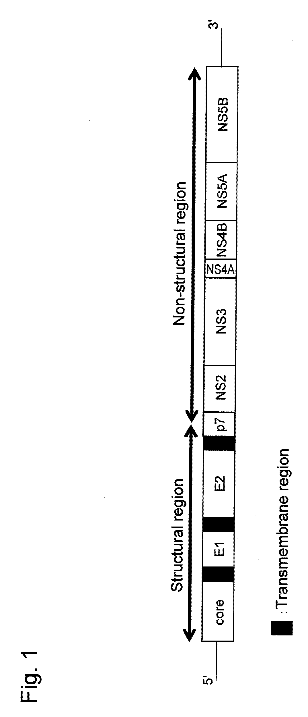
TECHNICAL FIELD[0001]The present invention relates to an antibody binding to envelope protein 2 of Hepatitis C virus and a method for identifying the genotype of Hepatitis C virus using the antibody.BACKGROUND ART[0002]Hepatitis C virus (which may be also referred to as “HCV” hereinafter) is a major causative virus of non-A and non-B hepatitis, which infects mainly via transfusion and sexual contact (Choo et al., Science, Vol. 244: 359-362, 1989). It has been estimated that there are 2,000,000 or more HCV carriers in Japan including those who show no hepatitis symptoms (virus carriers), and there are 170,000,000 or more HCV carriers in the world. The major causes for the increasing number of HCV carriers are the fact that the chronicity rate of hepatitis due to HCV infection is as high as 70% to 80%, and the fact that effective antiviral agents other than interferons do not exist.[0003]Pathological conditions exhibited by half or more of chronic hepatitis C patients will almost cert...

Claims


Application Information

Patent Timeline
IPC IPC(8): C12Q1/68C07K16/08
CPCC07K16/109G01N2333/18G01N33/56983C07K2317/34
Inventor WAKITA, TAKAJIAKAZAWA, YUKONAKAMURA, NORIKO
Owner TORAY IND INC
Learn More