Laser scanning cytometry mediated analysis of therapeutic efficacy in tumors

a technology of laser scanning and cytometry, applied in the field of cancer diagnosis and treatment, can solve the problems of large technical training, increased growth and metastasis in other parts of the body, and body does not acquire resistance to anti-angiogenic compound treatment,

Inactive Publication Date: 2011-09-29
BOARD OF RGT THE UNIV OF TEXAS SYST
View PDF1 Cites 2 Cited by
  • Summary
  • Abstract
  • Description
  • Claims
  • Application Information

AI Technical Summary

Problems solved by technology

However, once tumors acquire the capacity induce angiogenesis and produce their own blood vessels, they may grow and metastasize to other regions of the body.
Also, the body does not acquire resistance to treatment with anti-angiogenic compounds as it does with chemotherapeutic compounds.
The method requires considerable technical training to obtain reliable and reproducible results, and inter-investigator variability is a significant problem.
Difficulties in reproducing the method have been reported by several groups (Wiedner, 1995).
Additionally, the process of preparing specimens for histology and counting vessels is time consuming.
Therefore, the application of this technique has been limited generally to research purposes.
This can cause problems, especially if practitioners need to know the efficacy of treatments that have substantial adverse side effects.
Because previous studies of angiogenesis have identified tumor blood vessels by staining them with antibodies to endothelial cell antigens (CD31, CD34), it has been difficult to directly evaluate potential toxicity to adjacent normal tissue.
While determination of apoptosis and vascular parameters of tissue are known in the art, A single method in which all of these parameters are determined is not available.

Method used


Image

  • Laser scanning cytometry mediated analysis of therapeutic efficacy in tumors
  • Laser scanning cytometry mediated analysis of therapeutic efficacy in tumors
  • Laser scanning cytometry mediated analysis of therapeutic efficacy in tumors

Examples


example 1

ROCEDURES

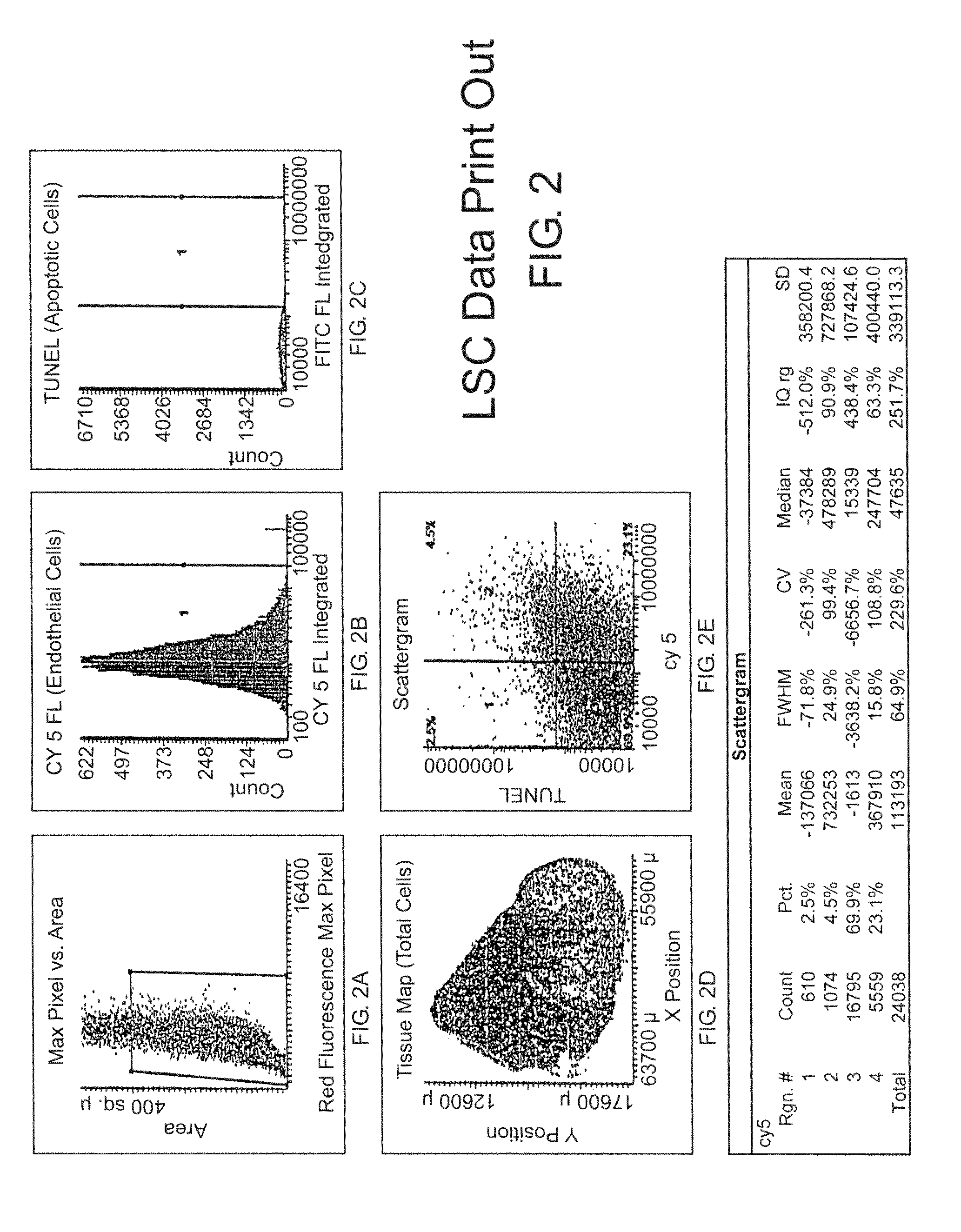
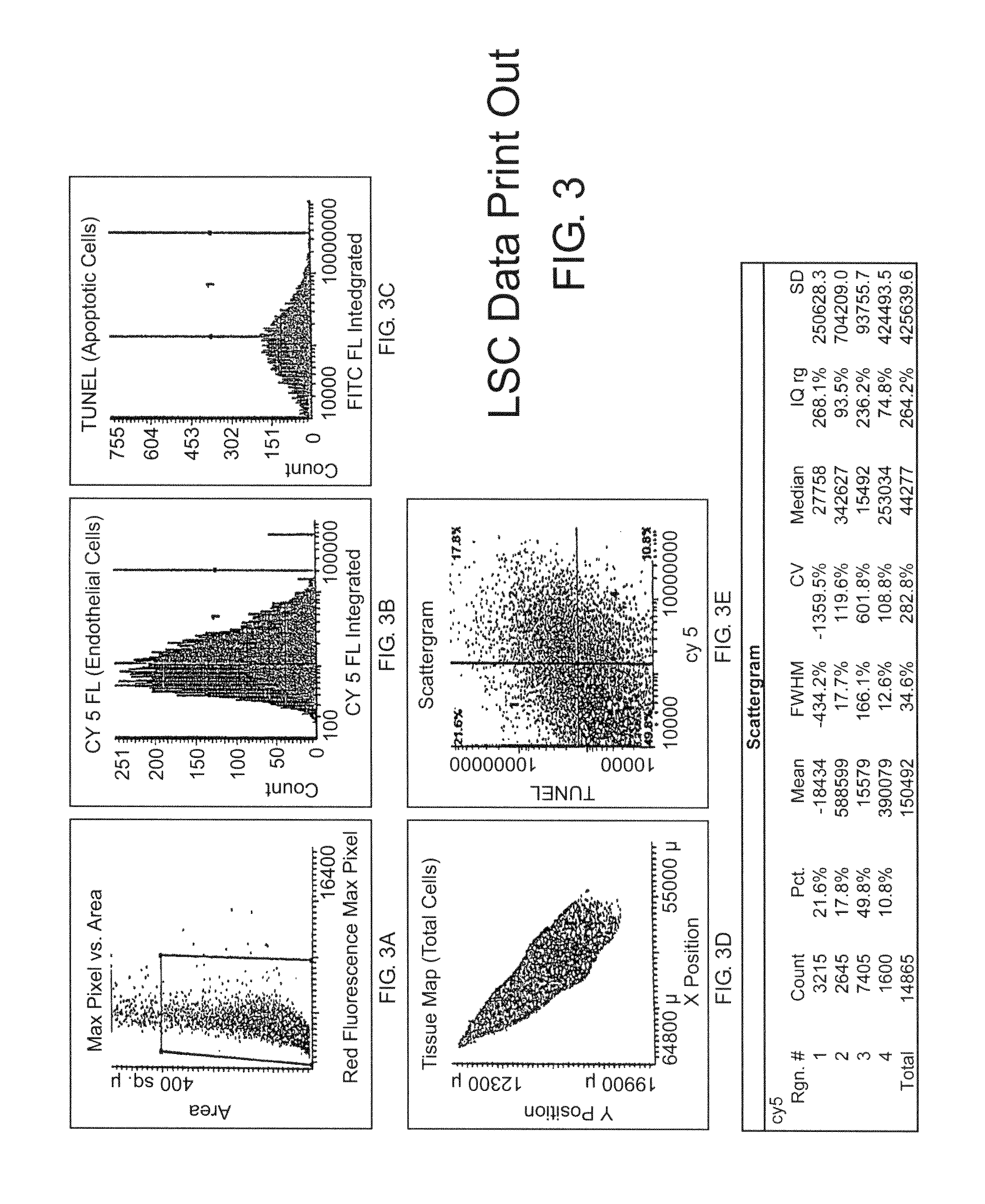
[0159]Tumor specimens or patient biopsies are obtained prior to and several time points post-treatment such as 1, 3, and 7 days or more. Tissue sections are stained using an immunofluorescent double-labeling technique which permits the simultaneous detection of endothelial cells and fluorescent markers of apoptosis such as TUNEL or caspase activity. The tissue sections are scanned by the LSC and cell data is collected by the instrument's software. For the first scan, the Wincyte software is setup by the user to contour on the cell nuclei based on propidium iodide staining. Contouring on the cell nuclei provides the total cell number of the tissue specimen and creates a ‘tissue map’ which permits selective gating of the tumor cells. In addition, contouring on the cell nuclei permits the software to identify endothelial cells (based on CD31 and / or CD34, Pharmingen, Inc., fluorescent staining) and / or TUNEL positive cells (based on fluorescent TUNEL, Promega, Inc.). This data p...

example 2

ORESCENCE DOUBLE STAINING

[0160]Formalin-fixed, paraffin embedded sections (5 μm) are deparaffinized in xylene, rehydrated in alcohol, and transferred to phosphate-buffered saline (PBS). Antigen retrieval is performed with 200 μg / ml proteinase-K for 30 minutes at room temperature. Frozen tissue sections (8 μm) are fixed by sequential incubation with cold acetone, acetone plus chloroform (1:1), and acetone (5 minutes each). Although frozen sections are usually easier to stain, both frozen and paraffin sections are used because paraffin sections are more convenient for clinical analysis. Sections are washed three times with PBS (5 minutes each) and incubated for 20 minutes at room temperature with protein blocking solution containing 5% normal horse serum and 1% normal goat serum. The blocking solution is removed and the sections are incubated for 24 hours at 4° C. with a 1:400 dilution of rat monoclonal anti-mouse CD31 antibody. Staining conditions may have to be optimized for some of...

example 3

NNING CYTOMETRY

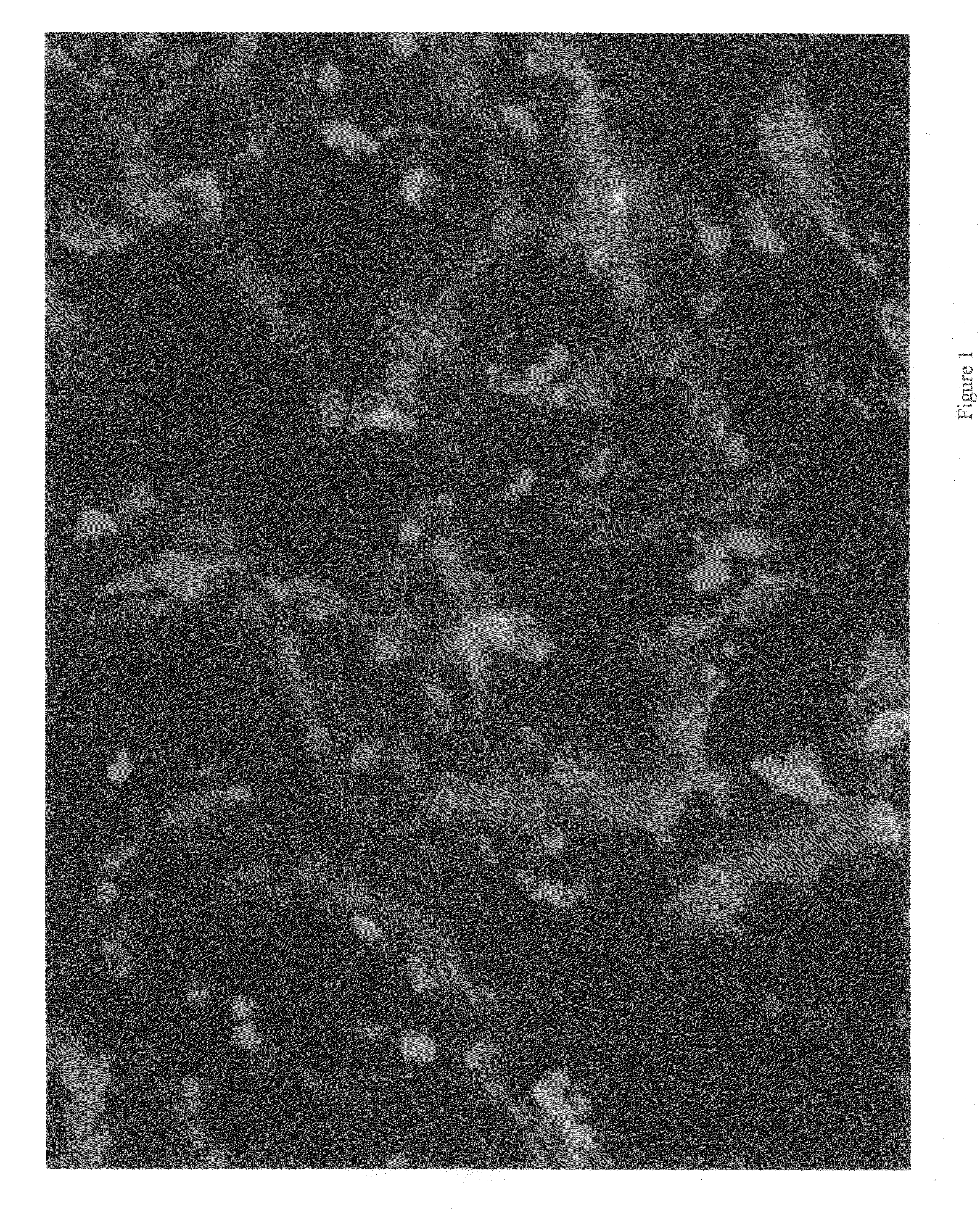
[0163]Immunofluorescence microscopy is performed on an epifluorescence microscope equipped with narrow bandpass excitation filters mounted in a filter wheel to individually select for green, red, and blue fluorescence. Images are captured using a cooled CCD camera and SmartCapture software. Images are further processed using Adobe Photoshop software.

[0164]The LSC (CompuCyte, Corporation, Cambridge, Mass.) is an instrument designed to enable fluorescence-based quantitative measurements on tissue sections or other cellular preparations at the single cell level. The instrument consists of a base unit containing an Olympus BX50 fluorescent microscope and an optics / electronics unit coupled to argon and HeNe laser support elements and a computer. Tissue sections that are mounted on a solid surface (i.e., a glass microscope slide) are interrogated by a 5 μm-diameter argon laser that repeatedly scans along a line as the surface is moved past it on a computer-controlled motori...


PUM

PropertyMeasurementUnit
sizeaaaaaaaaaa
diameteraaaaaaaaaa
timeaaaaaaaaaa

Abstract

This invention describes the use of a laser scanning device, for example a laser scanning CYTOMETRY (LSC), with a double-fluorescent labeling technique as a quantitative method that can be used to objectively and accurately measure endothelial cell death, endothelial tumor cell death and blood vessel density of tumor tissue. These parameters can be used as markers of efficacy in tumors treated with anti-angiogenic or traditional therapies and can distinguish patients who respond to these drugs from those who do not.

Description

[0001]This application claims benefit of priority to U.S. Provisional Application Ser. No. 60 / 246279 filed Jun. 6, 2000, the entire contents of which are incorporated by reference.BACKGROUND OF THE INVENTION[0002]I. Field of the Invention[0003]The present invention relates generally to the field of cancer diagnosis and treatment, and more specifically relates to the use of laser scanning cytometry for the determination of endothelial cell death, endothelial tumor cell death and tumor blood vessel density. This method can be used as an indicator for therapeutic response and for determining treatment protocols.[0004]II. Description of Related Art[0005]Angiogenesis, or new blood vessel growth, has become a topic of major prominence in both the scientific literature and the popular press over the past several years. Public interest centers on scientific studies and clinical trials now underway that have demonstrated that inhibitors of angiogenesis can substantially reduce or even stop g...

Claims


Application Information

Patent Timeline
Patent Type & Authority Applications(United States)
IPC IPC(8): G01N33/567C12Q1/02C12Q1/37G01N33/50
CPCG01N33/5011G01N2510/00G01N33/5088
Inventor DAVIS, DARREN W.MCCONKEY, DAVID J.
Owner BOARD OF RGT THE UNIV OF TEXAS SYST
Why Eureka
Try Eureka