Methods for stabilizing a bioprosthetic tissue by chemical modification of antigenic carbohydrates

a bioprosthetic tissue and antigenic carbohydrate technology, applied in the field of bioprosthetic implants, can solve the problems of limiting the antigenicity of bioprosthetic tissues, affecting the stability of bioprosthetic tissues, so as to improve stability, durability, and/or performance

Active Publication Date: 2011-12-22
EDWARDS LIFESCIENCES CORP
View PDF6 Cites 13 Cited by
  • Summary
  • Abstract
  • Description
  • Claims
  • Application Information

AI Technical Summary

Benefits of technology

[0009]Methods are provided herein for improving the stability, durability, and / or performance of a xenographic bioprosthetic tissue implant by chemically modifying antigenic carbohydrates within the bioprosthetic tissue.

Problems solved by technology

A primary limitation of bioprosthetic implants made from animal tissues is the occurrence of hyperacute rejection reactions in transplant recipients.
Although chemical fixation can considerably limit the antigenicity of bioprosthetic tissues, fixed tissues, particularly glutaraldehyde-fixed tissue, suffer from several drawbacks.
For example, the protective effects of glutaraldehyde fixation tend to deteriorate over the lifespan of bioprosthetic implants due to the labile Schiff Base cross-links, resulting in increased immunogenicity and impaired long-term stability and performance.
In addition, glutaraldehyde treatment renders bioprosthetic tissues more susceptible to calcification, particularly when an implant remains in place for an extended period of time (e.g., more than ten years) due to their high levels of residual aldehyde groups.
Structural valve deterioration (SVD) is the most common cause for early valve explanation, with tissue calcification the leading cause of failure in bioprosthetic implants.
Like conventional glutaraldehyde fixation, the methods result in residual reactive aldehyde groups, which serve as potential calcium binding sites and thus destabilize the tissue by ultimately compromising the biomechanical properties of the material.
Enzymatic approaches suffer from the general high cost of enzyme preparations and the fact that the large size of alpha-galactosiadase and other enzymes prevents these protein structures from penetrating deeply into tissues, such as the extracellular matrix of pericardial bioprosthetic tissues.
Thus, enzyme-based treatments do not eliminate all of the epitopes targeted by an enzyme, particularly in the interior of a bioprosthetic implant.
In addition, alpha-galactosiadase and other enzymes are specific for particular epitopes (e.g., α-GAL in the case of alpha galactosiadase), making it highly difficult to limit the antigenicity of tissues containing multiple and / or unknown epitopes.
The enzymatic removal of cellular components and tissue structures can also degrade the biomechanical properties of the tissue.
Moreover, these enzyme treatments cannot be used with glutaraldehyde-fixed tissue since the enzyme's protein structure will react with the residual aldehydes and become covalently bound to the material.
The result is an increase in foreign proteins and further degradation in tissue performance.

Method used


Image

  • Methods for stabilizing a bioprosthetic tissue by chemical modification of antigenic carbohydrates
  • Methods for stabilizing a bioprosthetic tissue by chemical modification of antigenic carbohydrates
  • Methods for stabilizing a bioprosthetic tissue by chemical modification of antigenic carbohydrates

Examples


example 1

Tissue Pre-Treatment

[0123]Prior to chemically modifying the antigenic carbohydrates in a xenographic bioprosthetic tissue, the tissue may optionally be pre-treated by exposure to cross-linking agents and / or surfactants. The following non-limiting procedure sets forth one potential tissue pre-treatment protocol that produces fixed tissues. Those skilled in the art will appreciate that various alternative methods, chemical compounds, or solutions may be substituted for those indicated.

Step 1: Harvest / Prepare Biological Tissue

[0124]A desired biological tissue is harvested (surgically removed or cut away from a host animal) at a slaughterhouse, placed on ice, and transported to the location at which the bioprosthesis will be manufactured. Thereafter, the tissue is typically trimmed and washed with a suitable washing solution, such as a saline solution, sterile water, or a basic salt solution. For example, harvested tissues can be rinsed, washed, and / or stored in a phosphate or non-phosp...

example 2

Selective Chemical Modification of Antigenic Carbohydrates

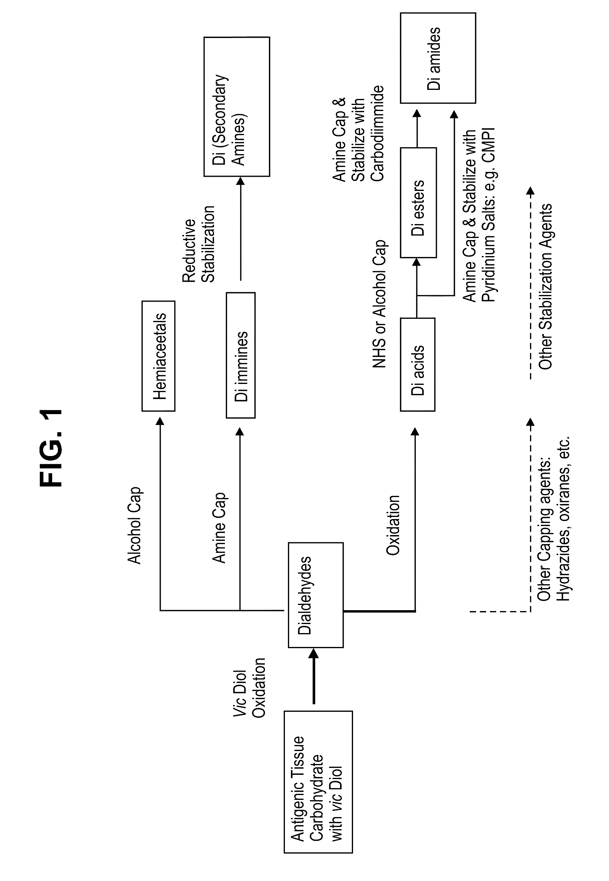
[0129]Chemical modification of antigenic carbohydrates in a xenographic bioprosthetic tissue, as described herein, may be performed whether or not the tissue is pre-treated. The following non-limiting procedure sets forth methods for chemically modifying select antigenic carbohydrates in either scenario.

[0130]After the bioprosthetic tissue has been rinsed and stored, the tissue is preferably immersed in isotonic buffered saline solution containing a periodate oxidizing agent, such as sodium periodate, at a concentration of about 20 mM for a period of about 20 minutes at room temperature with constant agitation.

[0131]After treatment with the periodate oxidizing agent, the tissue is rinsed extensively in 20% ethanol to completely remove the periodate, preferably in a vessel allowing a large solution to tissue volume ratio to create a favorable gradient for solute diffusion.

[0132]The tissue is then immersed in a solution contain...

example 3

Treatment of Un-Fixed Tissue with Periodate

Tissue Treatment

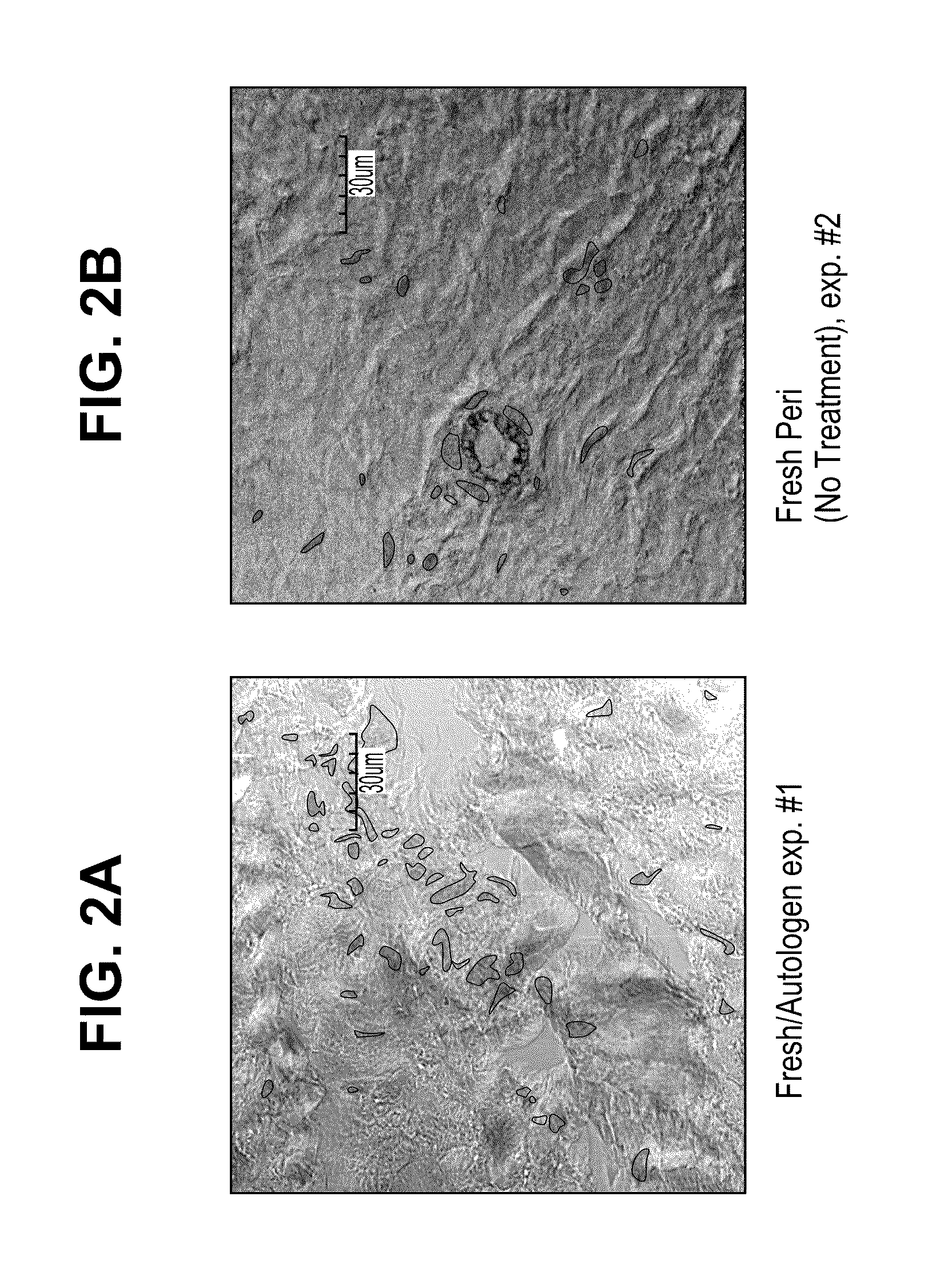
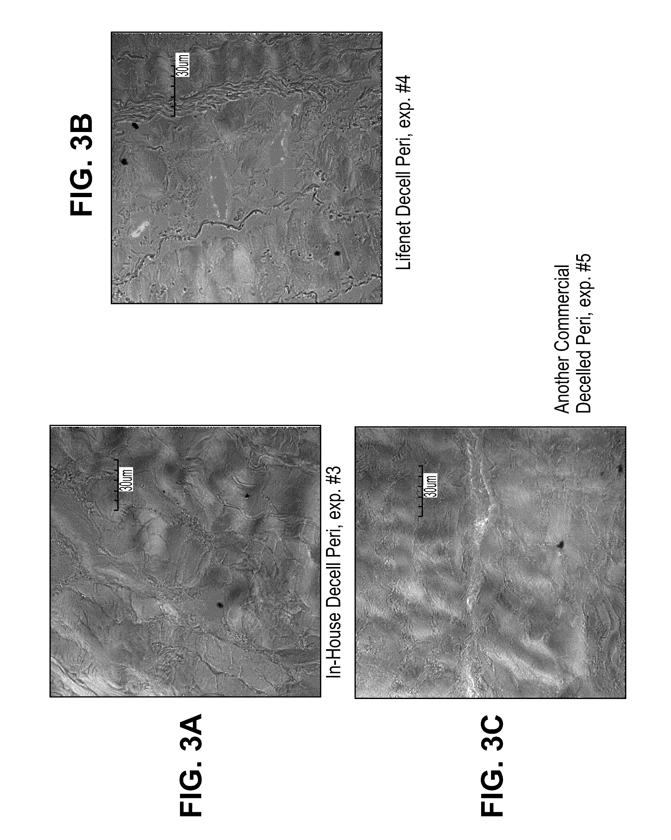
[0134]Bovine pericardial tissue (National Beef, Item # 192769001, WO# 58745266) was treated to mask antigens by the following procedure. Tissue was soaked in a phosphate buffer containing 10 mM ethanolamine (Alfa Aesar, #36260) with pH 7.0±0.5 or 10 mM taurine with 7.0±0.5 pH (Sigma, #T0625). In both treatment groups, sodium periodate (Sigma, #311448) was added to yield a 20 mM solution with 7.0±0.5 pH. Tissue from the two groups was incubated in one of three ways: 1) shaking at 4° C. for 18 hours (New Brunswick Scientific, Innova 4230, refrigerated incubator / shaker) 2) shaking at room temperature for 3 hours (VWR, Model 1000, orbital shaker) and 3) shaking at 37° C. for 30 min. (VWR, Model 1570, orbital shaker / incubator). After treatment the tissue was rinsed thoroughly in 0.9% saline (Baxter, #2F7124). The tissue was then incubated in ethanolamine and sodium borohydride (Sigma, #452882) for 1 hour at room temperature while...


PUM

PropertyMeasurementUnit
timeaaaaaaaaaa
temperatureaaaaaaaaaa

Abstract

Methods are provided herein for modifying antigenic carbohydrate epitopes within a xenographic bioprosthetic tissue by oxidation of vicinal diols to form aldehydes or acids and subsequence reductive amination of aldehydes to form stable secondary amines, or amidation or esterification of acids to form stable amides or esters. Advantageously, methods provided herein mitigate the antigenicity of the bioprosthetic tissue while leaving the overall tissue structure substantially undisturbed, and thereby enhance the durability, safety and performance of the bioprosthetic implant.

Description

RELATED APPLICATIONS[0001]The present application claims priority under 35 U.S.C. §119(e) to U.S. provisional application No. 61 / 355,948 filed Jun. 17, 2010.FIELD[0002]Methods are provided herein relating to the field of bioprosthetic implants, and more particularly to the treatment of bioprosthetic tissues to decrease post-implantation antigenicity and calcification in host subjects.BACKGROUND[0003]A primary limitation of bioprosthetic implants made from animal tissues is the occurrence of hyperacute rejection reactions in transplant recipients. Such reactions are driven largely by the presence of antigenic carbohydrate epitopes within implanted tissues, the most common of which is the α-GAL glycoprotein epitope: D Galactose (α 1-3) Galactose (β1-4) N acetyl Glucosamine-R-motif (α-GAL) is found on vascular endothelial tissues of all species with the exception of old world monkeys, great apes, and humans. The presence of α-GAL on harvested animal donor tissues elicits an immediate a...

Claims


Application Information

Patent Timeline
Patent Type & Authority Applications(United States)
IPC IPC(8): A61K35/12C25B7/00A61P9/00A61K35/34C12S3/24
CPCA61L27/3687A61L27/50A61L2430/40A61L2430/20A61L27/54A61P9/00
Inventor DOVE, JEFFREY S.TOD, TARA J.
Owner EDWARDS LIFESCIENCES CORP
Why Eureka
Try Eureka