Micropatterned co-culture systems as infectious disease analysis platforms

a co-culture system and infectious disease technology, applied in the field of micropatterned co-culture systems as infectious disease analysis platforms, can solve the problems of difficult realization of goals, severe side effects of ribavirin, and inability to cure many infected patients, and achieve the effect of improving the signal-to-noise ratio

Inactive Publication Date: 2011-12-29
THE ROCKEFELLER UNIV +1
View PDF3 Cites 5 Cited by
  • Summary
  • Abstract
  • Description
  • Claims
  • Application Information

AI Technical Summary

Benefits of technology

[0014]FIG. 4 shows the effect of dose and time of incubation with initial viral inoculum on the production of HCV particles in the culture medium of microscale human liver tissues. Cultures were infected with HCV as stated in the caption of FIG. 3; however, the initial viral inoculum was a) diluted up to ⅛ the original stock, and b) left on the cultures for 8, 16 or 24 hours, followed by washing of the cultures 3 times with fresh culture medium lacking HCV. Luciferase activity was assessed 72 hours after infection.
[0015]FIG. 5a depicts infection of microscale human liver tissues

Problems solved by technology

Current drug therapies (pegylated interferon and ribavirin) have severe side effects and do not cure many infected patients even after lengthy (months to year) treatment regimens.
However, these goals have been difficult to realize partly due to the difficulties in maintaining sustained and reproducible infection of isolated primary human hepatocytes (main cell type of the liver) with HCV in vitro.
However, this cancer-derived cell line has abnormal repertoire and levels of liver-specific functions.
Monitoring molecular biology of the parasite in the liver is difficult using current methods, and there is no easy way to determine in cell culture whether the vaccine is properly arresting the parasite.
Historically cell culture techniques and tissue development failed to take into account the necessary microenvironment for cell-cell and cell-matrix communication as well as an adequate diffusional environment for delivery of nutrients and removal of waste products.
While many methods and bioreactors have been developed to grow tissue for the purposes of generating artificial tissues for transplantation or for toxicology studies, these bioreactors do not adequately simulate, in vitro, the mechanisms by which nutrients, gases, and cell-cell interactions are delivered and performed in vivo.
Thus, cell culture systems and bioreactors that d

Method used


Image

  • Micropatterned co-culture systems as infectious disease analysis platforms
  • Micropatterned co-culture systems as infectious disease analysis platforms
  • Micropatterned co-culture systems as infectious disease analysis platforms

Examples


example 1

Disease Platform for HCV Drug Development

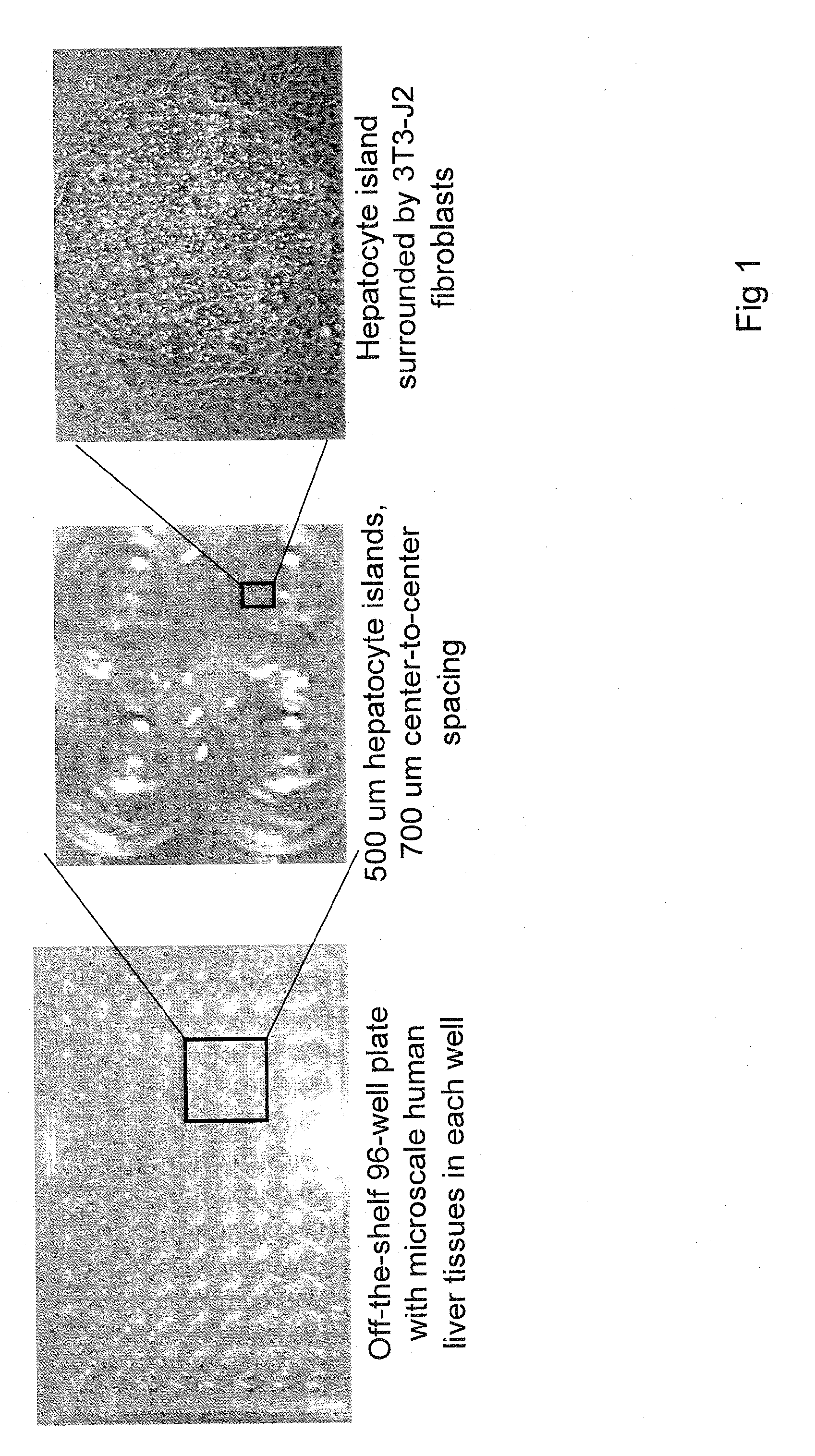
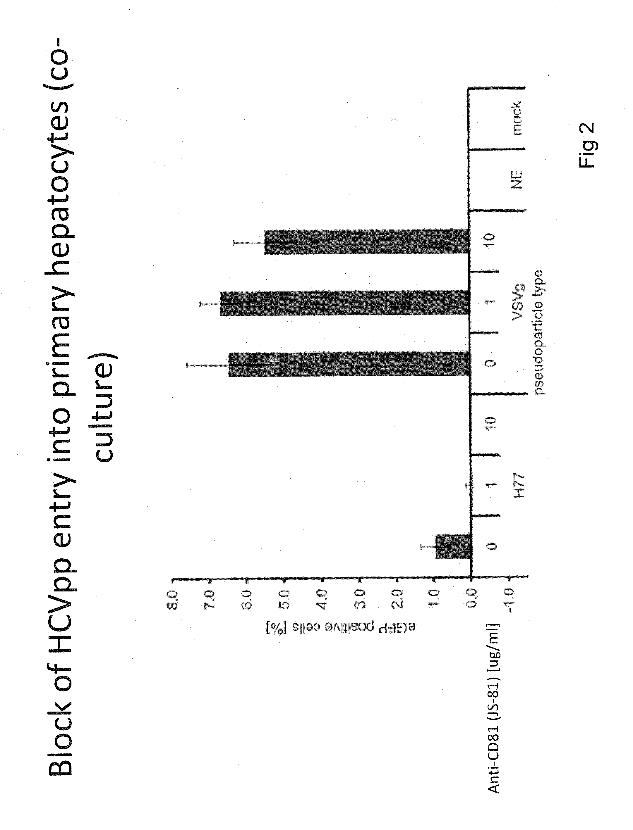
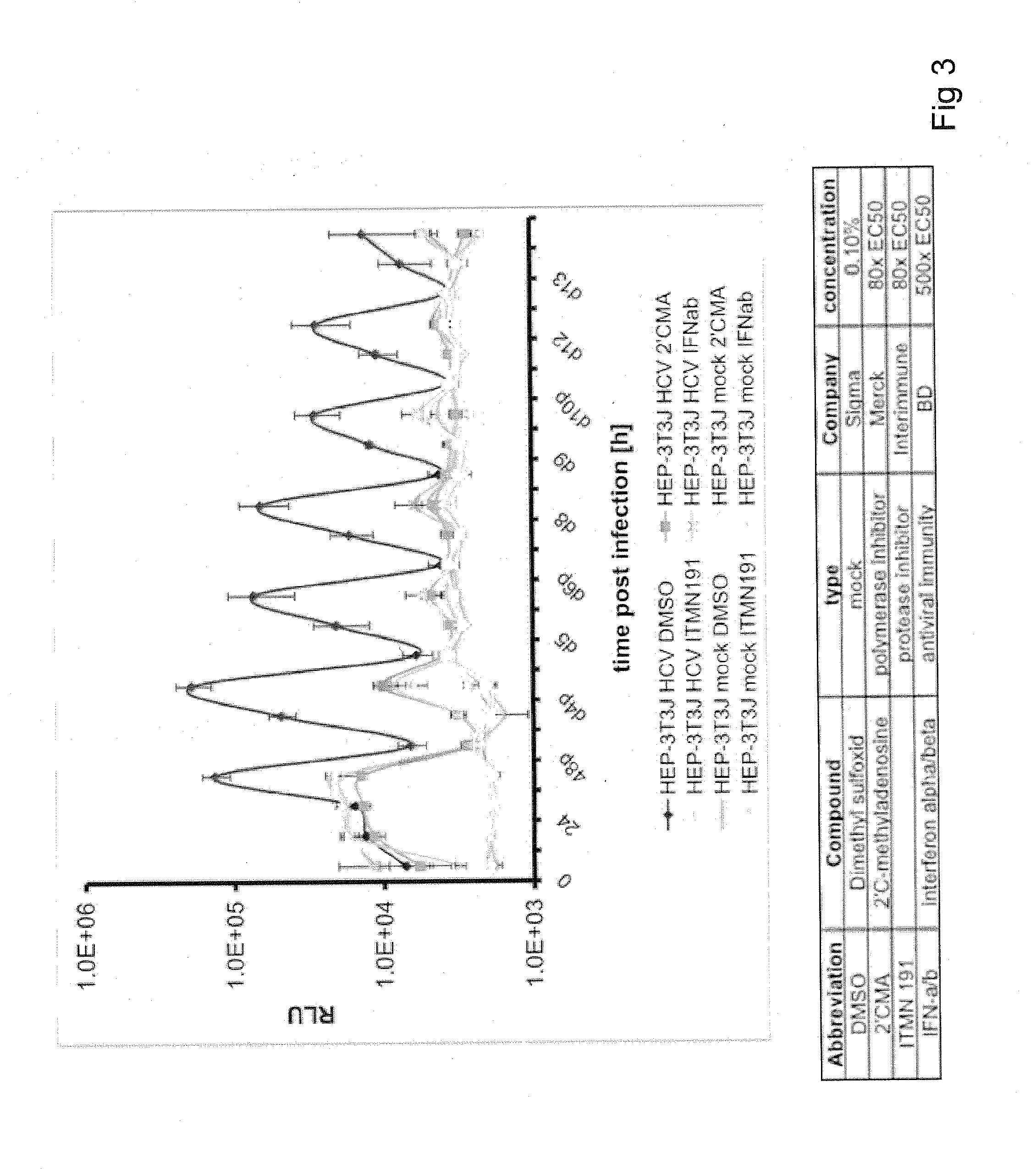
[0175]This Example presents the development and optimization of a disease platform for HCV drug development that couples micropatterned co-cultures of primary human hepatocytes and stromal cells in a multi-well format (FIG. 1) with highly sensitive viral reporter systems and optimized treatment protocols. The developed human liver platform can: a) Support entry of HCV-like particles via receptor-mediated processes (FIG. 2), and b) Produce and release infectious HCV particles into the culture supernatant for several weeks in vitro, and that such release can be blocked by treatment of cultures with small molecule inhibitors of viral proteins (FIG. 3). It was also found that infection of micropatterned co-cultures is dependent on the dose and time of exposure to the initial inoculum (FIG. 4), and magnitude of HCV infection in micropatterned co-cultures is 4-6 fold higher than in pure hepatocyte monolayers, the current gold standard in the field ...

example 2

A Fluorescent-Based Reporter for HCV Infection

[0177]A novel cell-based fluorescent reporter system has been developed to detect infection of unmodified HCV genomes. The example system described here uses an HCV-dependent fluorescence relocalization (HDFR) cassette that comprises a fluorescent protein (e.g. EGFP, mCherry or TagRFP), an SV40 nuclear localization sequence (NLS), and a C-terminal mitochondrial-targeting domain (IPS) derived from the interferon-beta promoter stimulator 1 protein, IPS-1. IPS-1 is a known cellular substrate for the HCV NS3-4A protease, and IPS-1 mutation C508Y has been shown to abolish cleavage. In HCV-infected cells the HDFR cassette is processed by the viral NS3-4A protease, resulting in translocation of the fluorescent protein from the mitochondria to the nucleus. Using a lentivirus based expression system to stably express the HDFR cassette, we have successfully established this system in the highly HCV permissive human hepatoma cell line, Huh-7.5 and ...

example 3

Use of HDFR to Detect Infection with HCVcc and Sera / Plasma from HCV+ Patients in MPCC

[0183]The cell-based fluorescent reporter system has recently proved particularly useful in studying HCV infection of primary cells. Traditionally, HCV-specific antigens have been extremely difficult to detect in infected primary cell cultures. This may be due to low permissiveness of these cells to infection, low intrinsic replication rate of this persistent virus, or high intrinsic liver cell fluorescence, preventing detection of weak signals. In contrast, the reporter system described here allows rapid detection and quantification of primary cell infection on a single cell basis. MPCC were transduced with a lentivirus encoding an mCherry-based HDFR cassette and subsequently infected with HCV cell culture virus (HCVcc). A flow diagram exemplifying this procedure is depicted in FIG. 10. Redistribution of mCherry fluorescence to the nucleus was detected in DMSO-treated MPCCs, but not in the presence...


PUM

PropertyMeasurementUnit
Fractionaaaaaaaaaa
Fractionaaaaaaaaaa
Adhesion strengthaaaaaaaaaa

Abstract

Cell cultures are provided that include a population of micropatterened hepatocytes and one or more non-parenchymal cell populations, where the hepatocytes are infected with a virus or parasite and include a reporter of virus or parasite infection. Methods of making and using the cell cultures are also provided.

Description

RELATED APPLICATIONS[0001]This application is related and claims priority to U.S. provisional application Ser. No. 61 / 078,683 filed Jul. 7, 2008 and U.S. provisional application Ser. No. 61 / 174,449 filed Apr. 30, 2009. The entire contents of each of the foregoing applications are incorporated herein by this reference.BACKGROUND[0002]More than 150 million people worldwide are infected with the Hepatitis C virus (HCV), and about 75% of those infected display chronic infection, which can lead to liver cirrhosis and hepatocarcinoma. Current drug therapies (pegylated interferon and ribavirin) have severe side effects and do not cure many infected patients even after lengthy (months to year) treatment regimens. Hence, there is an urgent need to better understand the variability in host response and propensity for chronic HCV infection, and then use such understanding to develop new classes of drugs that target specific pathways in the viral lifecycle. However, these goals have been diffic...

Claims


Application Information

Patent Timeline
IPC IPC(8): C12Q1/02G01N33/53C12Q1/68C12N5/071C12N5/077
CPCG01N33/5067C12N2535/10C12Q1/18G01N33/5014
Inventor RICE, III, CHARLES M.JONES, CHRISTOPHER THOMASPLOSS, ALEXANDERBHATIA, SANGEETA N.KHETANI, SALMAN
Owner THE ROCKEFELLER UNIV
Why Eureka
Try Eureka