

Bone-like prosthetic implants
a prosthetic and cartilage technology, applied in the field of bone-like prosthetic implants, can solve the problems of large considerable damage to the craniomaxillofacial bones, and significant morbidity and substantial pain of graft harvesting procedures, so as to improve the growth and/or viability, improve the media flow, and high density
- Summary
- Abstract
- Description
- Claims
- Application Information
AI Technical Summary
Benefits of technology
Problems solved by technology
Method used
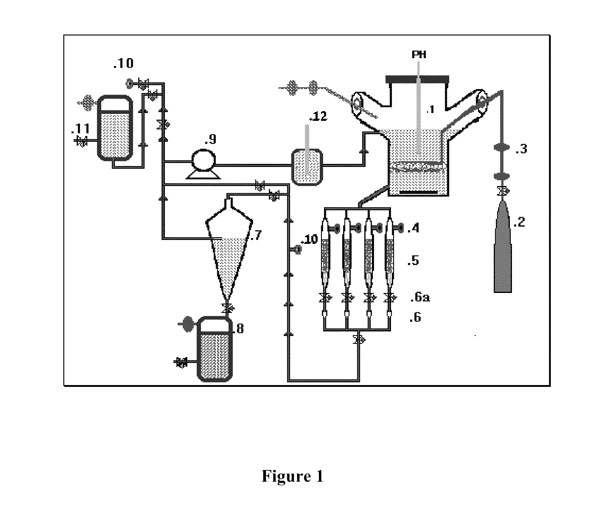
Image

Examples
example 1
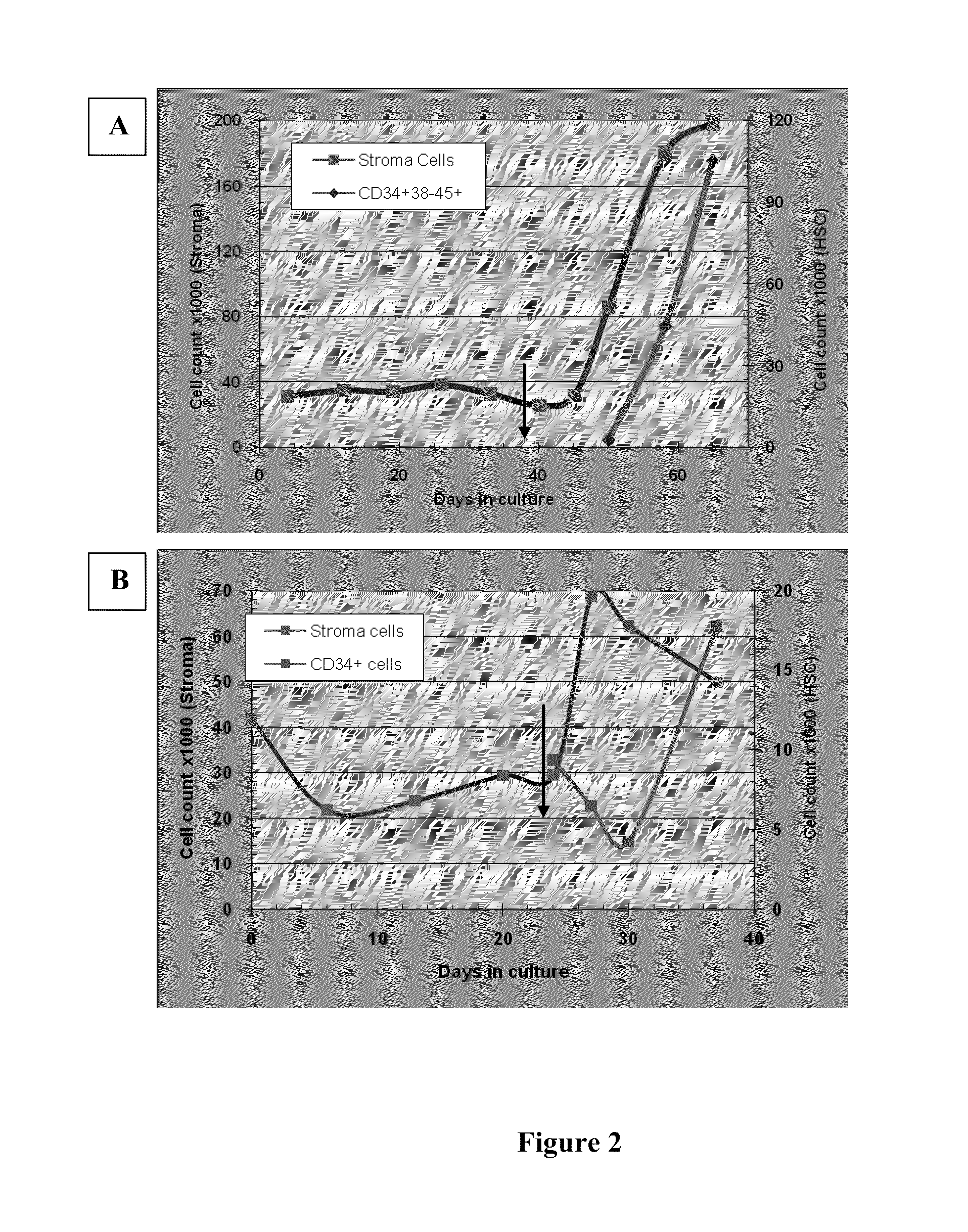
Growth of MSC and Osteoblasts 3-D Cultures in Flow System
[0129]Example 1 describes typical expansion experiments for co-culturing of these components, but with addition of the components at separate times.
[0130]In one experiment 30000 BM-derived MSCs were seeded on each carrier and the system was cultivated for 50 days. Results are shown in FIG. 2A. During the first six weeks, no cell expansion was noted but the following week was characterized by extensive cell proliferation. At the end of the 50-day period, expansion was measured at 2.8 folds (total of 85000 cells / carrier). 10000 CB derived CD34+ cells were then seeded onto the system of which early HSC (CD34+38-45+) counted for only 2500 cells (ratio of MSCs to HSC at time of co-culture establishment). Fourteen days later, at the time of harvest, MSCs cells were shown to expand by additional factor of 2. During the same period of time, HSC number in this system was increased by 42 folds.
[0131]In the second experiment, 42000 BM-de...
example 2
MSC Differentiation to Osteoblasts
[0141]This Example relates to 2D and 3D cultures of MSC cells which are then differentiated into preosteoblasts and osteoblasts. The 3D culture is similar to Example 1, except that the component cells are placed together simultaneously in culture.
[0142]Final bone tissue formation is executed in osteogenic culture differentiation medium composed of one or more of the following molecules in preferred concentration: dexamethasone (10-200 nM) (Sigma), sodium β-glycerophosphate(5-25 mM) (Sigma), 1,25 dihydroxycholecalciferol (calcitriol: 5-50 nM) (Sigma) and L-ascorbic acid-2-phosphate (10-500 uM) (Sigma).
[0143]FIG. 5 shows light microscopy image of MSC cultures, 7 days after inducing differentiation to osteoblasts.
[0144]FIG. 6 shows MSC differentiation to preosteoblasts and osteoblasts, and EDS spectrum analysis of elements in molar concentration in MSCs cultures after 3 growth weeks.
example 3
Co-Culture of Osteoprogenitor Cells and Endothelial Cells
[0145]Osteoprogenitor cells (OS) and Endothelial cells (C) were co-cultured on a 3-D scaffold for inducing osteogenesis in vitro.
[0146]Bone marrow MSCs—derived osteoprogenitor cells (OS) (50,000 cells / well of 24 wells culture plate) were either plated alone or co-cultured with Endothelial cells (EC) (2,000 cells / well of 24 wells culture plate), on 3-D hydrogel scaffold in {acute over (α)}-MEM medium containing osteogenic supplements, for up to 21 days. Endothelial cells were similarly cultured alone.
[0147]The effect of endothelial cells on the differentiation pattern of osteoprogenitor cells to the osteogenic lineage was followed by Alizarin Red S, staining for demonstration of calcium deposits and by osteocalcin inmmunostaining for demonstrating the synthesis of bone specific macromolecules.
[0148]The results in FIG. 7-9 demonstrate the difference in proliferative activity of these two cell types. Thus when cultured alone, the...
PUM
 Abstract
Description
Claims
Application Information