
Method for detecting afferent lymph vessel inflow regions and method for identifying specific cells

a lymph vessel and inflow region technology, applied in the field of detecting afferent lymph vessel inflow regions and a method for identifying specific cells, can solve the problems of poor shifting of lymph nodes to the lymphatic system, inability to observe lymph flow in real time, and escape of sentinel lymph nodes, so as to achieve enhanced detection accuracy, accurate measurement, and enhanced detection accuracy of specific cells

Active Publication Date: 2012-05-03
KONICA MINOLTA INC +1
View PDF2 Cites 5 Cited by
  • Summary
  • Abstract
  • Description
  • Claims
  • Application Information

AI Technical Summary

Benefits of technology

[0033]In accordance with the invention according to claim 1, the regions in the sentinel lymph node, in each of which the fluorescence intensity is high, are detected as the afferent lymph vessel inflow regions, and accordingly, the regions in the sentinel lymph node, where it is possible that the metastasis cancer cells can be present, can be detected accurately.
[0034]In accordance with the invention according to claim 2, the image acquired by the confocal fluorescence microscope for the single molecule observation is subjected to the image analysis, whereby the fluorescence intensity can be accurately measured for each of the plural regions.
[0035]In accordance with the invention according to claim 3, detection accuracy for the specific cells can be enhanced.
[0036]In accordance with the invention according to claim 4, detection accuracy for the T-lymphocytes can be enhanced.
[0037]In accordance with the invention according to claim 5, detection accuracy for the cancer cells can be enhanced.
[0038]In accordance with the invention according to claim 6, the regions where it is possible that metastasis cancer cells can be present in the sentinel lymph node can be detected accurately, and in addition, the detection accuracy for the specific cells can be enhanced.

Problems solved by technology

For example, in the case of the coloring matter, there are problems, which are: (1) in the case where dirt of anthracosis or the like is nonspecifically present in a lymph node in a living body, it becomes impossible to make evaluation as to whether or not the lymph node concerned is the sentinel lymph node; (2) an area from which the lymph node is to be dissected is contaminated, resulting in disturbance to the surgery; (3) the patient has allergy (anaphylactic reaction) to the coloring matter; and the like.
Meanwhile, in the case of the RI colloid, there are such problems, which are: (1) a particle diameter of the colloid is several hundred nanometers to several ten micrometers, which is larger in comparison with those of the coloring matter (with several nanometers) and quantum dots (with several ten nanometers), and the colloid is poor in shifting to a lymphatic system, and accordingly, such a lymph flow cannot be observed in real time; (2) since resolution is low, there is an apprehension that the sentinel lymph node may escape detection in the case where the sentinel lymph node is located in the vicinity of a part into which the colloid is to be locally injected (shine-through phenomenon); (3) the RI colloid is regulated with regard to use thereof, and it is difficult to expand adaptable facilities thereof; and the like.
Moreover, in comparison with a cancer of a region having a relatively simple lymph flow, such as abreast cancer and a skin cancer, in a digestive system cancer in which the lymph flow is multi-directional and complicated, it is difficult to observe the lymph flow in real time, and accordingly, application of such a sentinel lymph node biopsy method is delayed.
However, since only a tiny part of the extirpated lymph node tissue just can be observed, there is a risk that minute metastasis may escape detection.
However, in the multiple staining method, as in the usual staining method, there is an apprehension that the metastasis cells may escape detection at a stage of creating a tissue slide.
Moreover, the RT-PCP method has such shortcomings that the inspection is cumbersome and requires a time though the detection capability for the cancer cells is high, and the RT-PCR method concerned is not suitable for rapid diagnosis during the surgery, that only information in which the whole of the tissue is averaged is obtained, that it becomes impossible to make a pathological sample since it is necessary to inspect the whole of the tissue in order to surely find the minute metastasis, and the like.

Method used


Image

  • Method for detecting afferent lymph vessel inflow regions and method for identifying specific cells
  • Method for detecting afferent lymph vessel inflow regions and method for identifying specific cells
  • Method for detecting afferent lymph vessel inflow regions and method for identifying specific cells

Examples


Embodiment Construction

[0098][Device Configuration]

[0099]A description is made below of embodiments of the present invention with reference to the drawings.

[0100]First, a description is made of a configuration of devices.

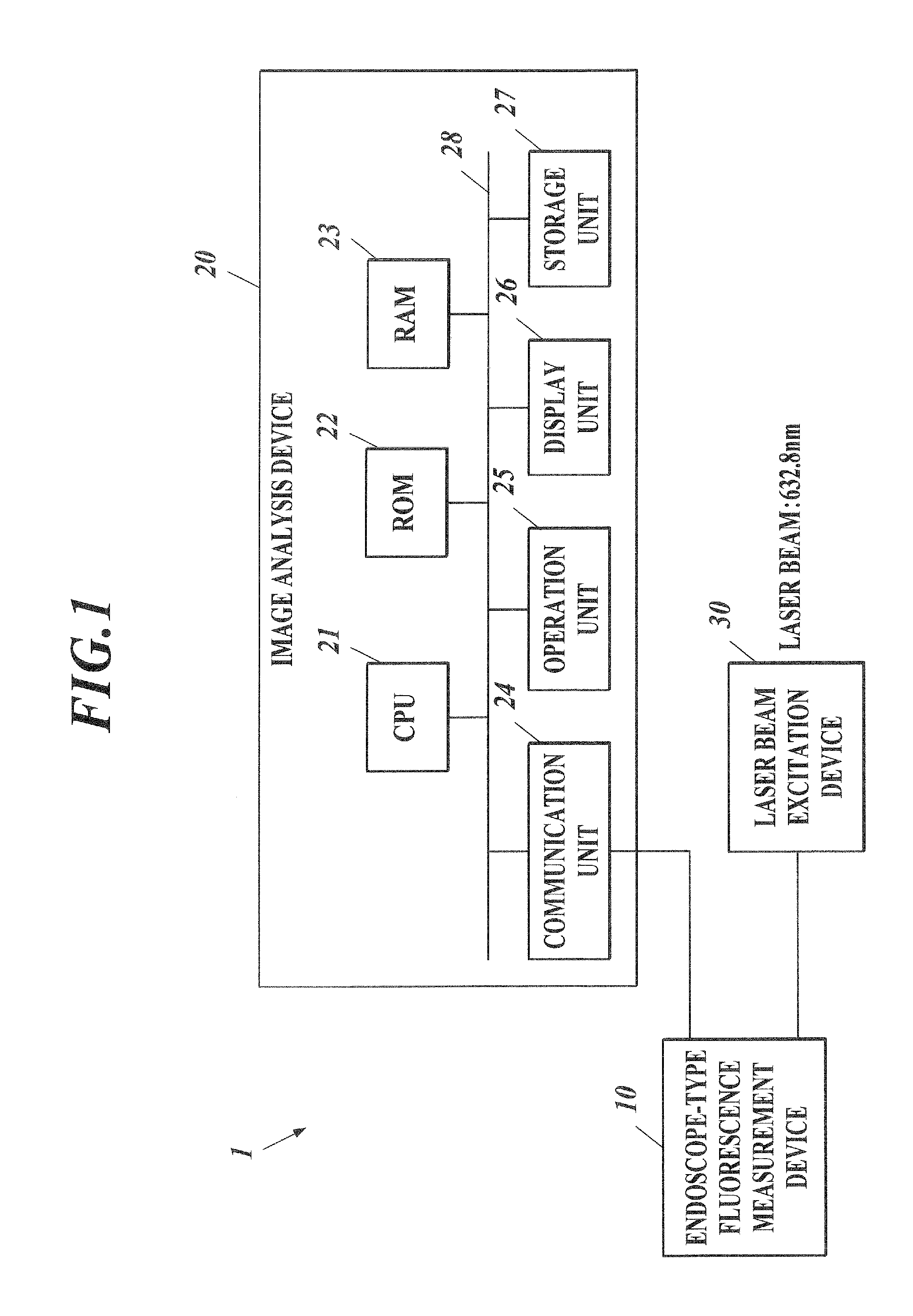
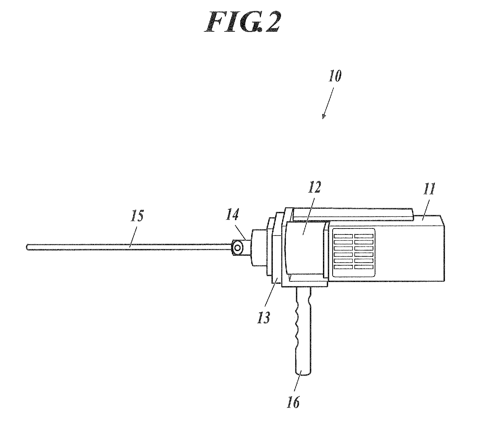
[0101]FIG. 1 shows a configuration of a fluorescence measurement system 1. As shown in FIG. 1, the fluorescence measurement system 1 includes: an endoscope-type fluorescence measurement device 10; an image analysis device 20; and a laser beam excitation device 30. The fluorescence measurement system 1 is a system that performs fluorescence analysis in a surgical field during surgery, and is used in the event of specifying a position of a sentinel lymph node from a living body into which quantum dots are injected as a tracer.

[0102]The quantum dots are clusters of a semiconductor with a diameter of 15 to 20 nanometers (nm), and are particles having characteristics to emit various types of fluorescence in response to a particle diameter thereof. Each of the quantum dots is composed of: a cor...


PUM

PropertyMeasurementUnit
particle diameteraaaaaaaaaa
diameteraaaaaaaaaa

Abstract

Regions where metastatic cancer cells can exist are detected with high accuracy in a sentinel lymph node. Quantum dots are injected into the vicinity of a cancer in a living body, thereby identifying the location of the sentinel lymph node by means of fluorescence. Subsequently, the sentinel lymph node is extracted. With respect to the sentinel lymph node extracted with quantum dots injected, structural analysis is conducted by means of precision fluorescence measurement which uses a confocal fluorescence microscope for monomolecular observation. Specifically, the fluorescence intensity is measured with respect to each of multiple areas in the sentinel lymph nodes, and out of the multiple areas measured, one or more areas are detected as afferent lymph vessel inflow regions in descending order of fluorescence intensity.

Description

TECHNICAL FIELD[0001]The present invention relates to a method for detecting afferent lymph vessel inflow regions and a method for identifying specific cells.BACKGROUND ART[0002]Heretofore, in an excision surgery of a cancer, in many cases, not only a lesion part but also a lymph node to which metastasis of the cancer is suspected has been excised. In recent years, a sentinel lymph node biopsy has been performed in order to minimize the excision of the lymph node.[0003]The sentinel lymph node (SLN) is defined as a lymph node that first receives lymph flows from the respective internal organs. In the cancer surgery, such a sentinel lymph node theory is widely accepted that a radical lymph node dissection is unnecessary unless metastasis of the cancer to the sentinel lymph node is present. The sentinel lymph node biopsy makes it possible to reduce a wasteful surgical invasion, and contributes to establishment of order-made medical care in which a surgical method is finely selected in ...

Claims


Application Information

Patent Timeline
Patent Type & Authority Applications(United States)
IPC IPC(8): G01N21/64B82Y5/00
CPCA61K49/0017A61K49/0067B82Y5/00B82Y15/00G01N21/6458G01N33/57492G01N33/588G01N2201/0221A61B5/0071A61B5/41Y10T436/13A61B5/418
Inventor HIKAGE, MAKOTOGONDA, KOHSUKETAKEDA, MOTOHIROKAMEI, TAKASHIOHUCHI, NORIAKIGOUDA, HIDEKINAKANO, YASUSHI
Owner KONICA MINOLTA INC
Learn More